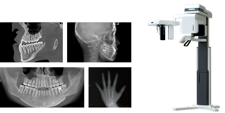
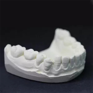
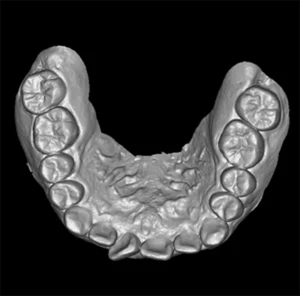
Option 1
Option 2
Option 3

12x10cm

8x8cm

5x8cm
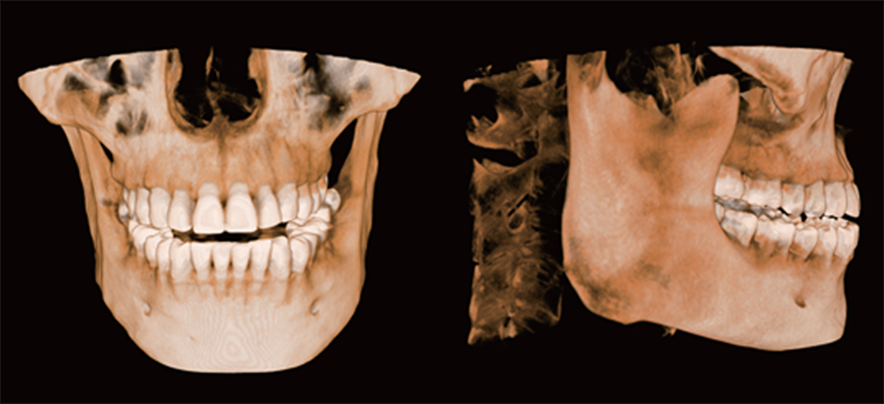
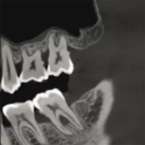
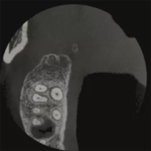
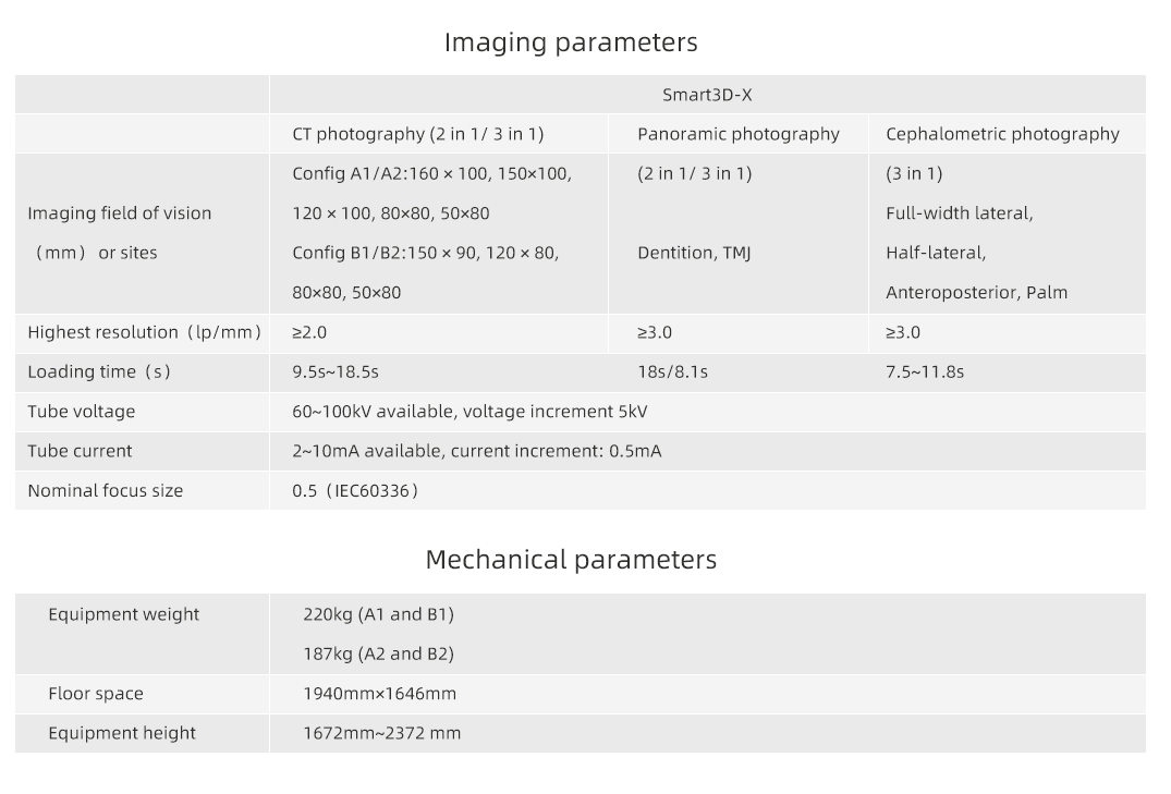
0.5mm small focus tube ensures outstanding image quality.
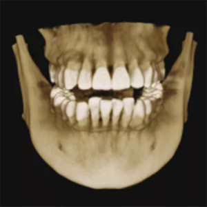
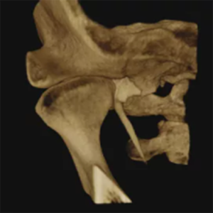
Resolution up to 2.0lp/mm, voxel size of 0.25~0.05 mm optional.
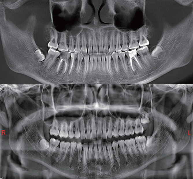

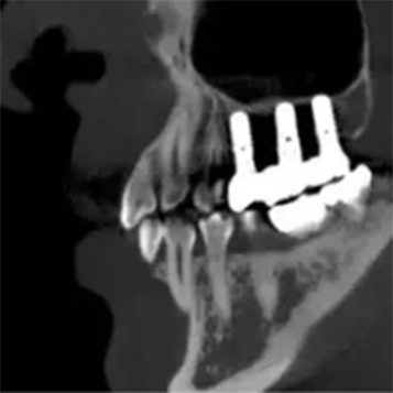
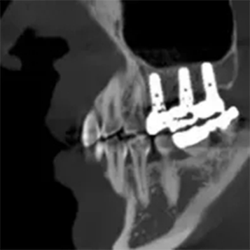

360°scan and 800 frame images with unique CT algorithms

QuartZ 4 scan platform, supporting flexible scan mode

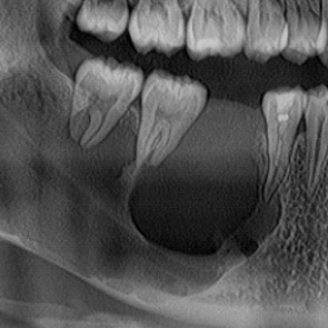
Multiple focus layers in panoramic imaging, fitting the patient's dental arch

Easy-to-target scan area

Six positioning lasers with face-to-face communication to posit precisely

X-type base is convenient for wheelchair-bound patients

10"LED touch screen

Storage box design

Voice reminder
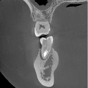
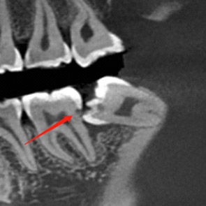
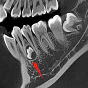
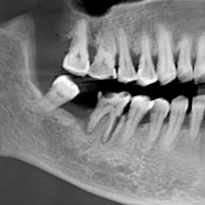
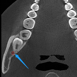
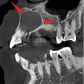
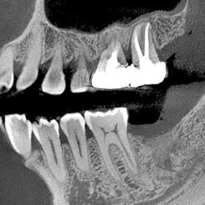
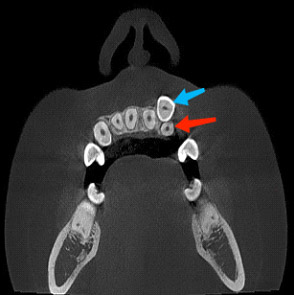
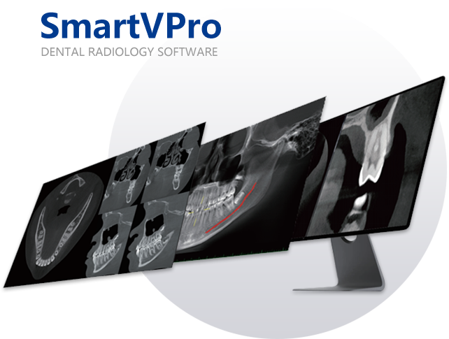
Axial, coronal and sagittal slices can be observed simultaneously. Besides, the slice in any direction is available. Buccolingual slices, distal and mesial sections were obtained to facilitate diagnosis.
Local fine reconstruction is conducted in the designated area.
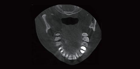
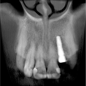
With the new T-MAR correction module for metal artifact removal, the system corrects metal artifacts intelligently. It avoids overmodification and saves the original clinical data.
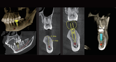
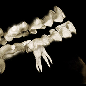
The bone and bone mass in the implant area will be evaluated by dental 3D images using HiRes3D. The neural tube will be highlighted automatically, which presents the relationship between the implant and the neural tube. This is a better way to approach a successful implant surgery.
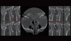
SmartVPro software has a visual pattern of comparing the left and right joints, allowing doctors to evaluate the diagnosis and treatment effect on temporomandibular joint diseases.
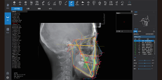
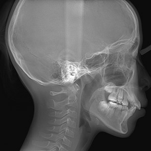
The neural network is trained by mega data, which automatically identifies orthodontic anatomical landmark points, draws anatomical structures and outputs measurement reports according to the selected measurement methods.
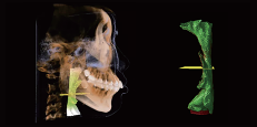
The airway is segmented automatically, which calculates the volume and the narrowest area of the airway.
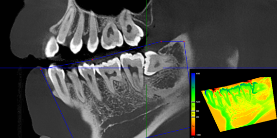
Used to assess bone mineral density in selected areas.
| Vendor | Medist |
|---|---|
| Type | Collection name |
| Color | Aliceblue, Antiquewhite, Azure |
| Weight | 5.52kg |
Size: 1
Digital BP apparatus with a stand for stable and precise measurementsReliable equipment for accurate blood pressure ....
Size: 1
1: Hydro-dermabrasion, applicable to normal or sensitive skin, Or skin with whelk, comedones, acne,etc.2: Cleaning ....
Size: 1
MeasurementsItem Weight200 GramsItem Dimensions14.5 x 9 x 4.5 centimetersNumber of Items5Features & SpecsFlavorLemon ....
Size: 1
Detoxifying agent Pharmachologic effect: Glutathione is a linear tripeptide with a sulfhydryl group, which includes L-gl ....
Size: 1
Technical Specification (1)Different applications based on Digital Pre Programs (2)A current module of 4-channel E ....
Size: 1
Effective smoke evacuation is crucial for maintaining a clear surgical field, minimizing odors, and reducing exposure to ....
Size: 1
The base is stable and not easy to tilt, ensuring your safety, ideal for the elderly, leg injury use, men, women.
Size: 1pcs
The goggles are sealed within clear plastic packaging, indicating they are new or unused. There are also elements o ....
Size: 1pcs
These kits are commonly used in healthcare settings for wound care and dressing changes, providing the necessary tools i ....
Size: 1pcs
Disposable hair nets are lightweight, single-use coverings designed to contain hair in various professional and hyg ....


Lorem ipsum dolor sit amet, consectetur adipiscing elit, sed do eiusmod tempor incididunt ut labore et dolore magna aliqua. Ut enim ad minim veniam.
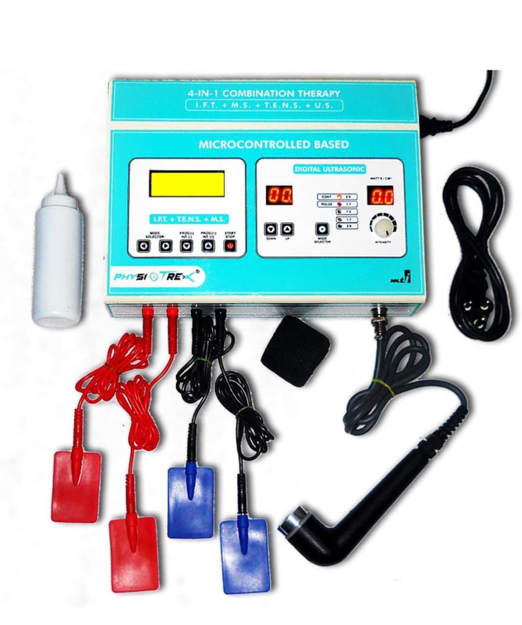 IFT WITH ULTRASOUND
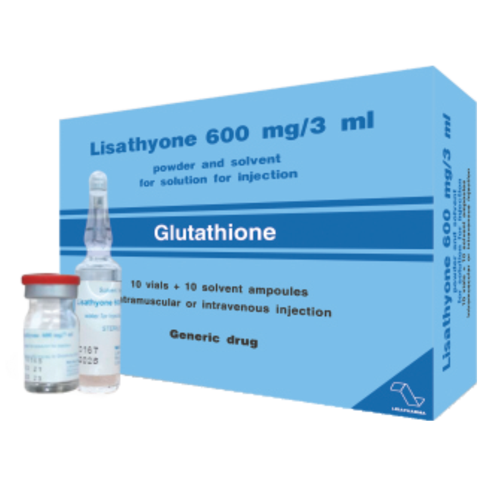 Lisathyone 600mg | DHA
 LEMONBOTTLE Ampoule Solution
 Hydra Dermabrasion Aqua Peeling Beauty Device - Scalp Cleanser
 Riester Digital BP Apparatus
.png) iCare HOME2 Tonometer
iCare HOME2 Tonometer
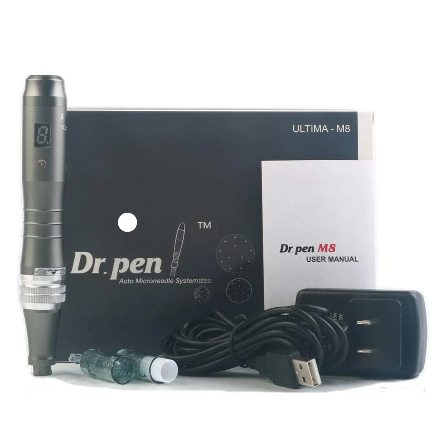 Dr. pen Ultima M8 Wireless/Wired Professional Derma Pen Electric Skin Care Set Microneedle Therapy System High Quality Beauty Machine
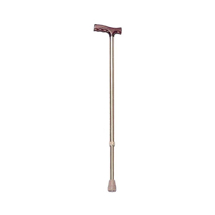 Walking Stick
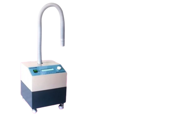 Smoke Evacuator
 Glutathione Injection Glutax 2000000gx 180W Whitening Products Injection
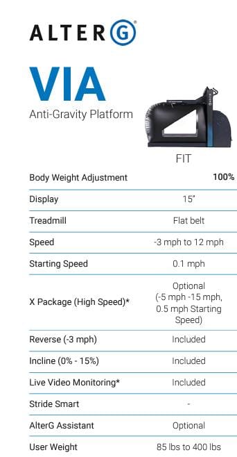 ALTER G FIT ANTIGRAVITY TREADMILL
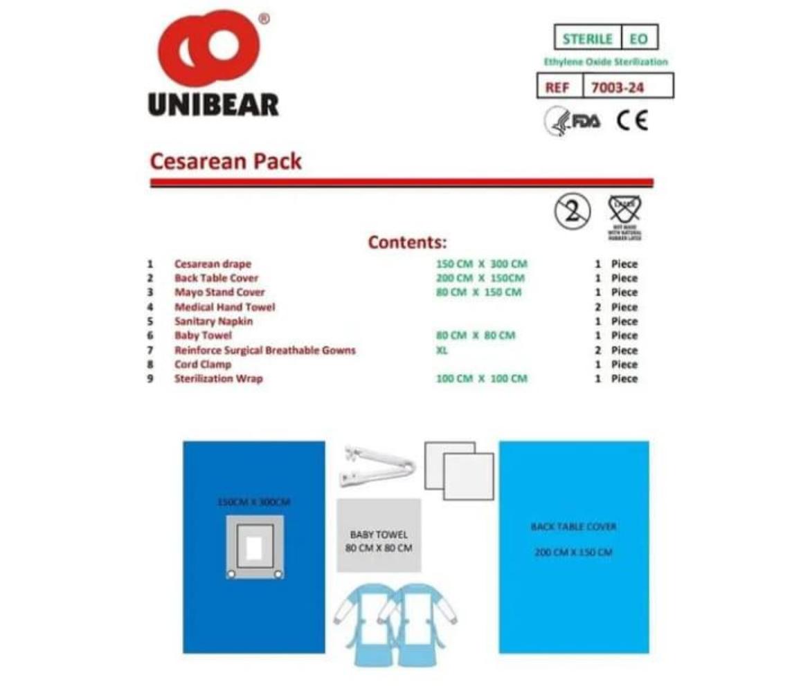 CESAREAN PACK
.jpeg) AED MACHINE - LIFELINE Defibtech US
AED MACHINE - LIFELINE Defibtech US
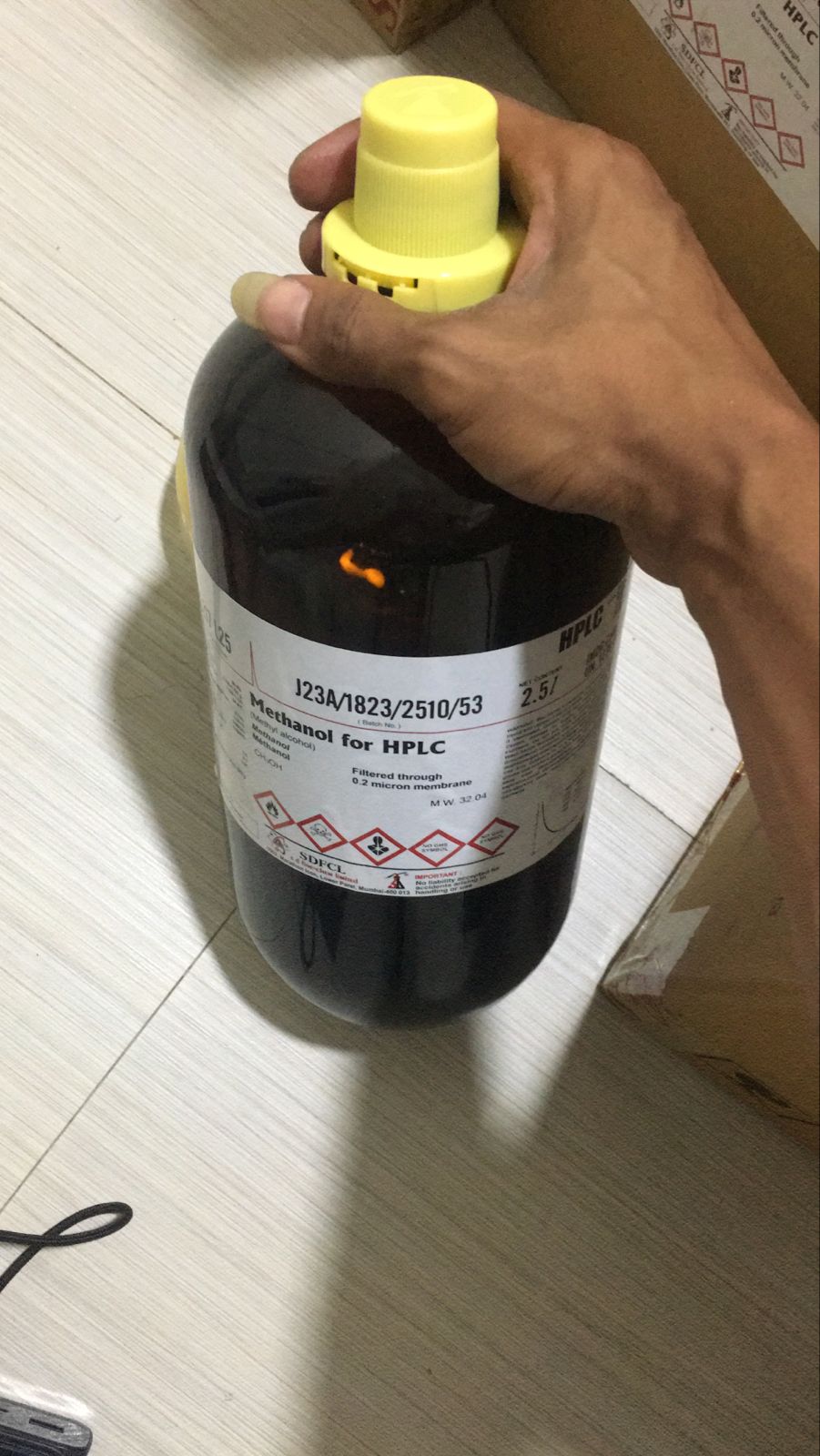 METHANOL FOR HPLC
 POTASSIUM SODIUM (+)-TARTRATE UR"UNIVERSAL REAGENT
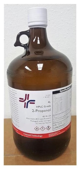 PROPAN-2-OL AR, ACS (2.5 LITRE)PB
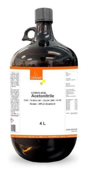 ACETONITRILE FOR HPLC & SPECTROSCOPY
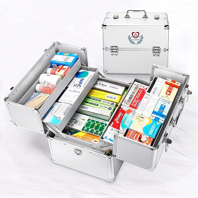 Medicine Storage Box
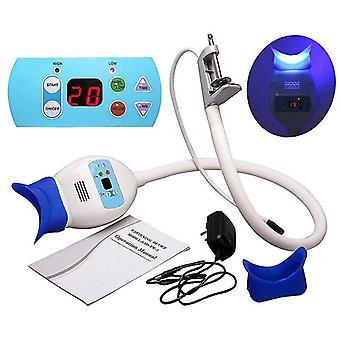 Teeth Whitening Lamp/ Whitening Machine
.webp) Dental Mobile Teeth Whitening Lamp
Dental Mobile Teeth Whitening Lamp
.webp) LCD Mercury Free Sphygmomanometer
LCD Mercury Free Sphygmomanometer
.png) Medical compressor nebulizer atomizer
Medical compressor nebulizer atomizer
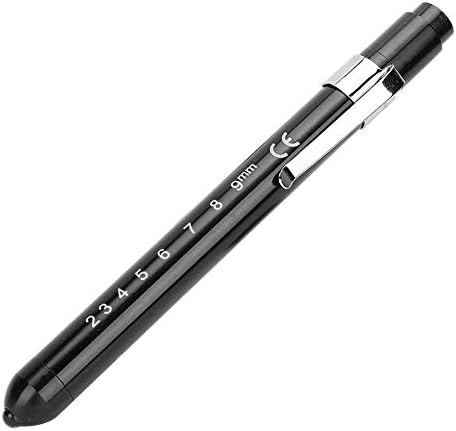 PENLIGHT LED
.jpeg) Sanakin therapy kit
Sanakin therapy kit
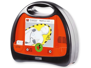 PRIMEDIC HEART SAVE AED - Defibrillator with lithium battery
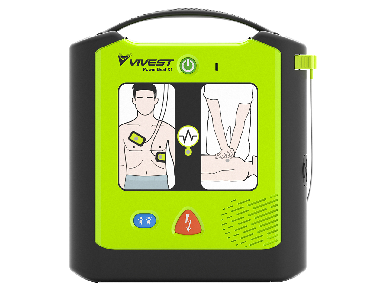 PowerBeat X1 AED - UK
.jpeg) Positive airway pressure device CPAP/ Auto CPAP
Positive airway pressure device CPAP/ Auto CPAP
.jpeg) EMPTY FIRST AID BOX
EMPTY FIRST AID BOX
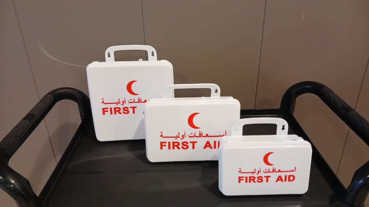 EMPTY FIRST AID BOX
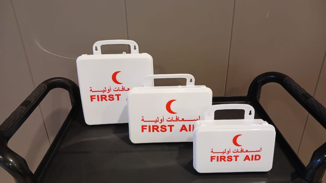 EMPTY FIRST AID BOX
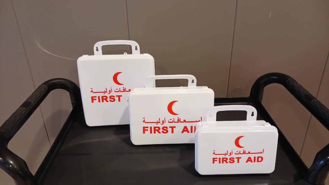 EMPTY FIRST AID BOX
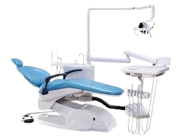 DENTAL CHAIR Model:WL-A1000
.jpeg) DENTAL CHAIR Model:WL-A3000 (cart)- Premium quality
DENTAL CHAIR Model:WL-A3000 (cart)- Premium quality
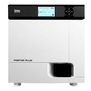 Fomos Autoclave - 22 ltr
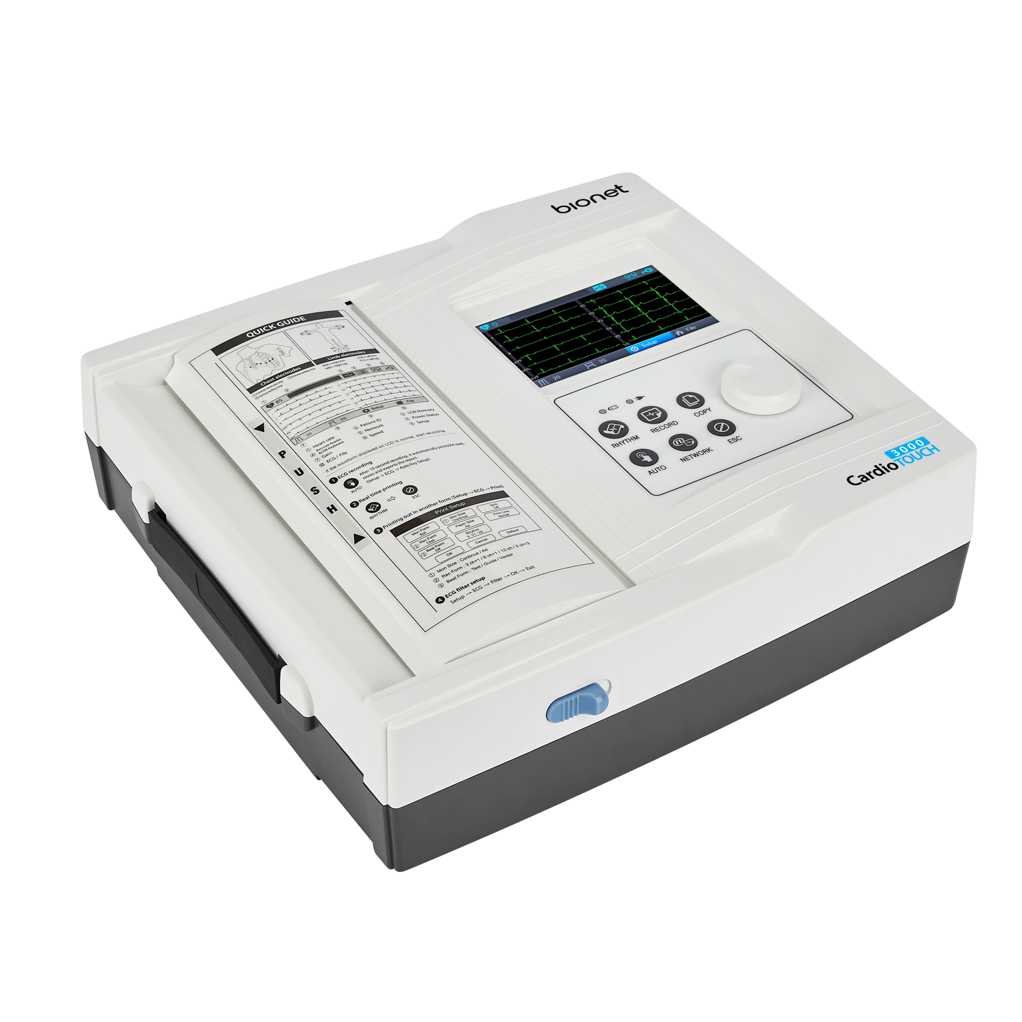 Bionet CardioTouch 3000 12 Channel ECG Machine
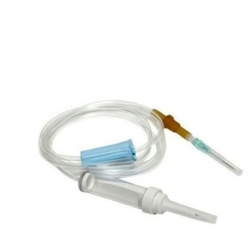 INFUSION SET
 Boost Oxygen Think Tank Large 10 L
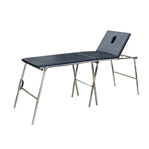 Foldable Examination Couch
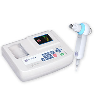 Helios 702 Portable Spirometer - INDIA
 ICE GEL (Physiotherapy)
 Ultrasound Gel
.jpeg) Portable Ultrasonic Bone Mineral Density Detector Bmd-A3
Portable Ultrasonic Bone Mineral Density Detector Bmd-A3
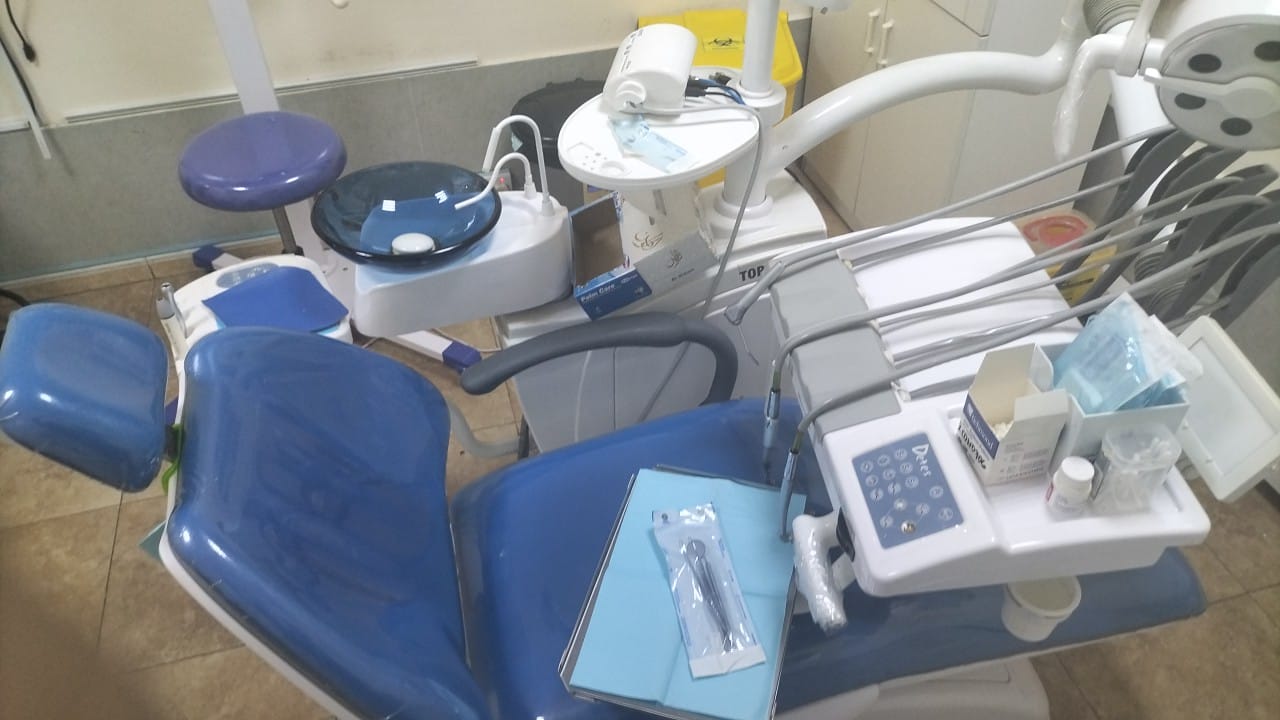 DENTAL CHAIR - USED
.jpeg) ECG 12 CHANNEL - MEDEXBIO
ECG 12 CHANNEL - MEDEXBIO
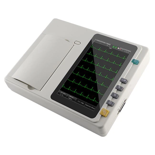 ECG MACHINE 6 CHANNEL - MEDEXBIO
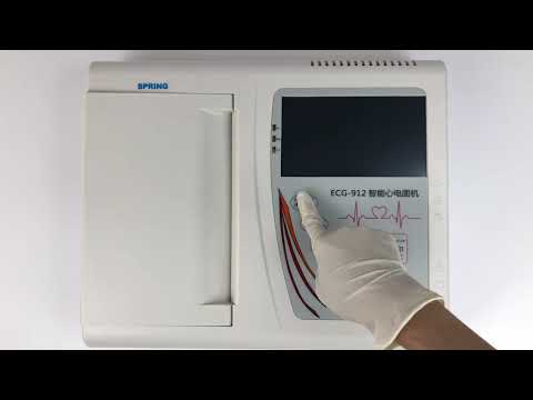 3 Channel ECG - MEDEXBIO
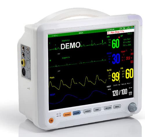 Patient Monitor - medexbio
.jpg) ENDO MOTOR & APEX LOCATOR - WOODPECKER
ENDO MOTOR & APEX LOCATOR - WOODPECKER
 EndoArt Action Gold Rotary Files SX to F3 - 31mm
 Endo Motor REBORN ENDO
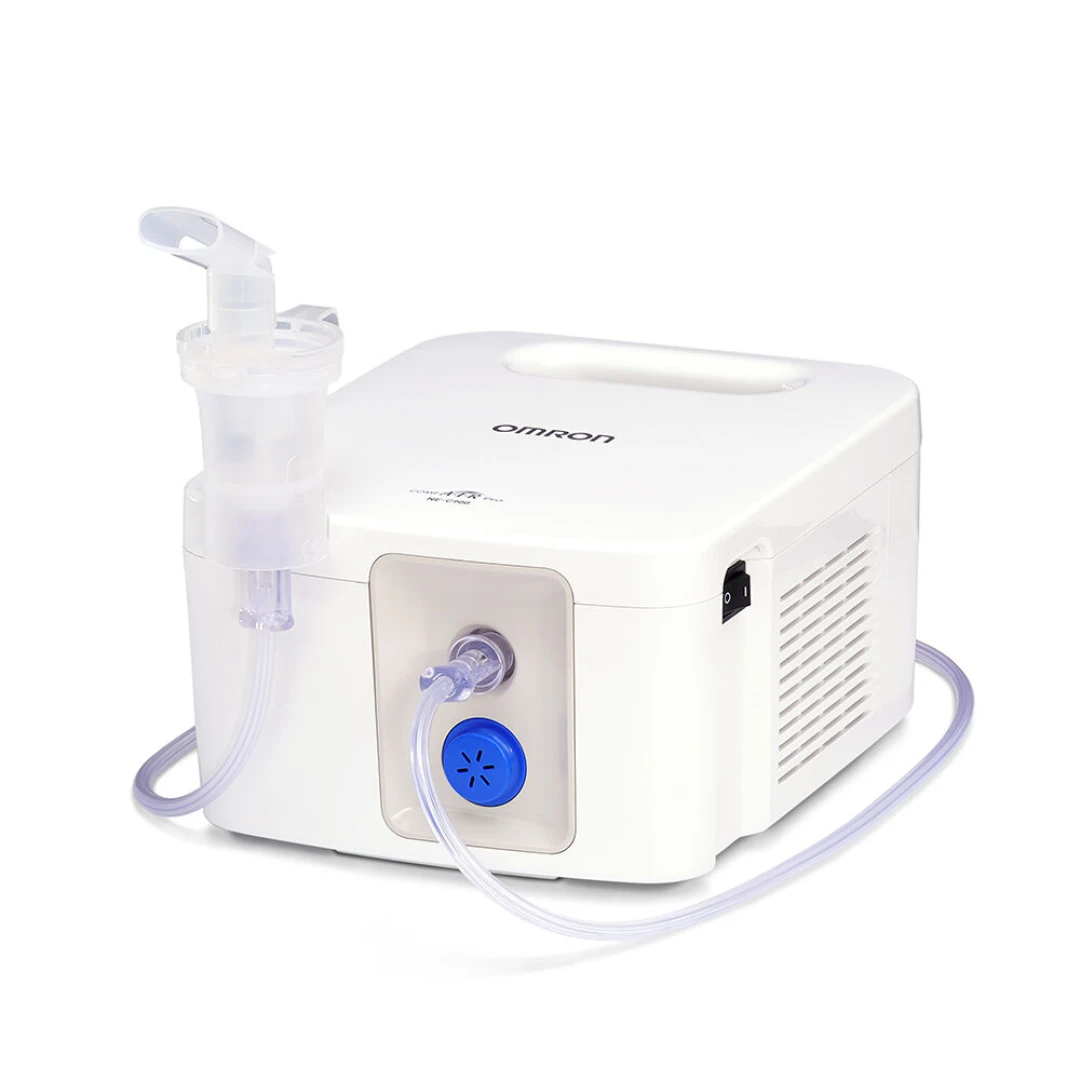 OMRON - NEBULIZER
.jpeg) DM500 Binocular Microscope
DM500 Binocular Microscope
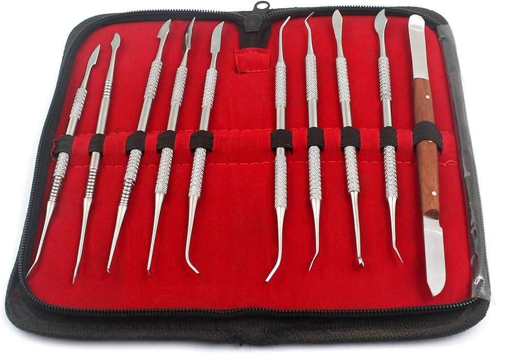 Dental Dentist Sculpture KIT 'Wax Carver Set,10 PCS/Set
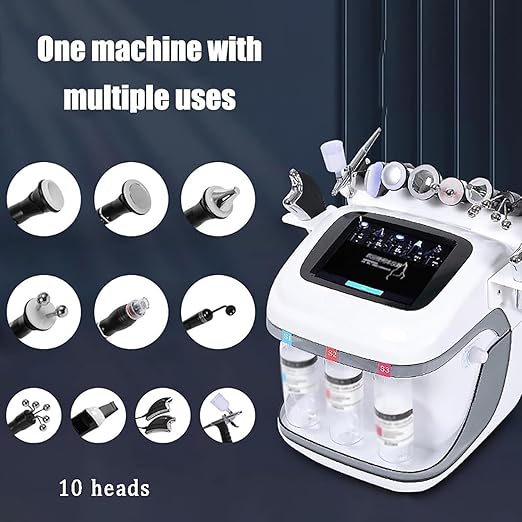 10 IN 1 DERMABRASION H2O2 HYDRAFACIAL BEAUTY MACHINE
 NE-C900 OMRON NEBULISER
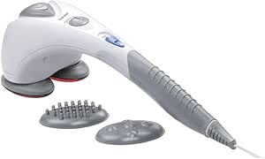 Beurer MG 80 Infrared Massager
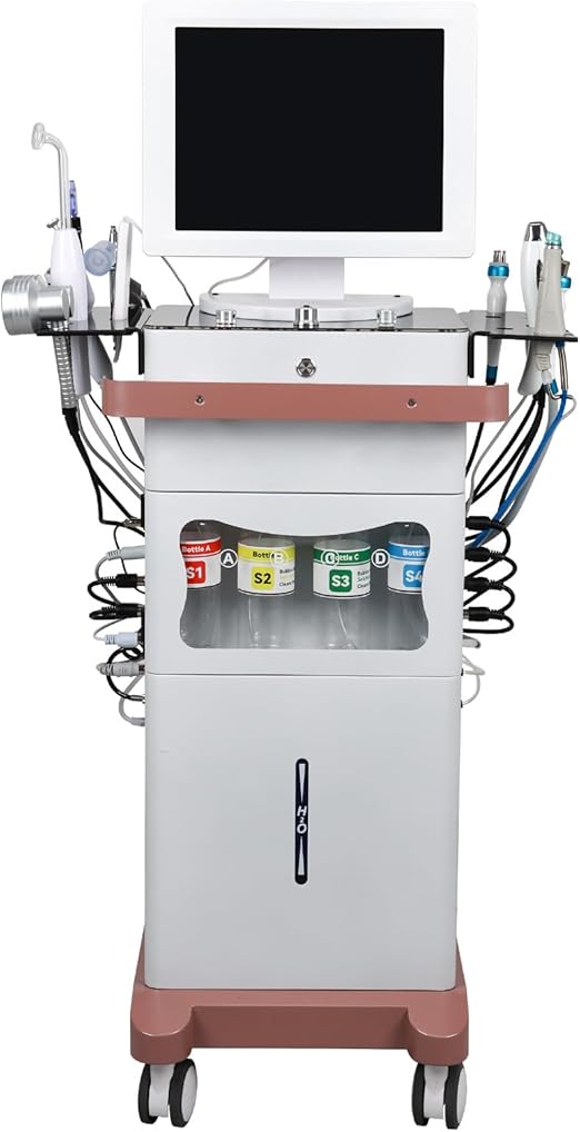 Hydrafacial Machine 15in1 Premium Beauty Multifunctional Hydrogen Oxygen Face Care Device
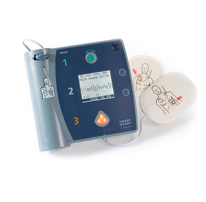 Philips HeartStart FR2 Plus AED
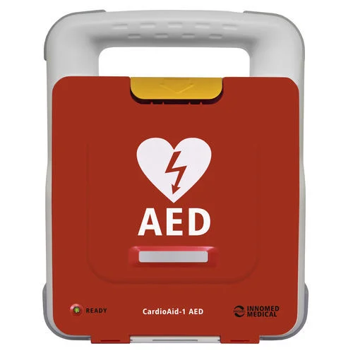 CardioAid-1 AED MACHINE - HUNGARY
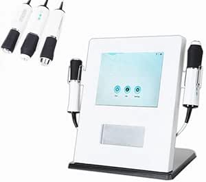 Pollogen 3 in 1 oxygeneo facial machine
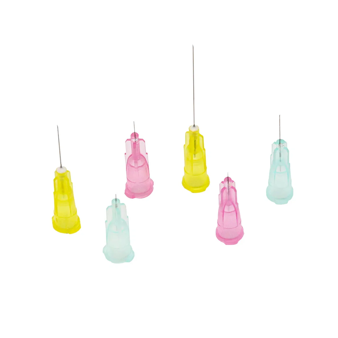 DermaTech (Korean) Meso Needles – Pack of 100
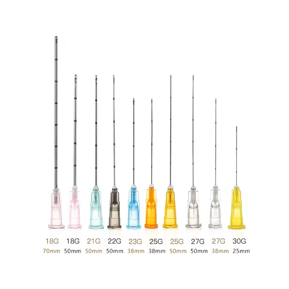 DermaTech Derma Cannula – Pack of 25 KOREAN
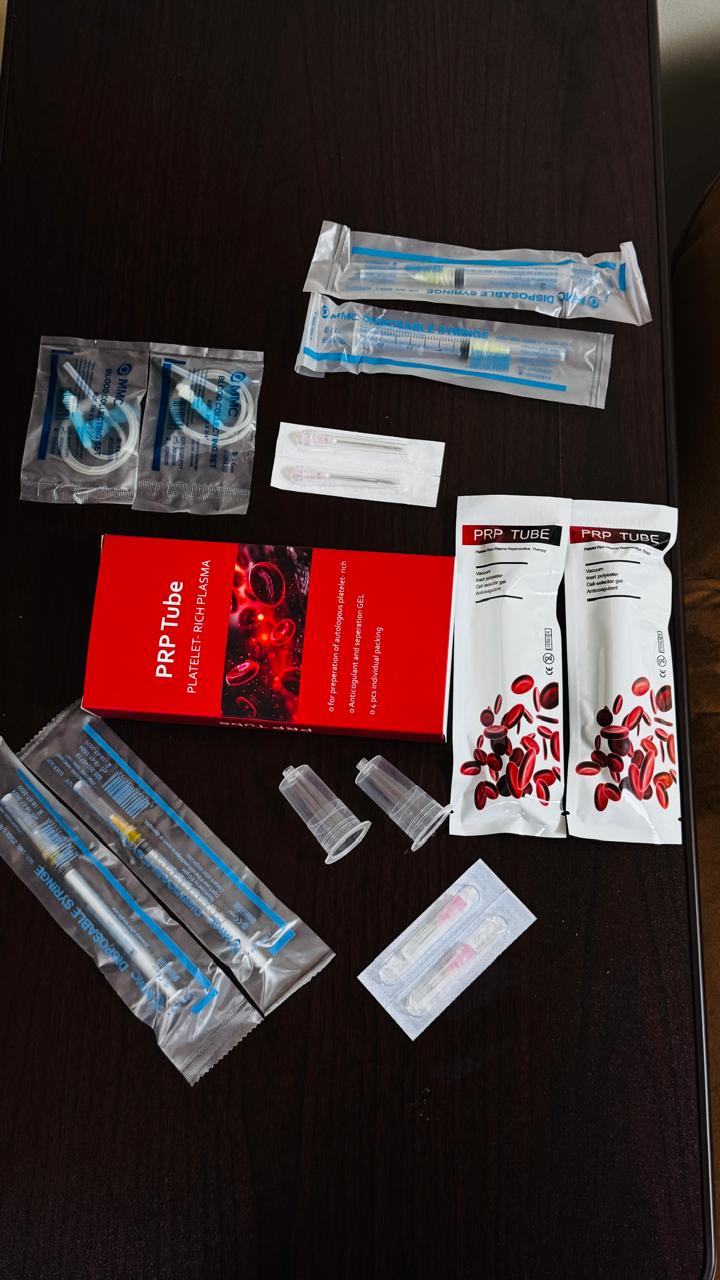 PRP KIT
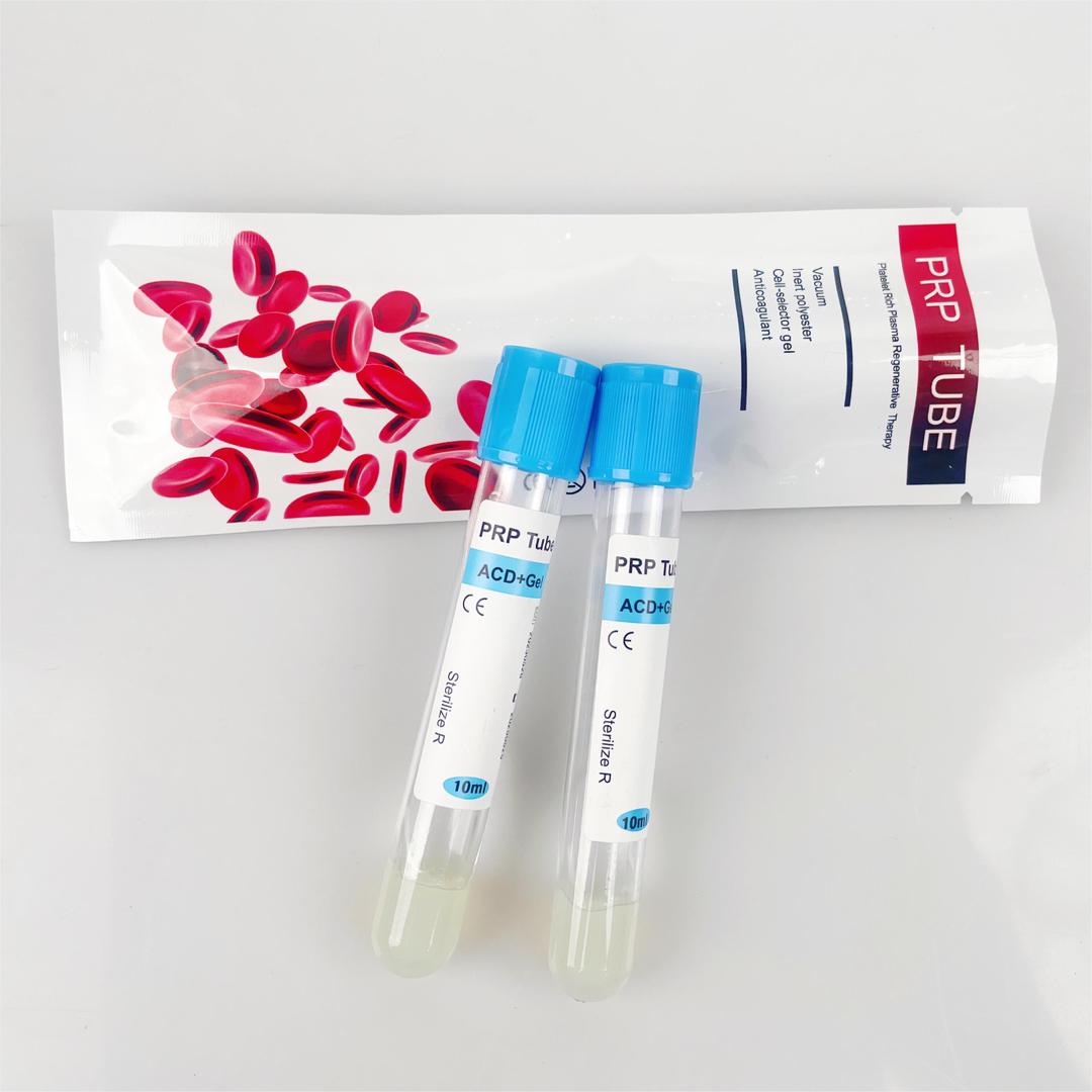 PRP TUBE
 1 BoX Aqua Skin Pure Gold Anti Aging Whitening Serum
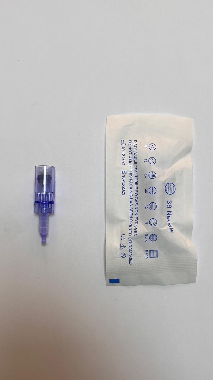 DERMA PEN CARTRIDGE
.webp) INSULIN PUMP
INSULIN PUMP
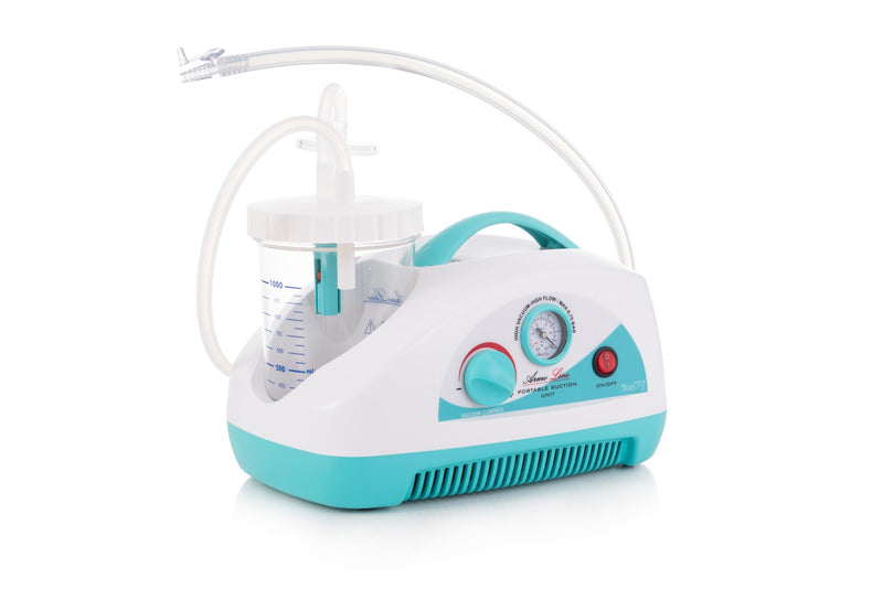 AL-03 ArmoLine Portable Suction Unit 30 L / min Flow Rate
 Nebulizer – Armoline – AL-50
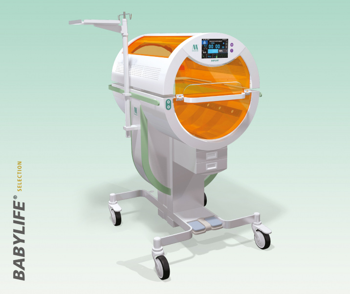 MEDICOR® BabyLife KLA-145LT
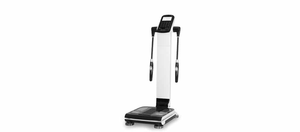 I20 Body Composition Analyzer
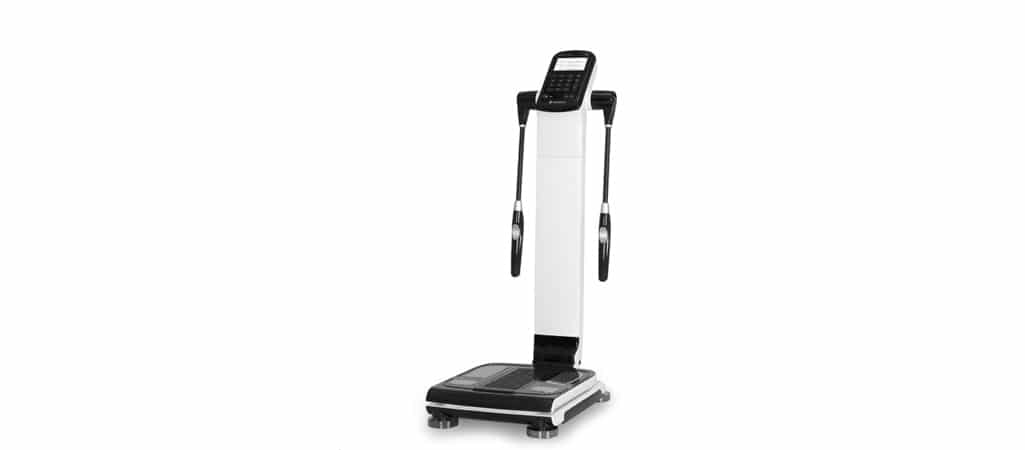 I20 Body Composition Analyzer
.jpeg) Aqua Solution S1
Aqua Solution S1
.jpeg) Respiratory Exerciser spirometer
Respiratory Exerciser spirometer
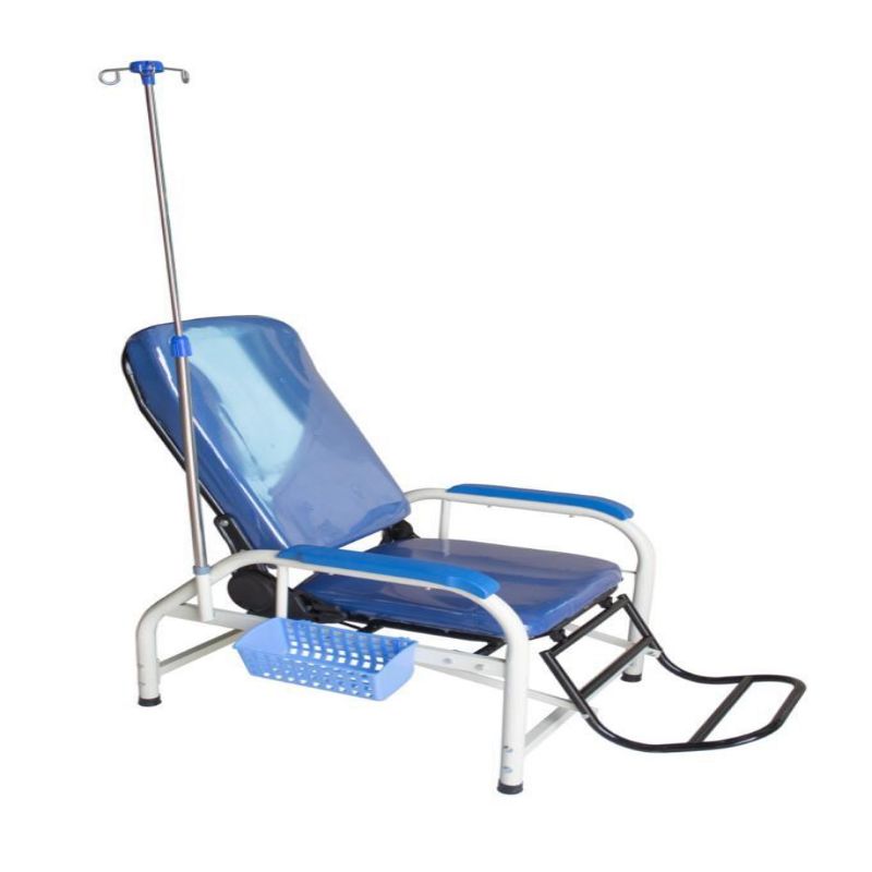 INFUSION CHAIR
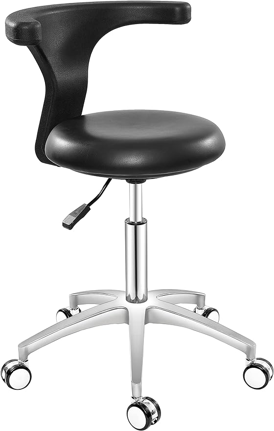 Doctor backrest stool
 EXAMINATION COUCH
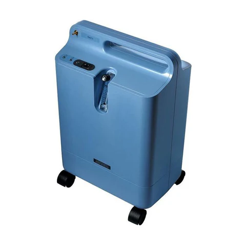 Oxygen Concentrator 5L - BARELY USED
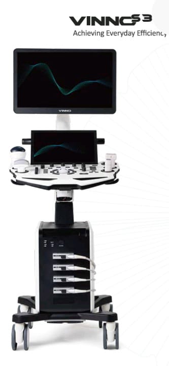 VINNO S300
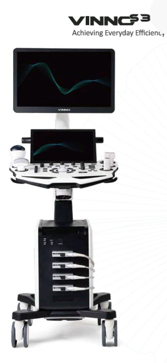 VINNO S300
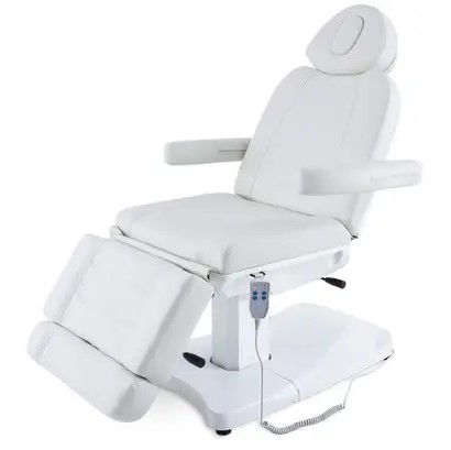 DERMA CHAIR 2 MOTOR
.jpeg) Chemistry Analyzer BS-240Pro Mindray
Chemistry Analyzer BS-240Pro Mindray
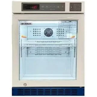 Laboratory Refrigerator 68L – BIOBASE
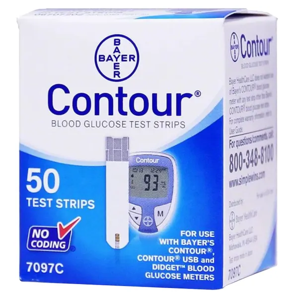 contour glucometer Strips
 DENTAL X RAY - MOBILE - MYRAY ITALIAN
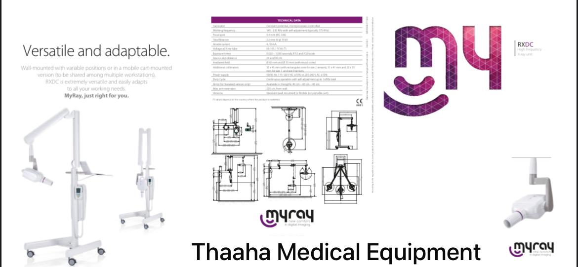 MOBILE DENTAL XRAY
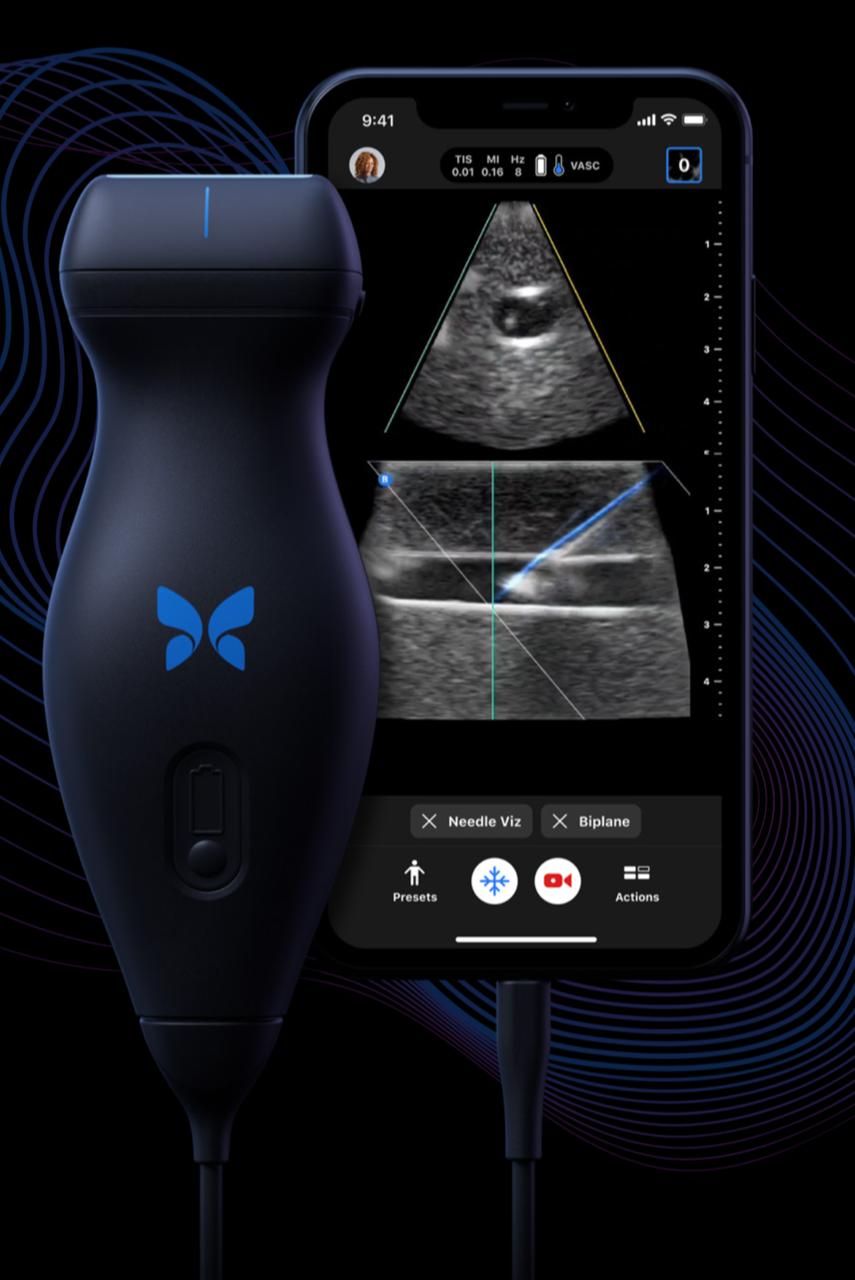 PORTABLE ULTRASOUND SYSTEM
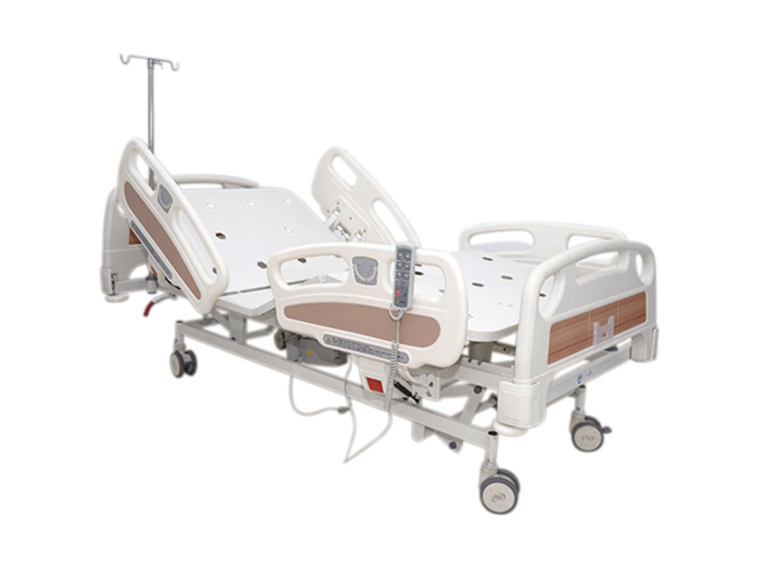 5 FUNCTION BED
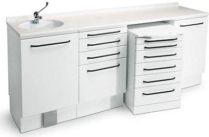 DENTAL CABINET
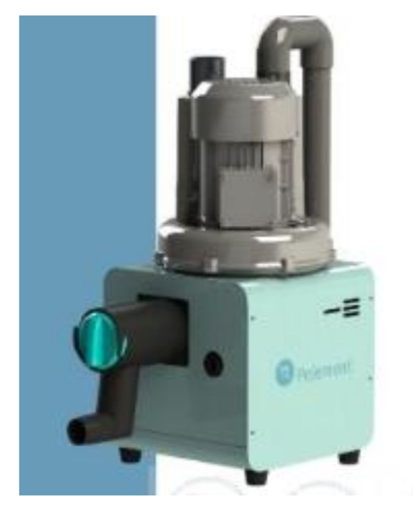 DENTAL SUCTION
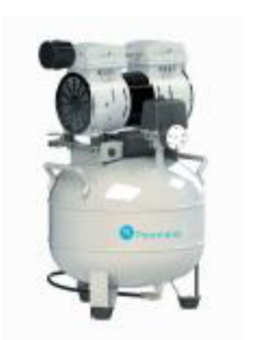 STORM OILLESS COMPRESSOR- PELEMENT
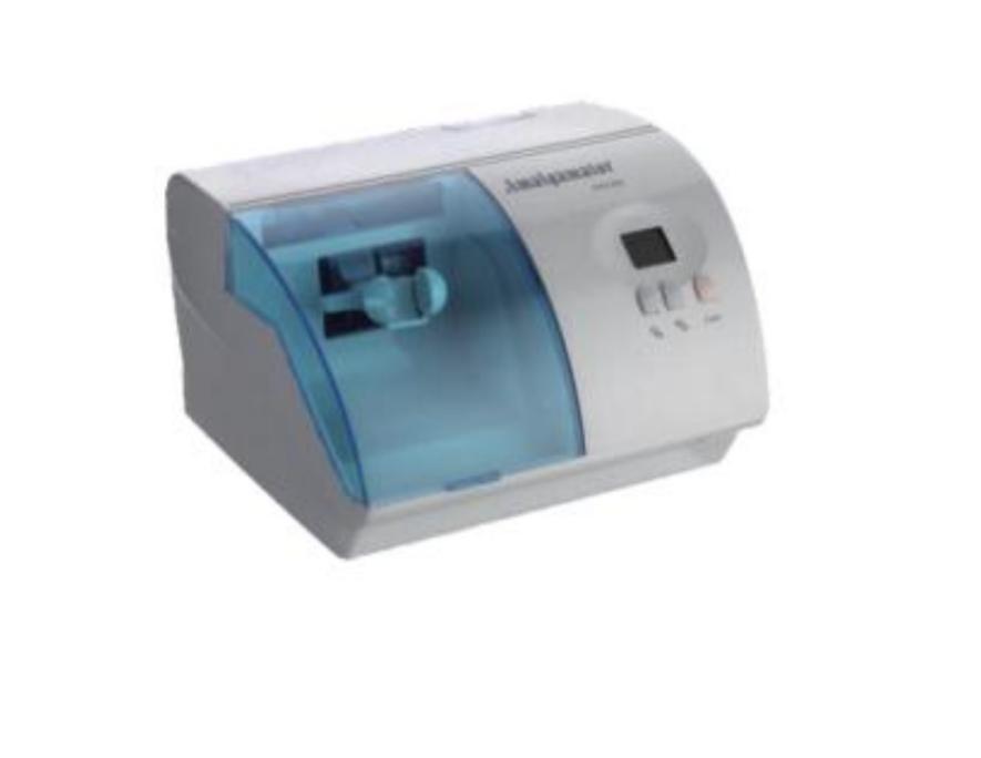 AMALGAMATOR
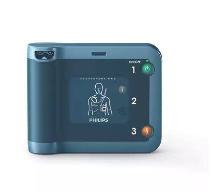 PHILIPS HEARTSTAT Frx
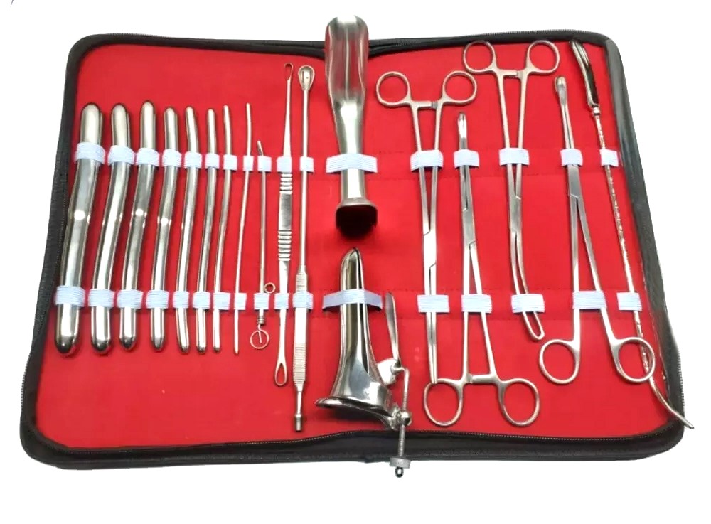 Gynecological Exam Kit
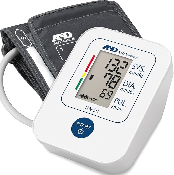 DIGITAL BP - AND MEDICAL
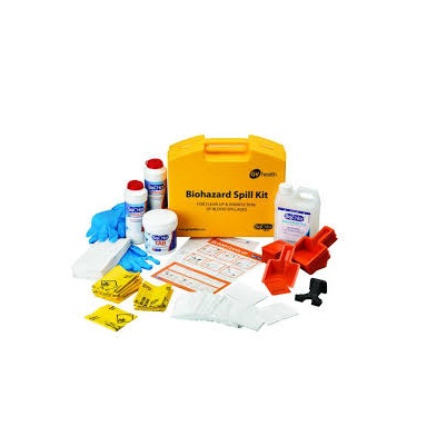 Biohazard Spill Kit
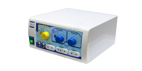 SSE 400 PLUS Cautery Electrosurgical Generator Unit Diathermy Machine ESU Set
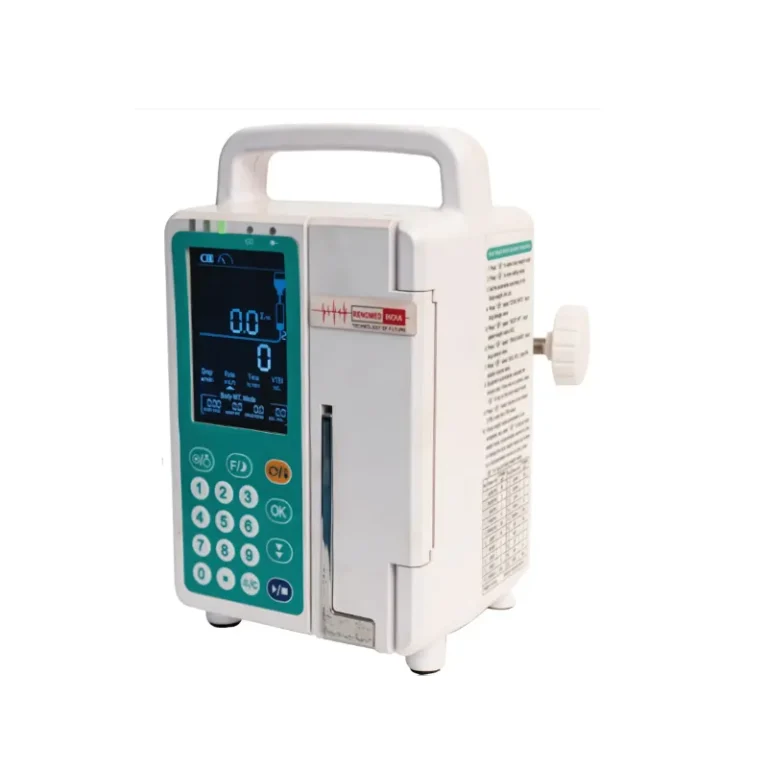 INFUSION PUMP
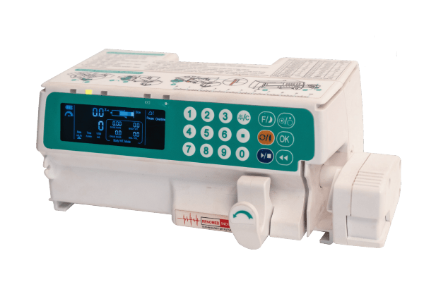 SYRINGE PUMP
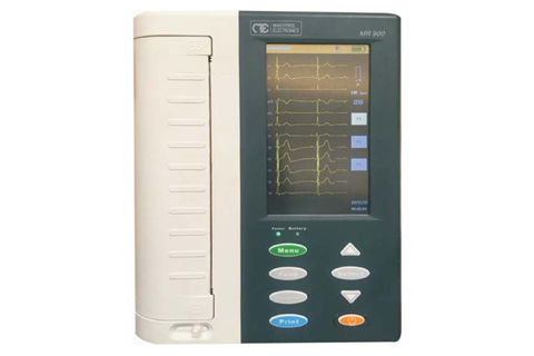 ECG MACHINE 12 CHANNNEL - INDIAN BRAND
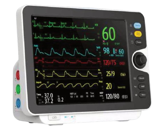 PATIENT MONITOR 5 PARMETERS
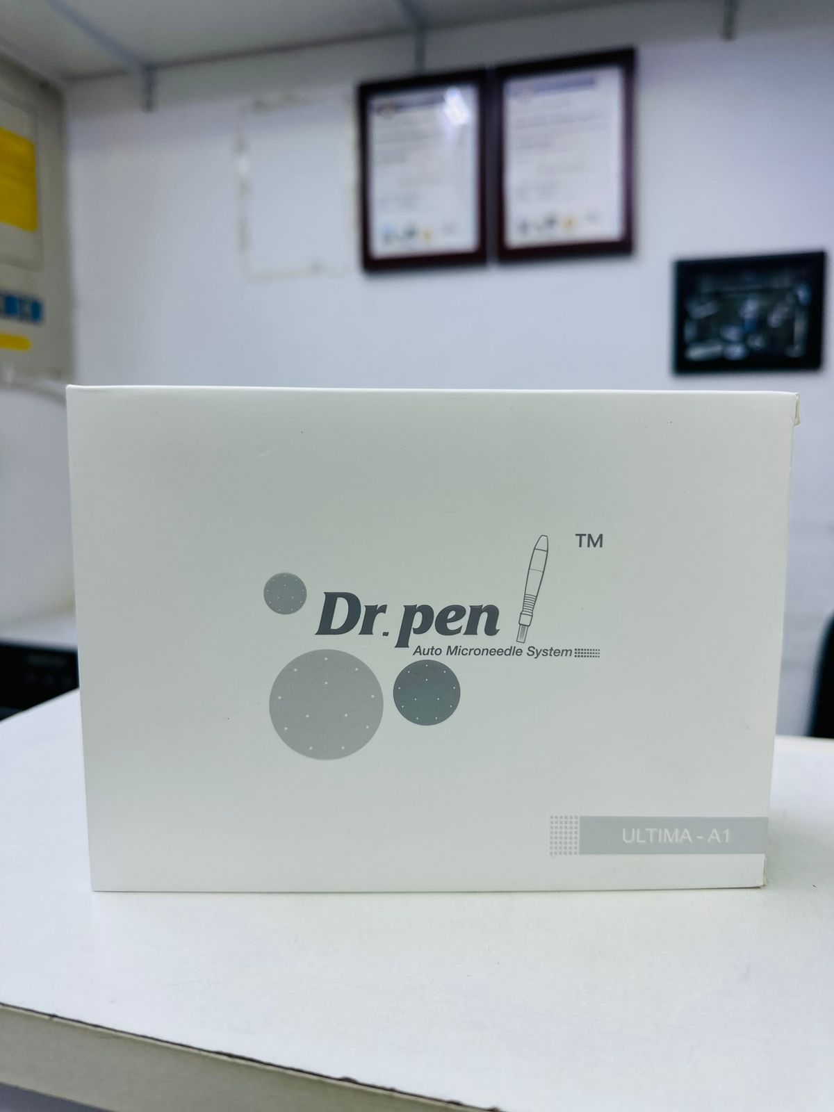 Dr pen
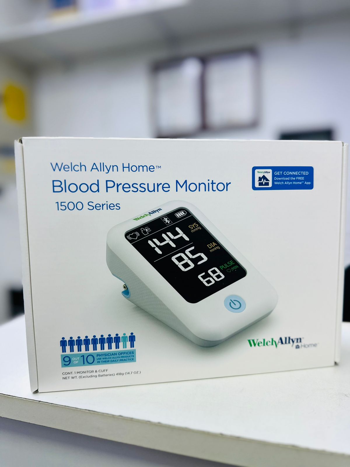 Welch Allyn BP
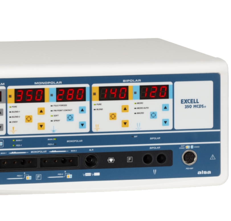 DIATHERMY UNIT
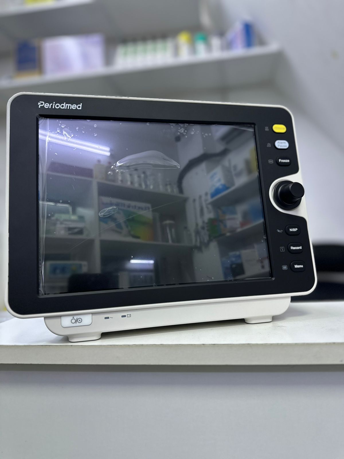 PATIENT MONITOR
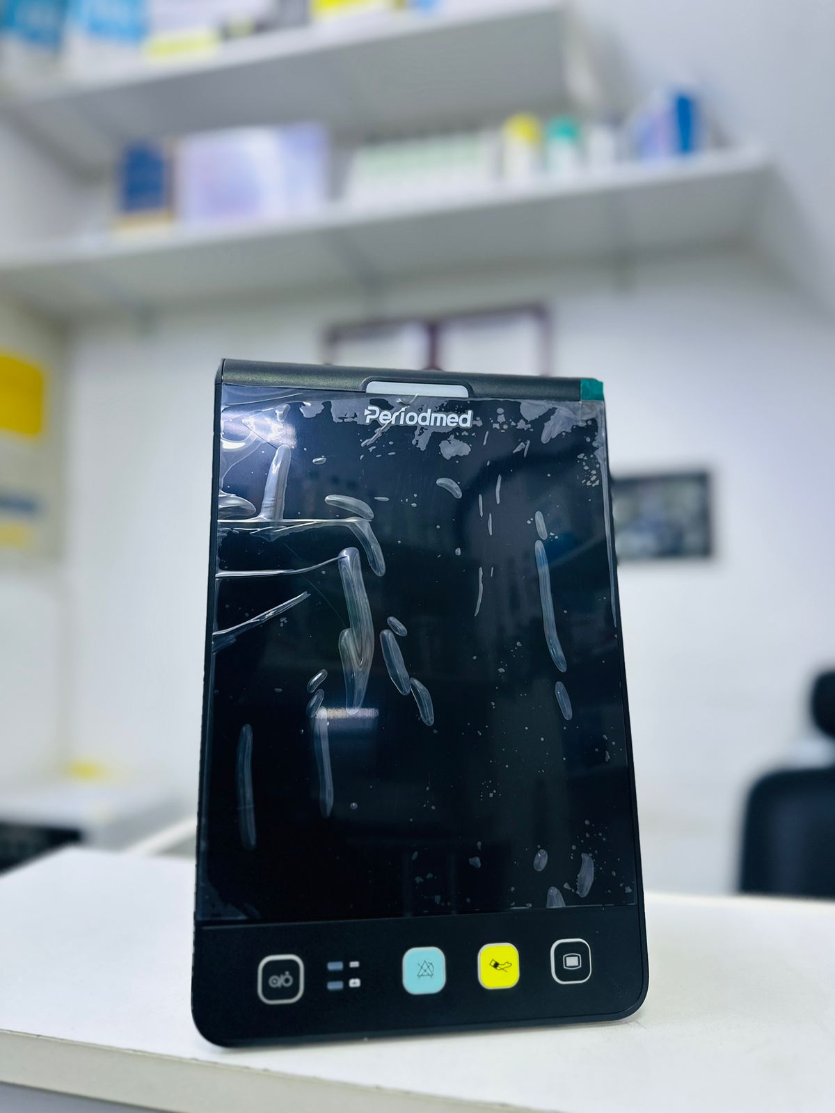 VITAL SIGN MONITOR
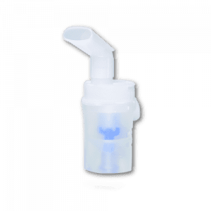 VVT KIT
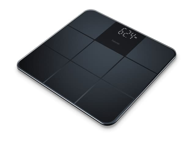 WEIGHING SCALE
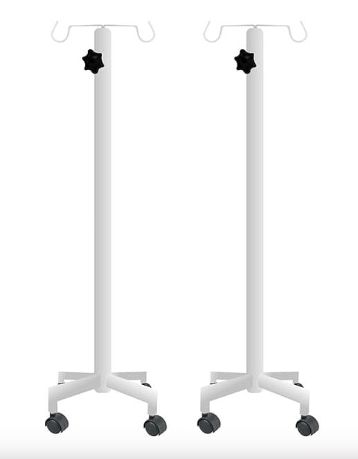 IV STAND
.png) Biofresh hand disinfectant gel 500ml
Biofresh hand disinfectant gel 500ml
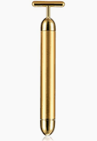 Energy beauty bar
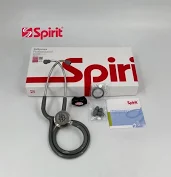 Spirit Stethoscope
 MICROTOME BLADE
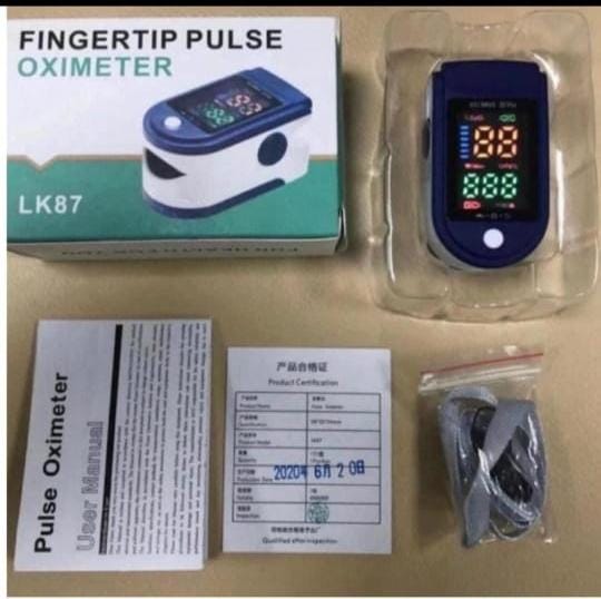 PULSEOXIMETER
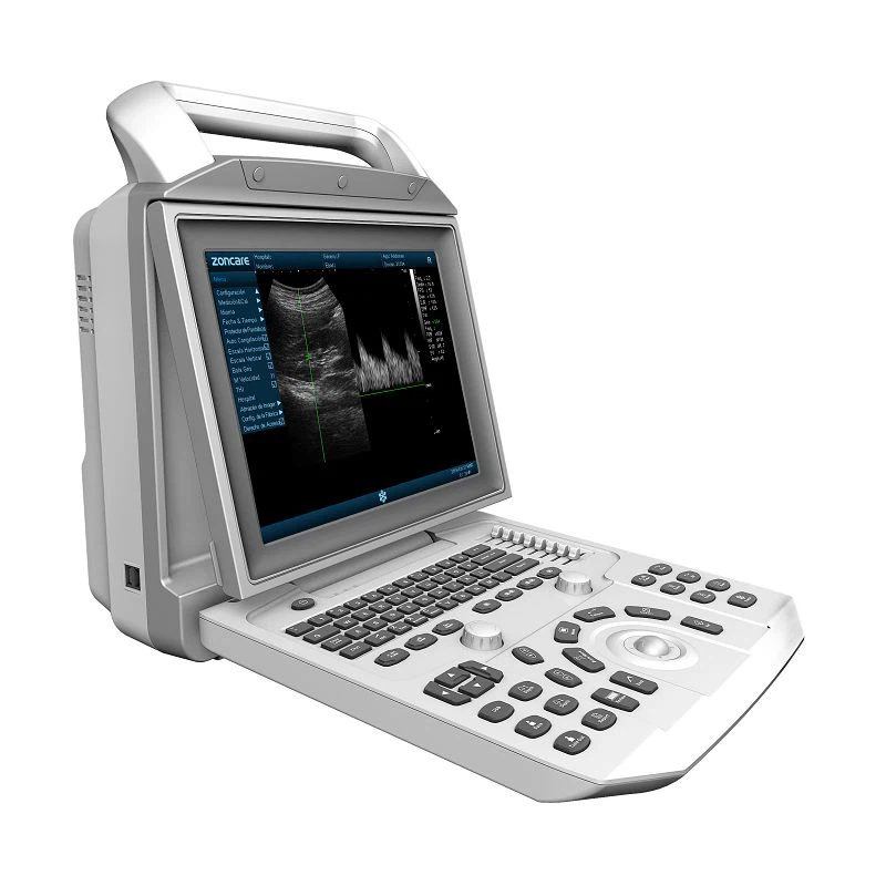 ULTRASOUND MACHINE I50 - ZONCARE BLACK AND WHITE
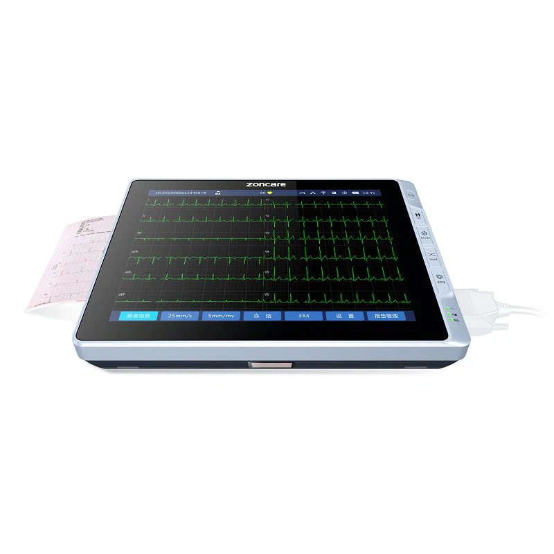 Digital 6 Channel ECG Machine
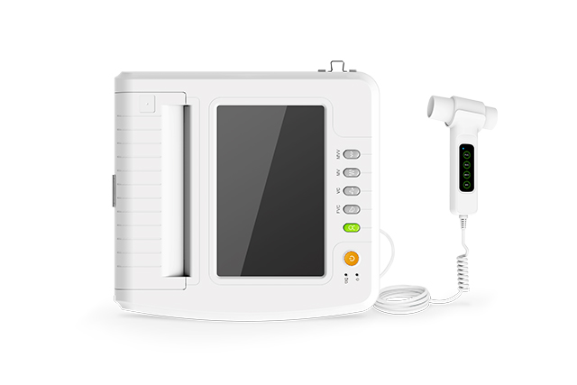 SP100B SPIROMETER
.jpeg) CMS800G Fetal Monitor
CMS800G Fetal Monitor
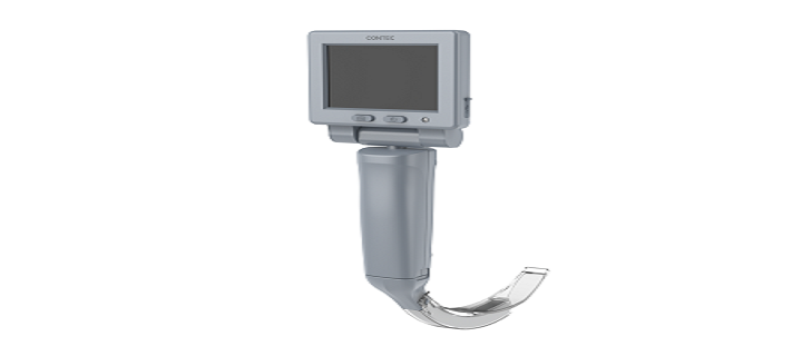 CMS-GS2 Video Laryngoscope
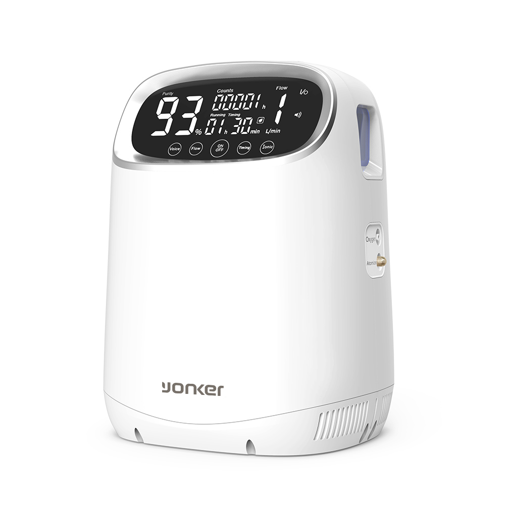 Oxygen Concentrator 3L
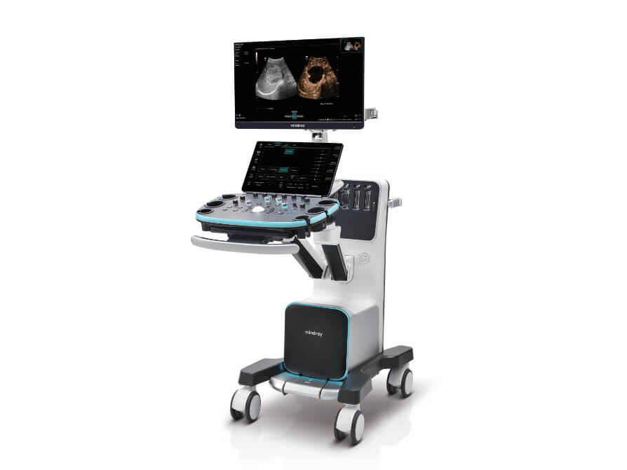 Mindray Resona I9 - 4 Probes
 Couch Roll
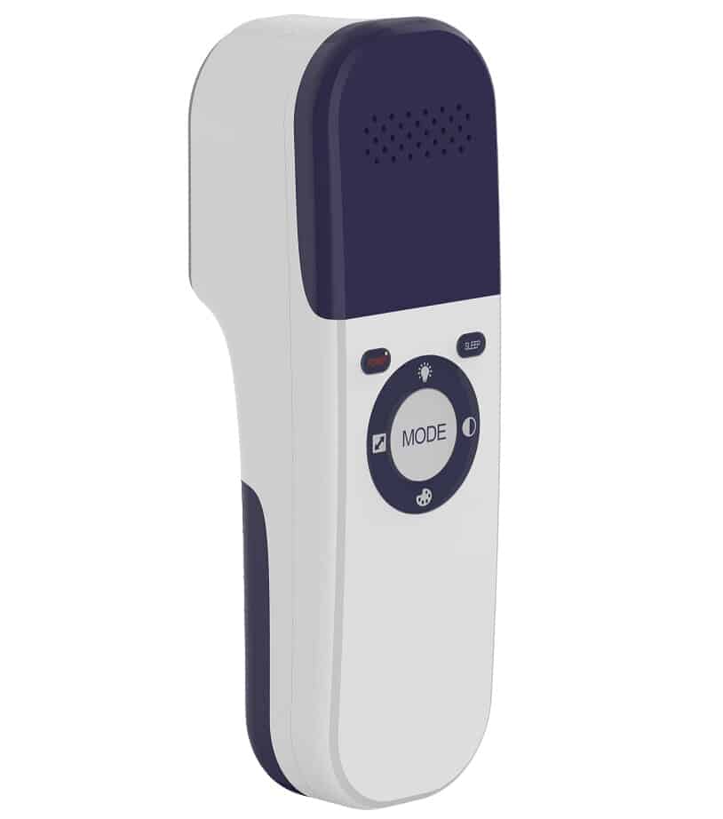 VEINFINDER 500
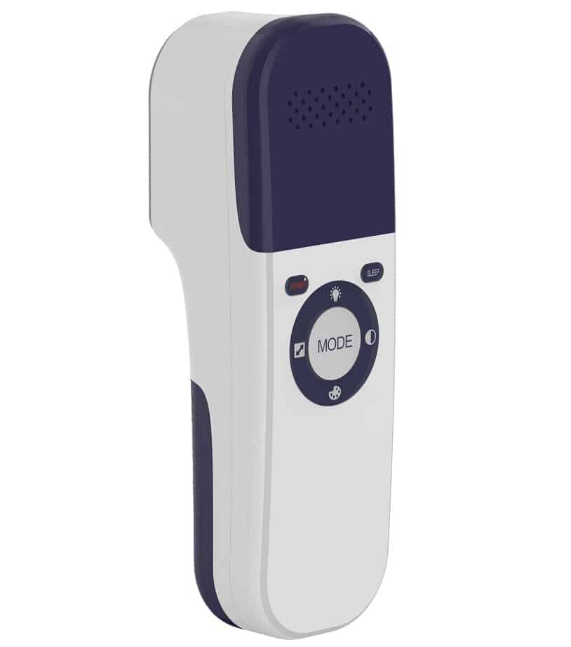 VEINFINDER 600
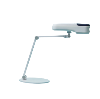 VEIN FINDER - Table Stand
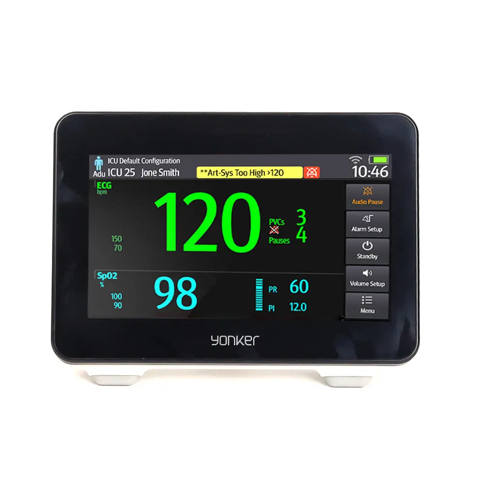 New E7 Portable Patient Monitor
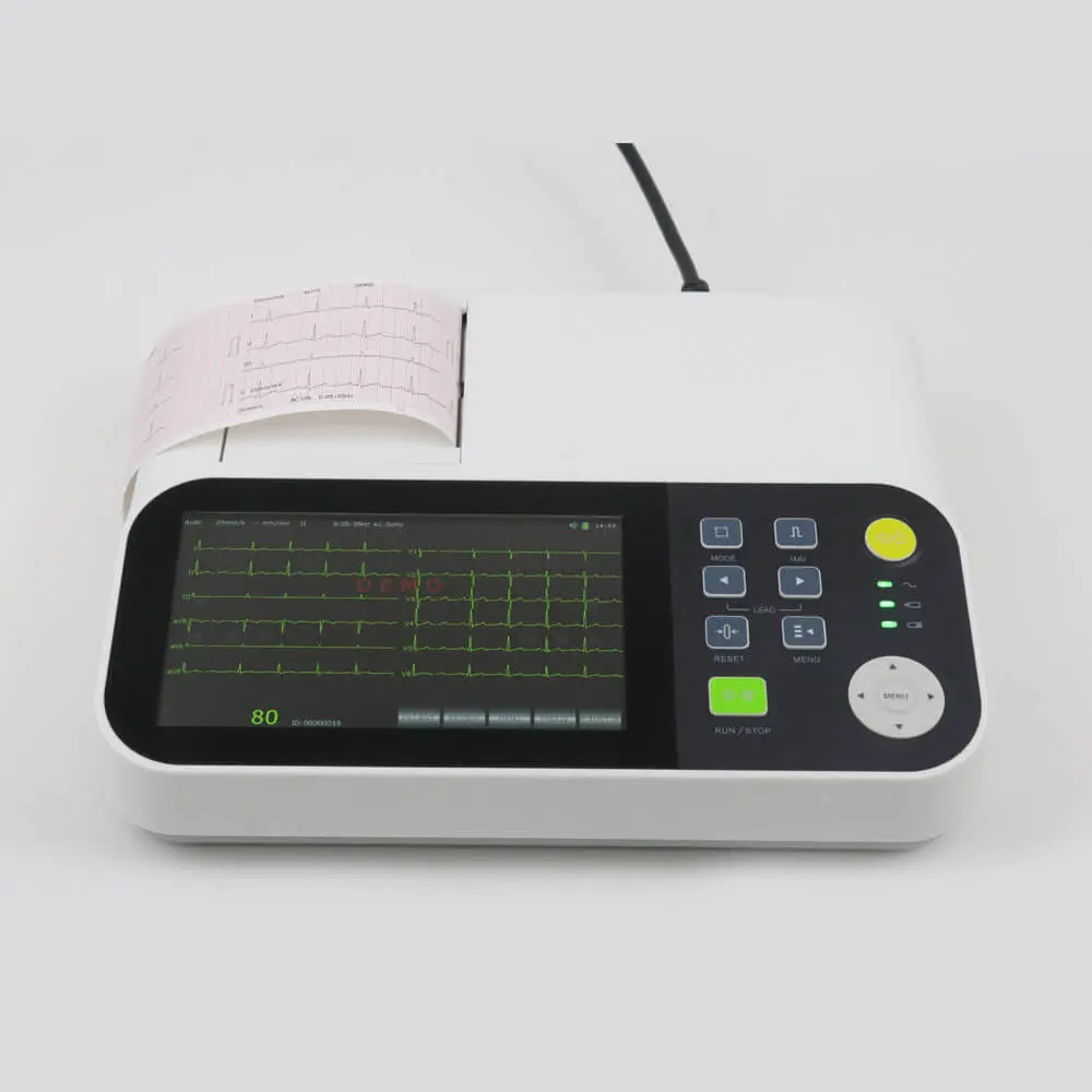 Yonker 7inch display 3 channel ECG Machine with touch screen
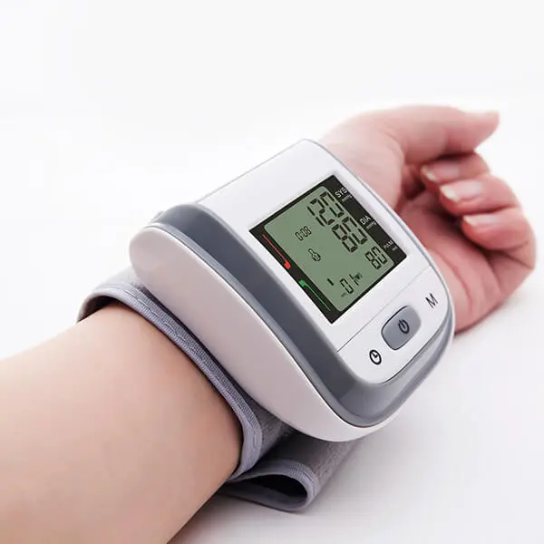 Yonker Smart Wrist Blood Pressure Machine
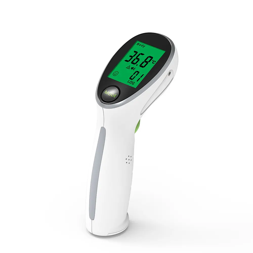 Yonker IRT2 Infrared Thermometer For Homecare
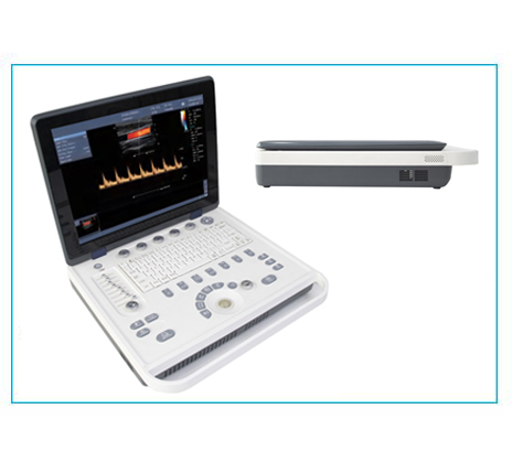 YK-UL8 _ Color Ultrasound Machine
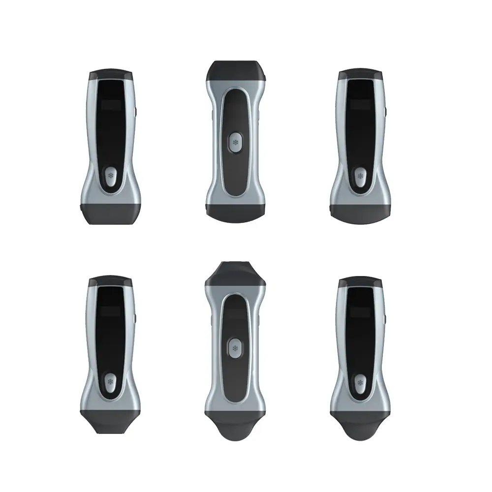 COLOR ULTRA SOUND 15 B
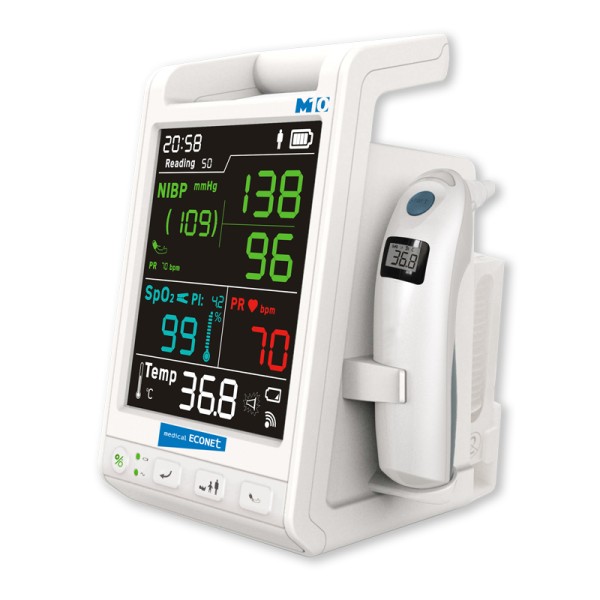 M10 vital signs monitor - Germany
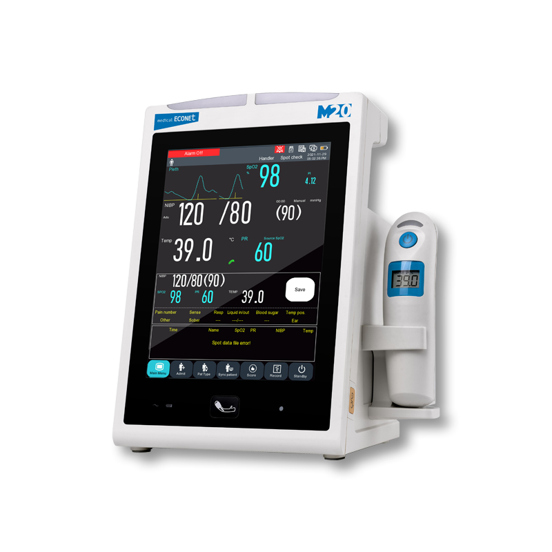 M20 vital signs monitor - GERMANY
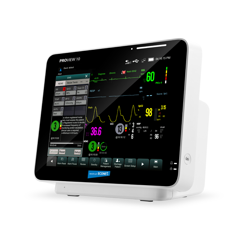 PROVIEW 10 PATENT MONITOR - GERMANY
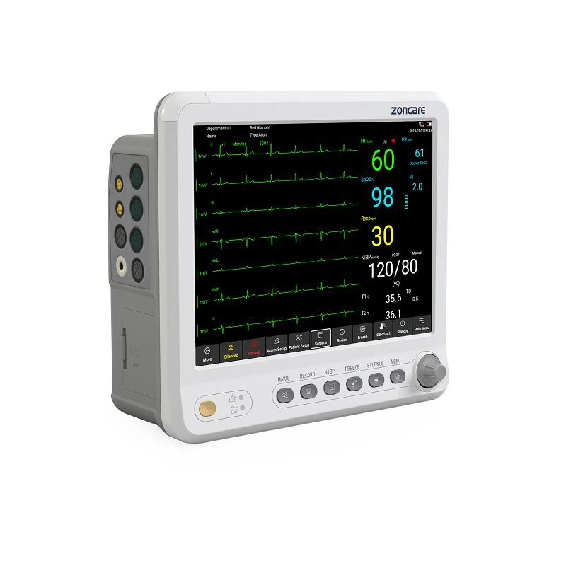 Patient Monitor for Adult Children Neonate
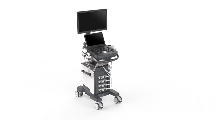 Compact Color Doppler Imaging Ultrasound - Zoncare viv20
.webp) 3D 4D Color Doppler General Imaging Ultrasound P7 - ZONCARE
3D 4D Color Doppler General Imaging Ultrasound P7 - ZONCARE
.webp) 3D 4D Color Doppler General Imaging Ultrasound P7 - ZONCARE
3D 4D Color Doppler General Imaging Ultrasound P7 - ZONCARE
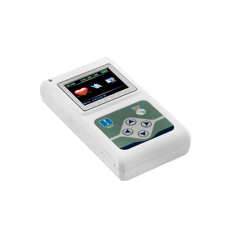 TLC9803 Dynamic ECG Systems
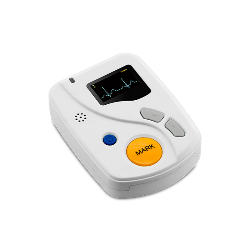 DYNAMIC ECG SYSTEM - TLC6000
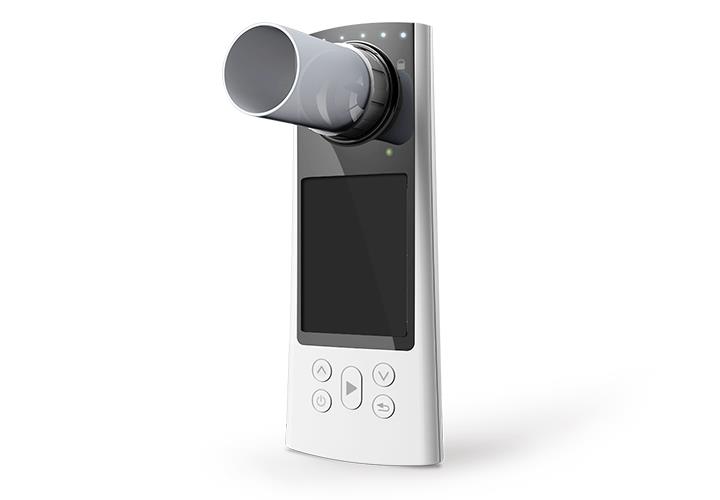 SP80B SPIROMETER
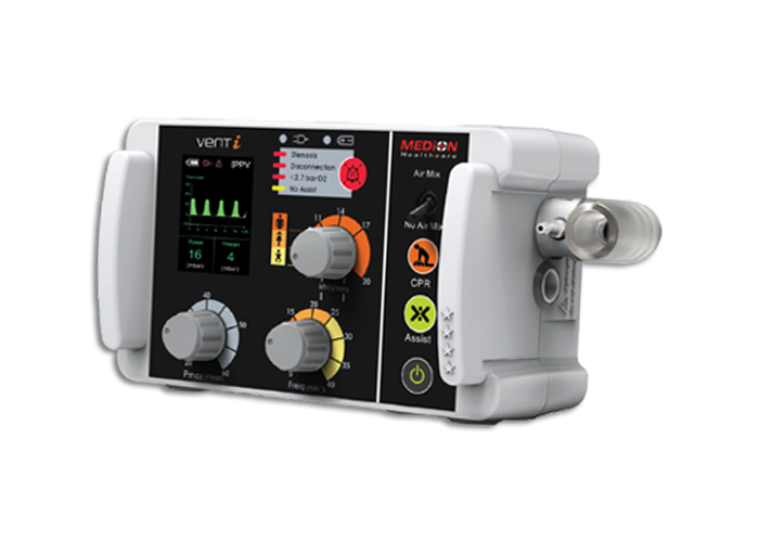 Transport Ventilators - vent I
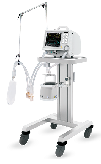 Portable ICU Ventilator Optima
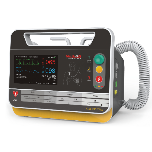 Defibrillator Machine
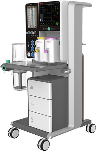 Anaesthesia Workstation Asteros Royale V-12
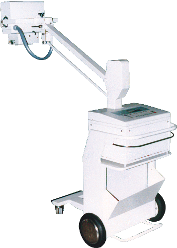 PORTABLE X RAY MACHINE - MOBILE
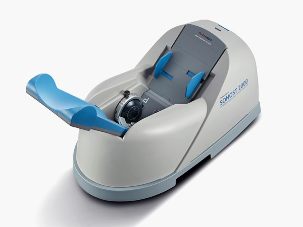 SONOST-2000
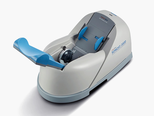 BONE DENSITOMETER SONOST-2000
 1 BoX Aqua Skin Pure Gold Anti Aging Whitening Serum
.jpeg) Excell 350MCD Electrocautery
Excell 350MCD Electrocautery
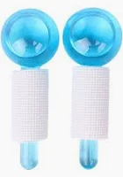 BEAUTY CRYSTAL BALL
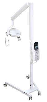 Acteon X-Mind DC Intraoral X-Ray (Mobile Base)
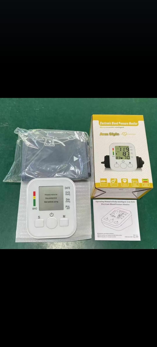 Blood Pressure Monitor
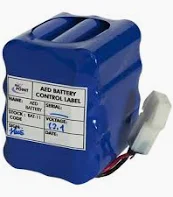 Lifepoint Pro AED Replacement Battery
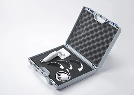 VLSCOPE Video Laryngoscope
.webp) Electric wheelchair
Electric wheelchair
.jpeg) SPO2 probe adult Clip type
SPO2 probe adult Clip type
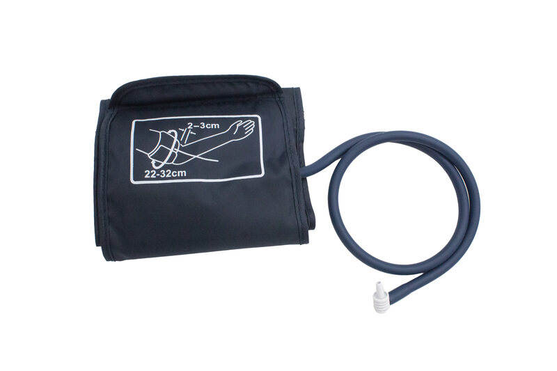 Blood Pressure Cuff
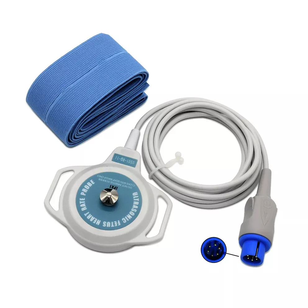 Fetal Monitor Fetal Toco Probe
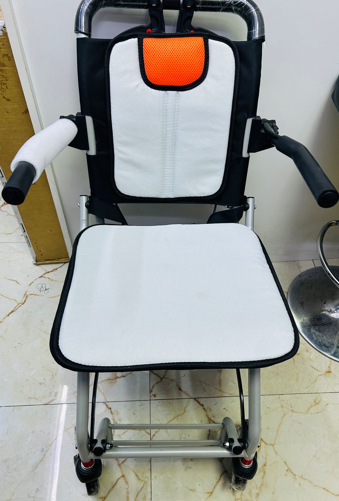 Manual Wheelchair
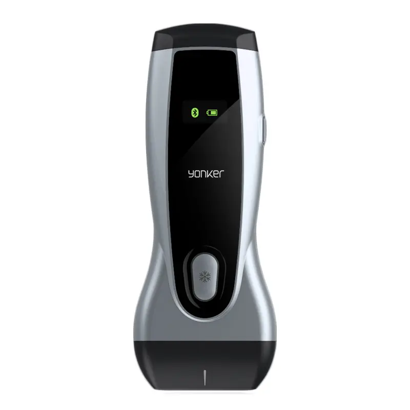 Wireless Ultrasound Machine
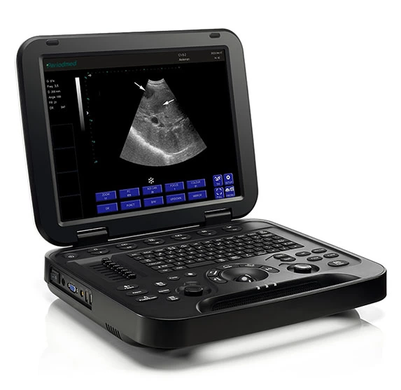 Diagnostic Ultrasound System PU-DL151A
 Braun Thermometer
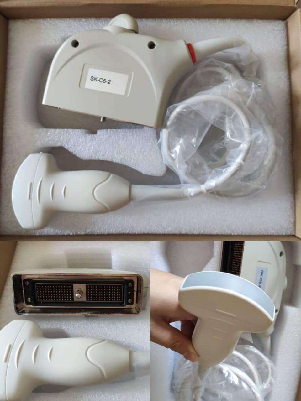 Mindray C5-2 New Convex Array Sensor Ultrasound Probe Ultrasonic Transducer
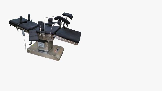 OT TABLE - PULSEMED
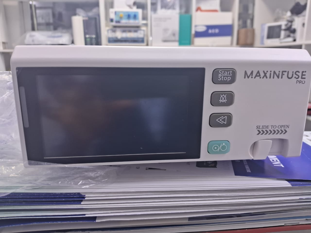 INFUSION PUMP - PULSEMEDUK
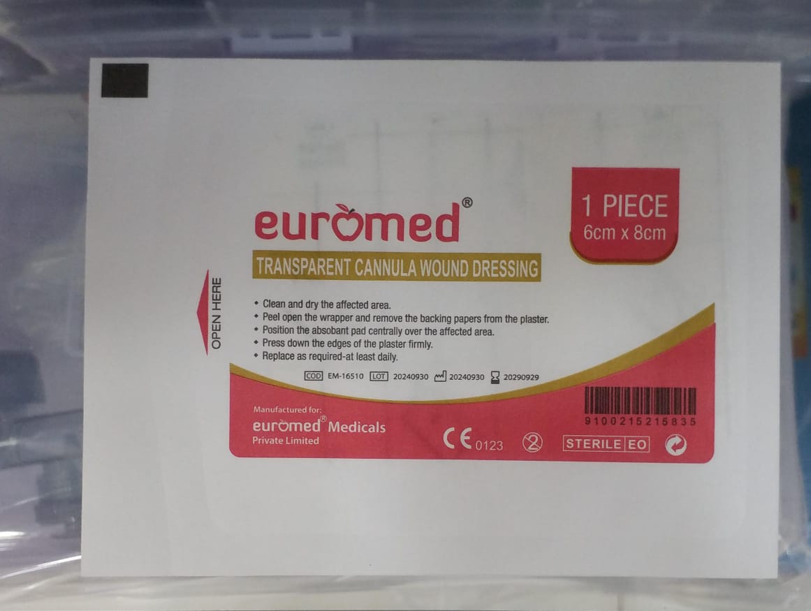 IV CANNNULA FIXING DRESSING
 ghtrfhytr.jpeg) MEDICAL EQUIPMENT - TRAININGS
MEDICAL EQUIPMENT - TRAININGS
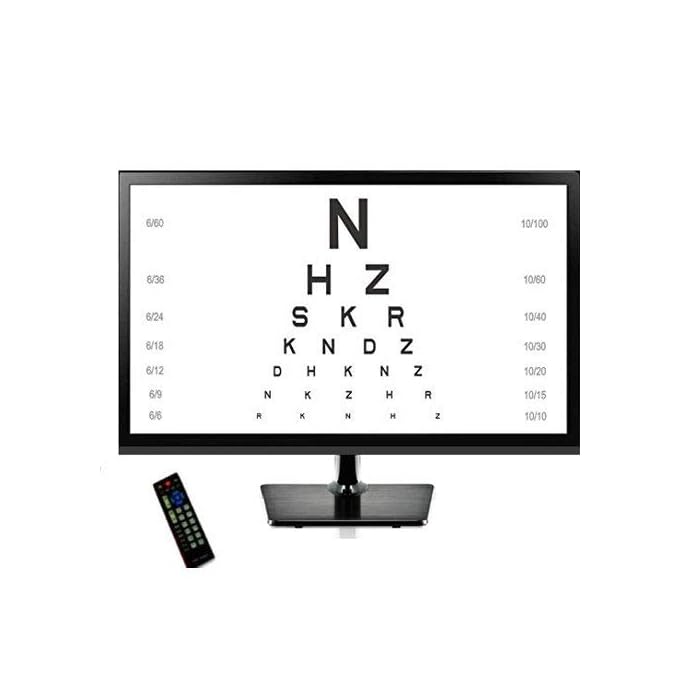 DIGITAL VISION CHART
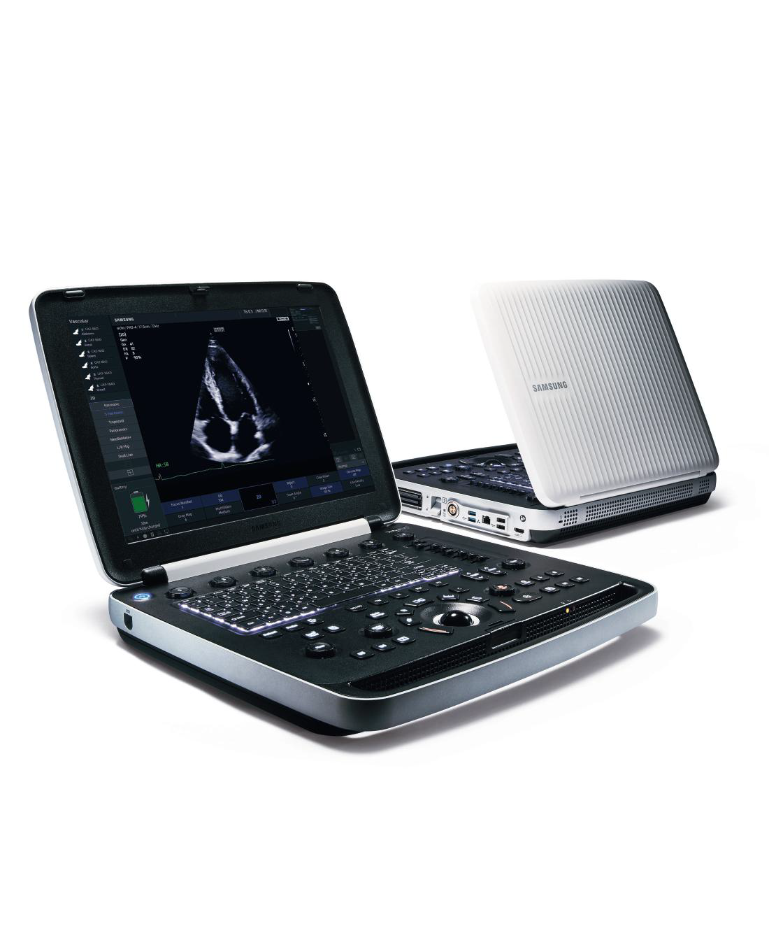 HM70 EVO SAMSUNG
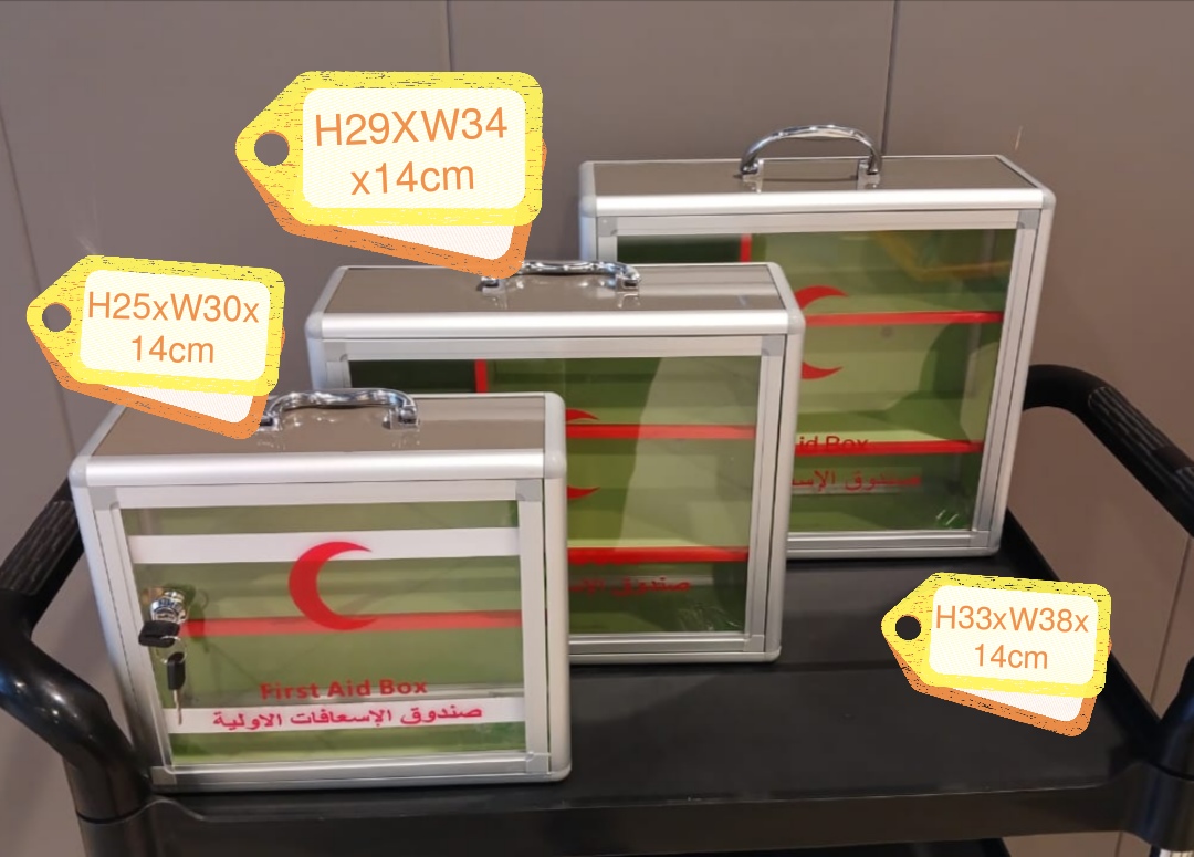 FIRST AID BOX METALLIC
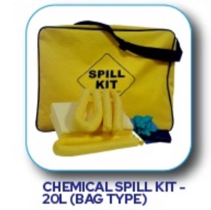 CHEMICAL SPILL KIT
 CHEMICAL SPILL KIT
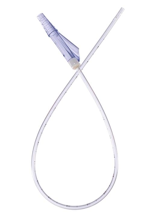 SUCTION CATHETER
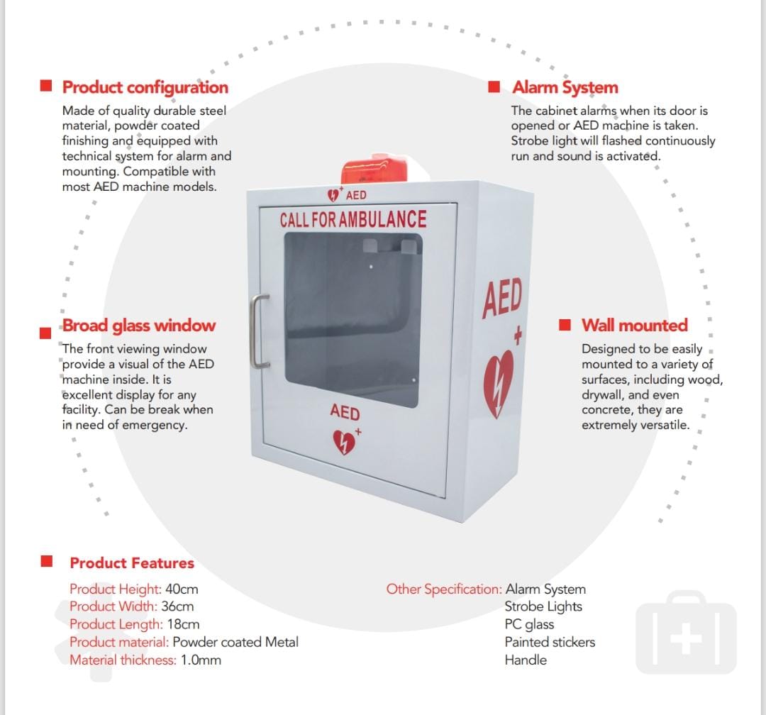 AED ALARM WALL CABINET
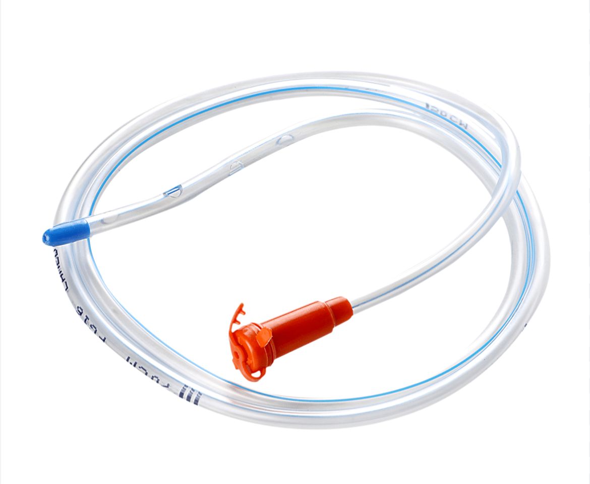 RYLE'S TUBE
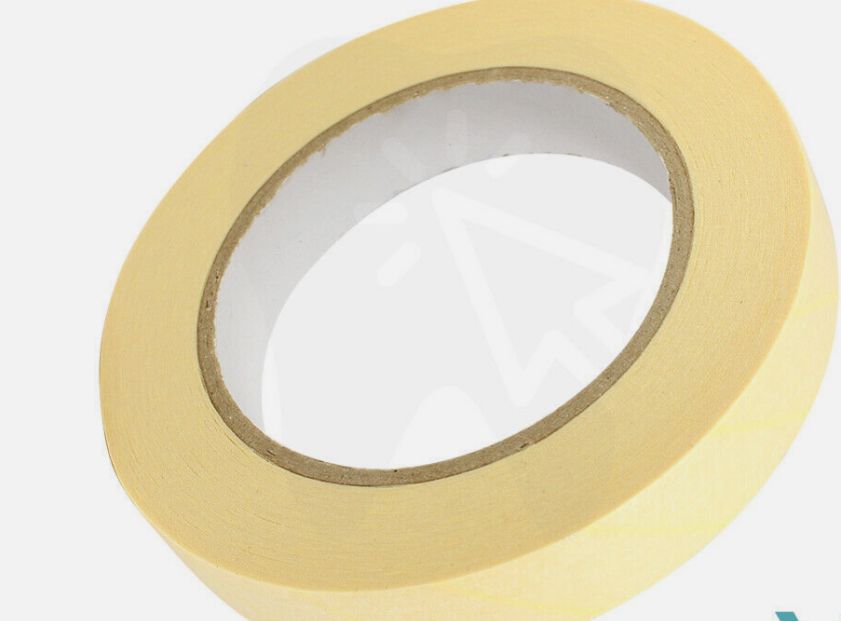 AUTOCLAVE TAPE - Steam Sterilisation Indicator Tape 25mm x 75m
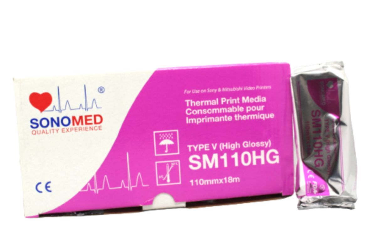 SONOMED Thermal Print Media SM 110 HG
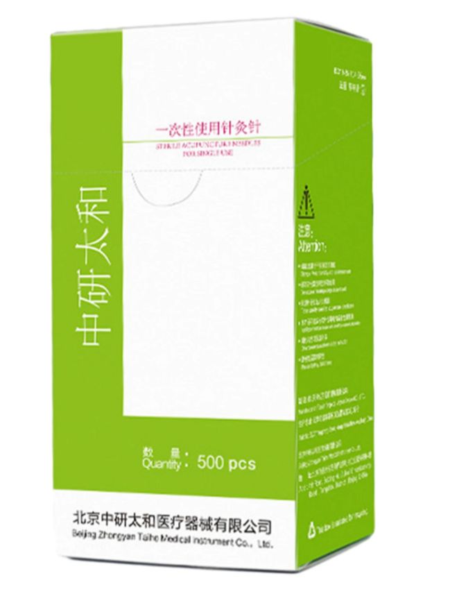 DISPOSABLE ACCUPUNCTURE NEEDLE
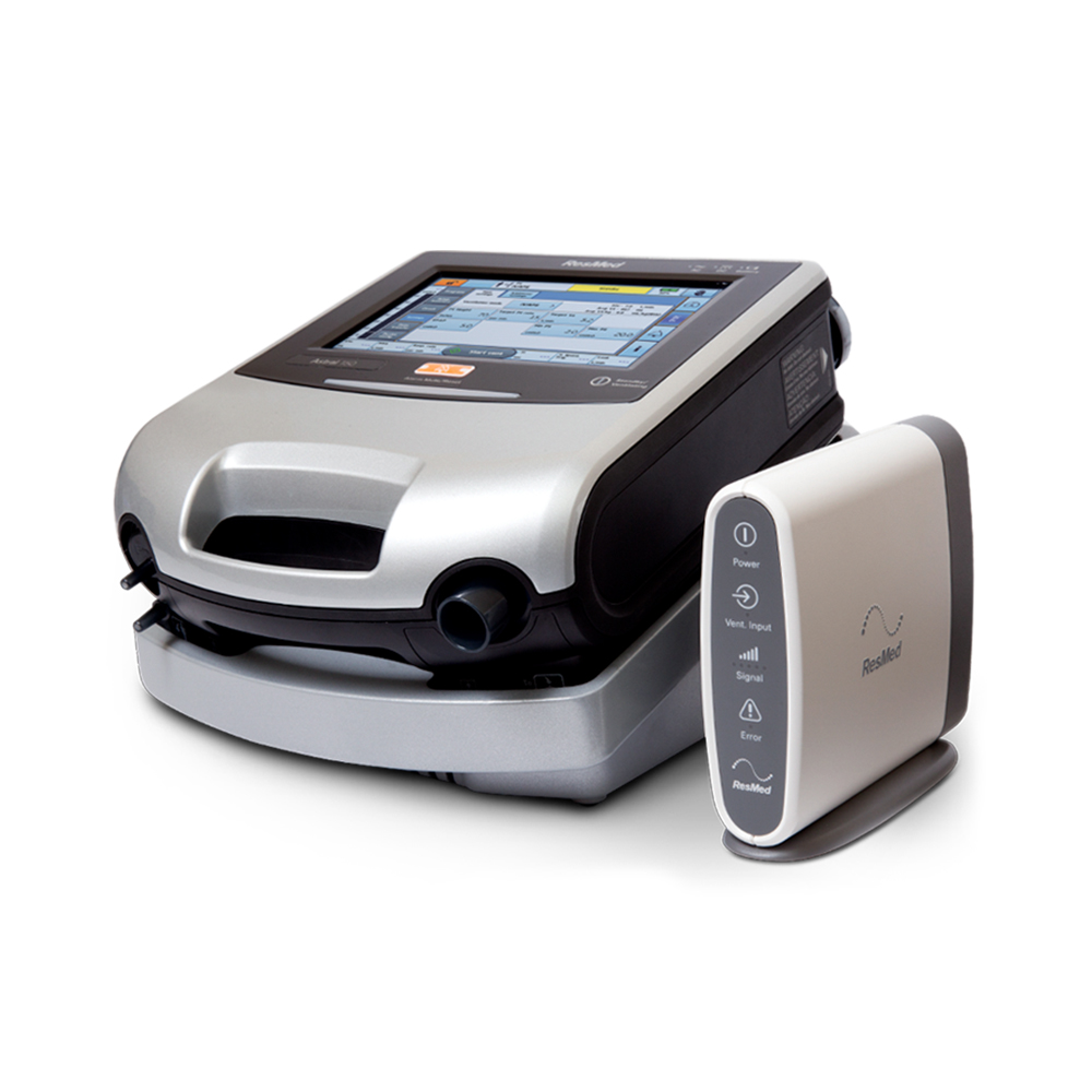 Astral Ventilator - Rental
 SUCTION MACHINE - RENTAL
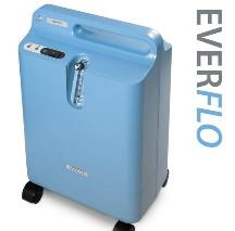 OXYGEN CONCENTRATOR - RENTAL
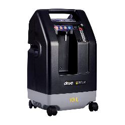 Oxygen Concentrator 10 Ltr - Rental
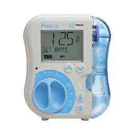 FreeGo Feeding Pump - - Rental
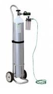 Oxygen Cylinder 10 Ltr - Rental
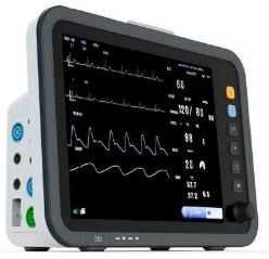 Patient Monitor - Rental
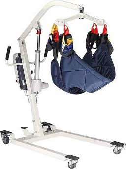 Portable Patient Hoist Lifter -Rental
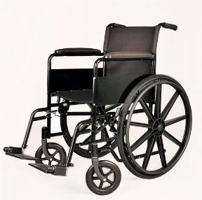 Basic Wheel Chair - Rental
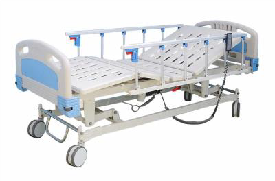 Electric Bed - Rental
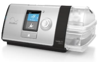 BiPAP ST - RENTAL
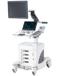 ARIETTA 50LE FUJIFILM ULTRASOUND
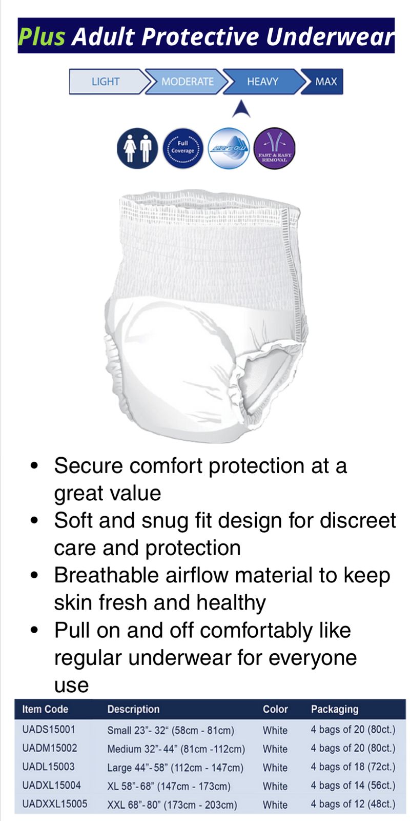 PROTECTIVE UNDERWEAR
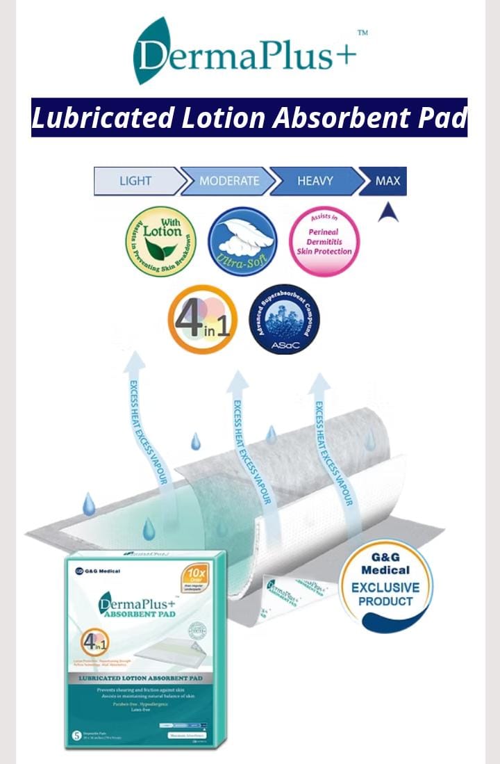 ABSORBENT PAD
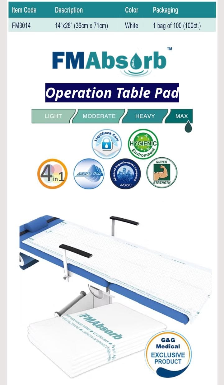 OPERATION TABLE PAD
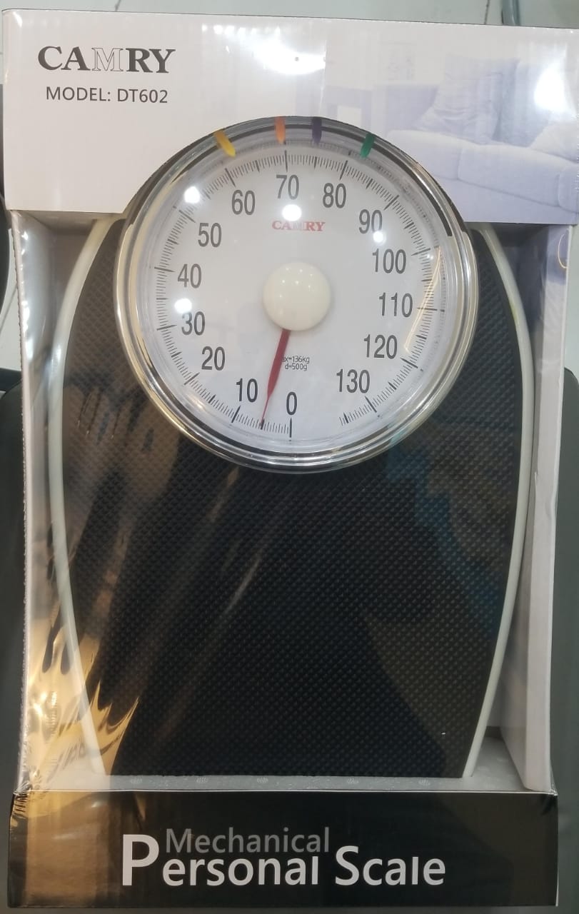 PERSONAL SCALE
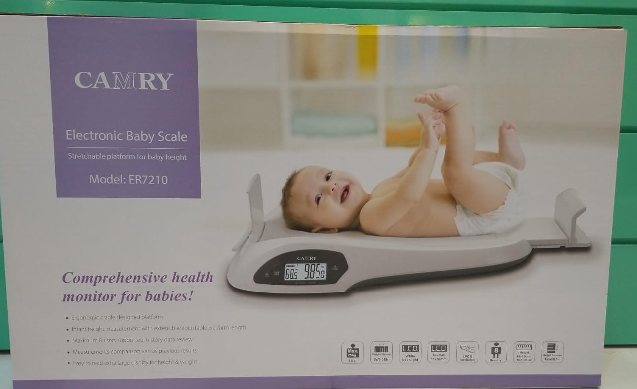 ELECTRONIC BABY SCALE
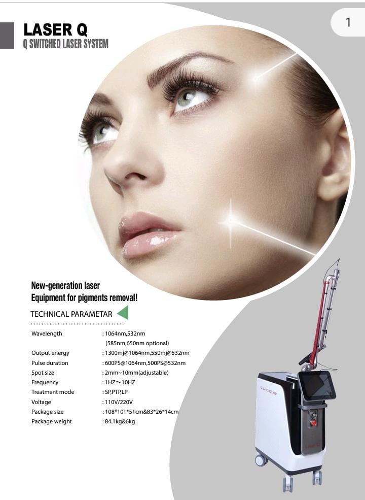 LASER Q MACHINE
.jpeg) HYDRAFACIAL MACHINE
HYDRAFACIAL MACHINE
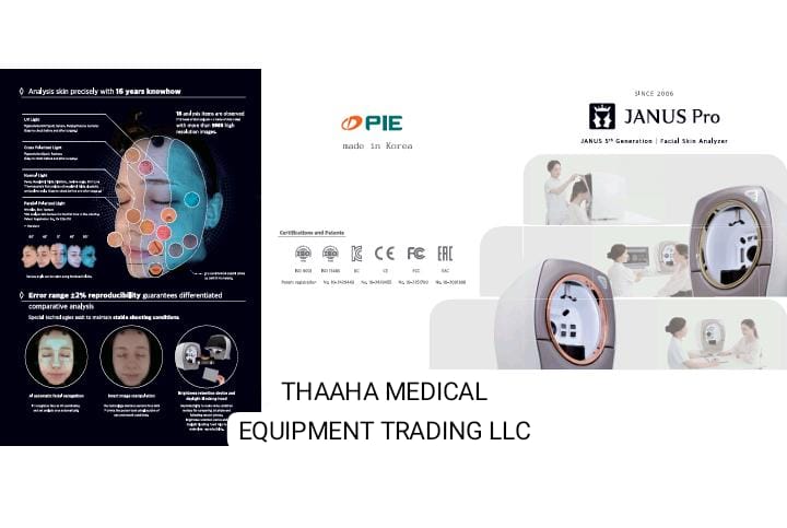 FACIAL SKIN ANALYZER JANUS PRO SUNLIKE
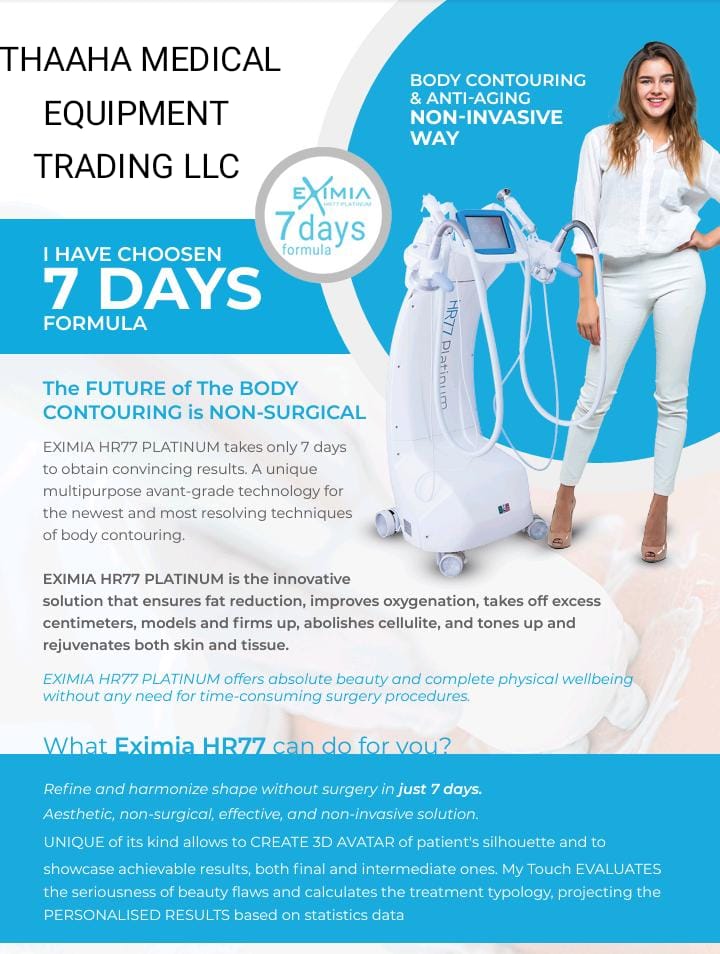 BODY CONTOURING MACHINE
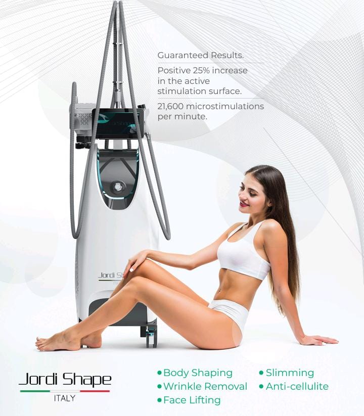 BODY SCULPTING MACHINE
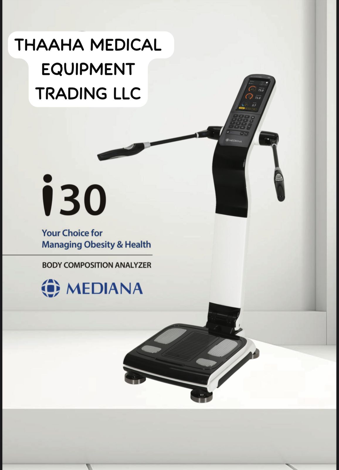 BODY COMPOSITION ANALYZER
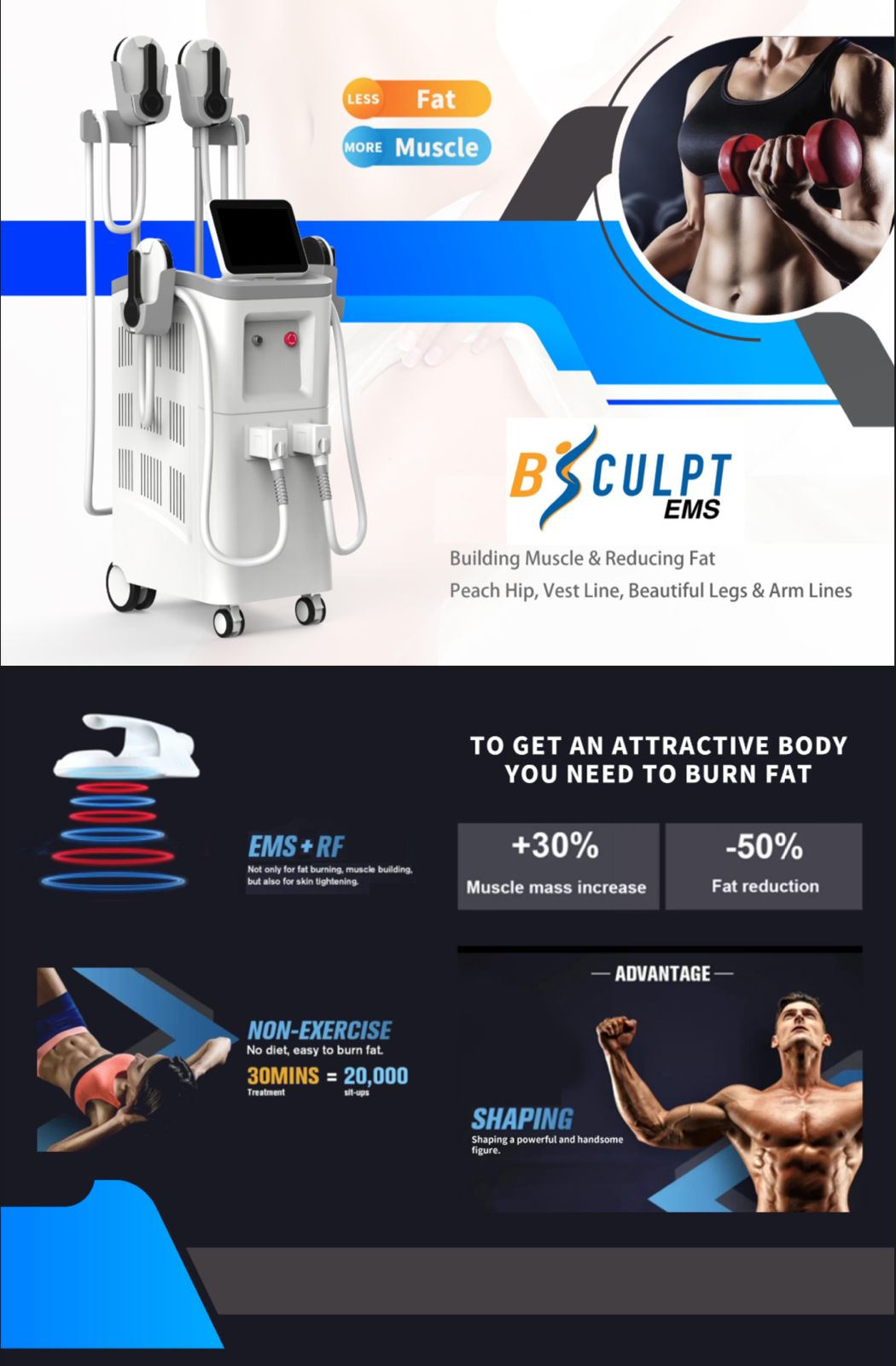 EMS FITNESS MACHINE
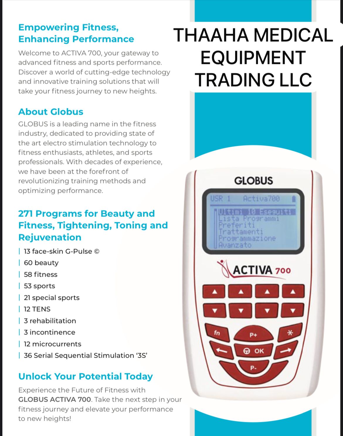 MUSCLE STIMULATOR
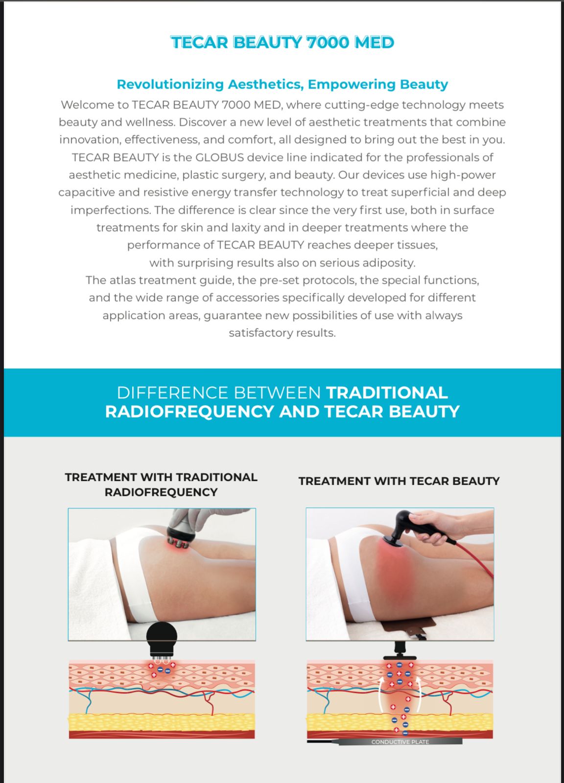 TECAR BEAUTY 7000 MED
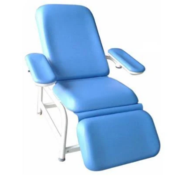 Blood donor chair
 PATIENT WARMER
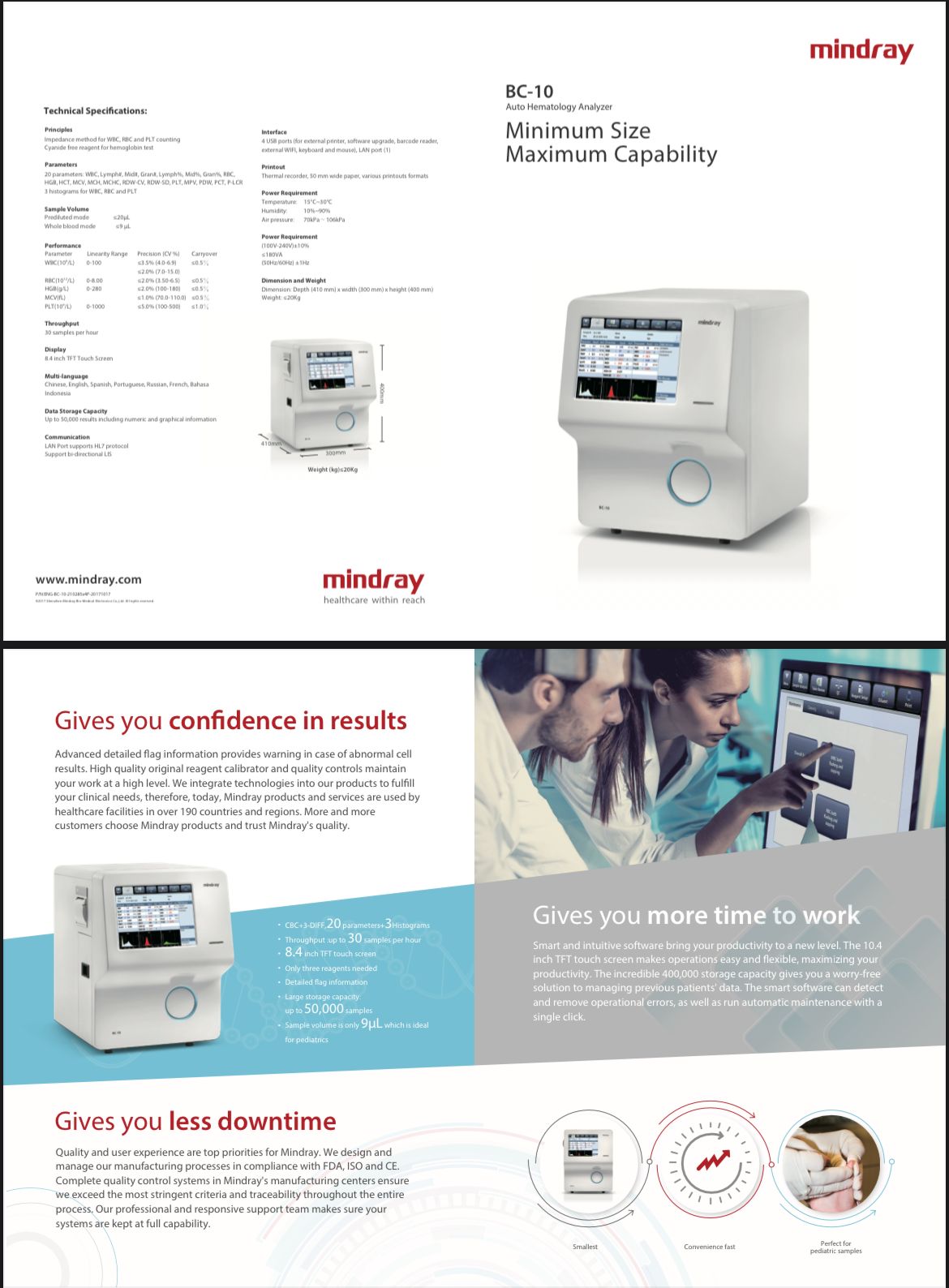 Auto Hematology Analyzer
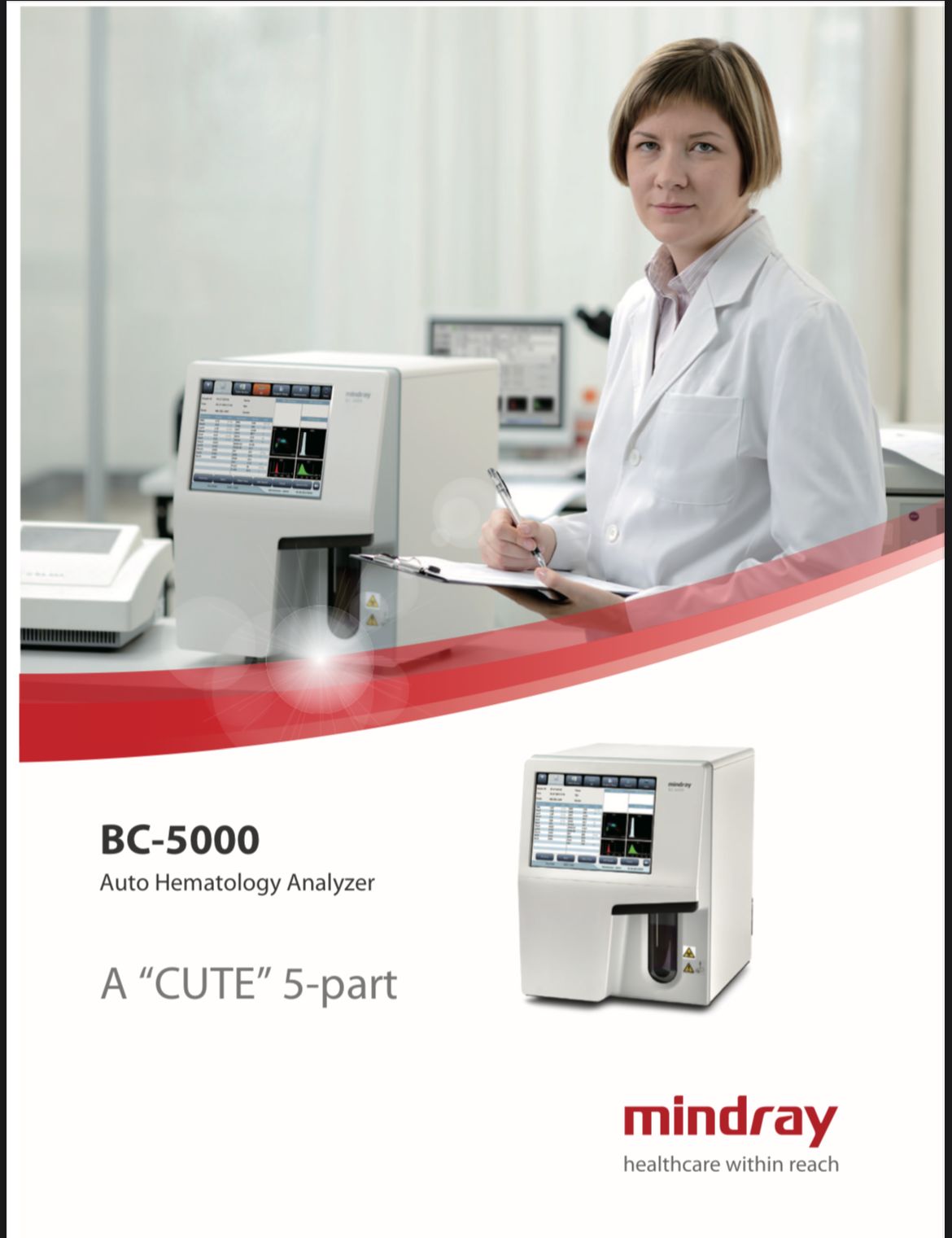 Auto Hematology Analyzer
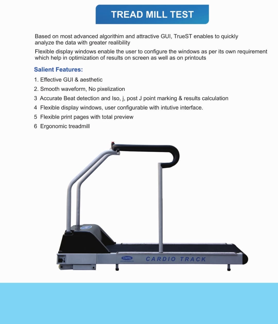 TREAD MILL
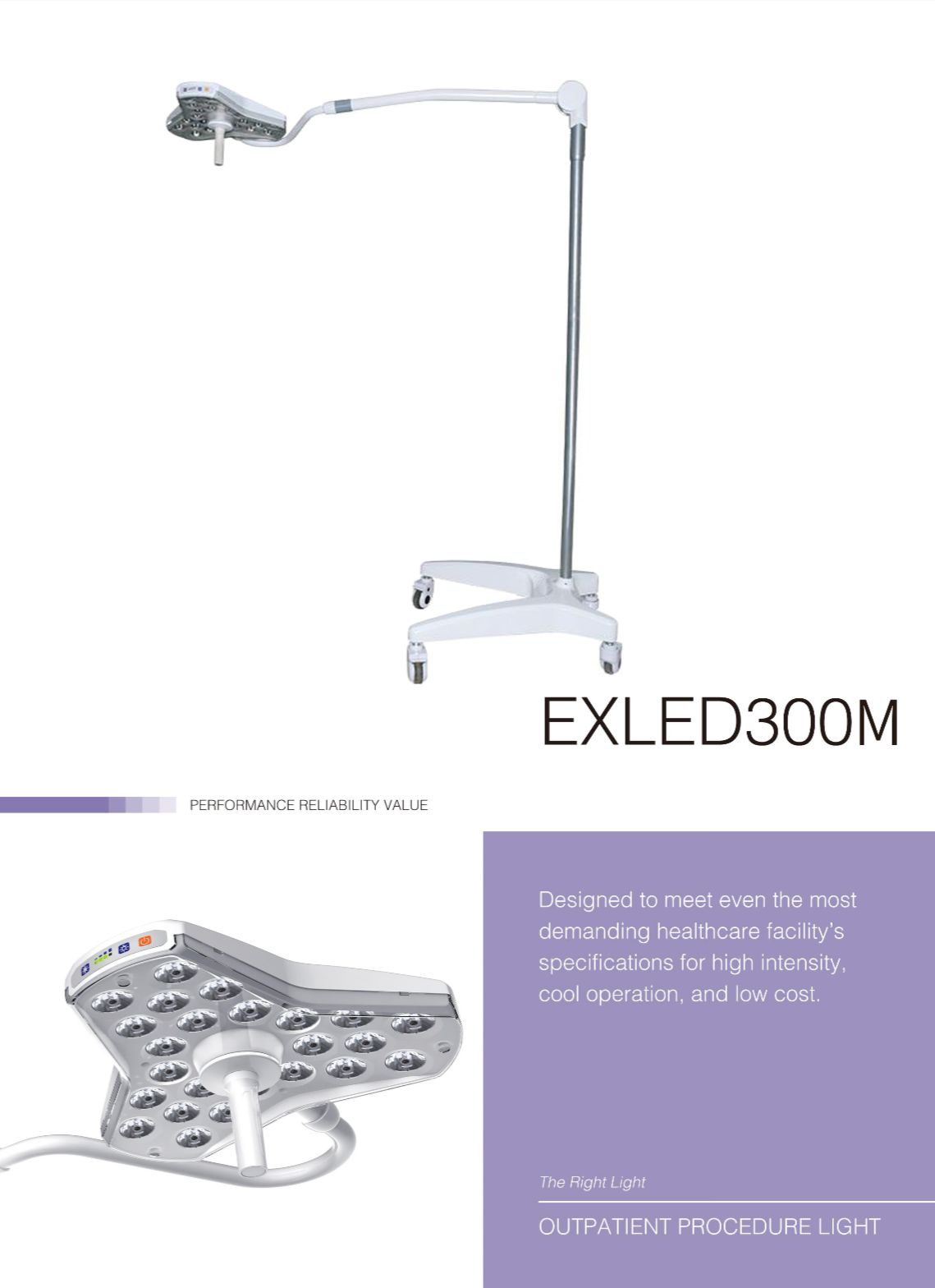 PROCEDURE LIGHT
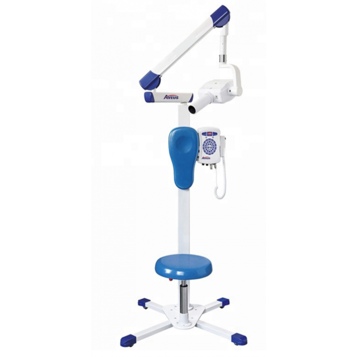 DENTAL X RAY - MOBILE
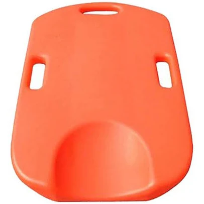 CPR board
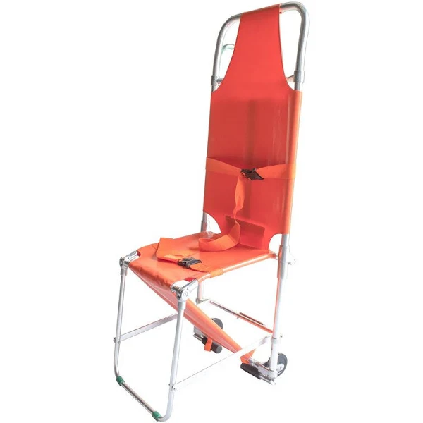 Chair Stretcher
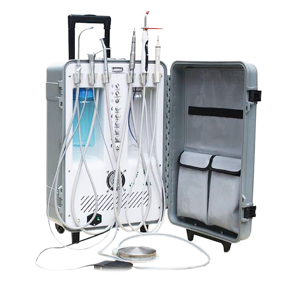 Portable Dental Unit
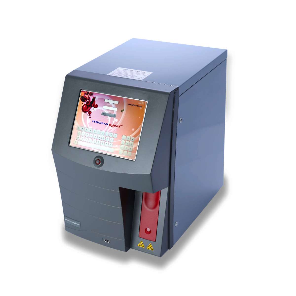 HEMATOLOGY ANALYZER
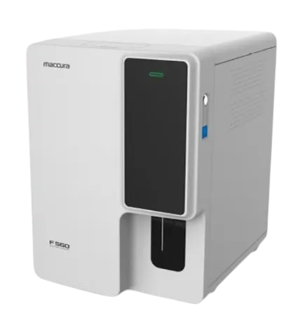 Automatic Hematology Analyzer
 Mindray Mindray Ultrasound Consona N6
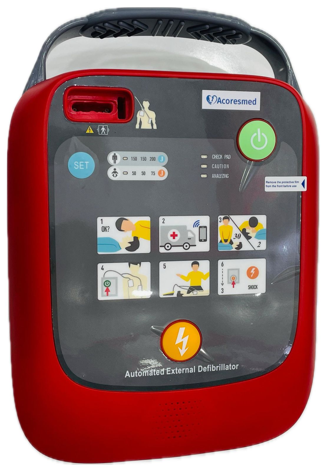 AED MACHINE A102
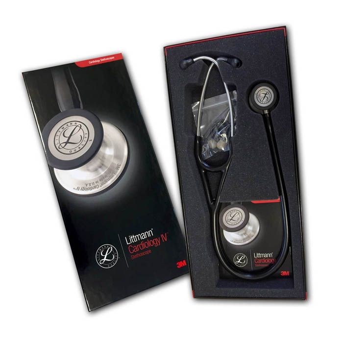 3M Littmann Cardiology IV Diagnostic Stethoscope
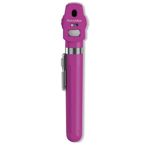 Welch Allyn Pocket Plus LED Ophthalmoscope
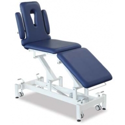 Couch 5 Section Electric
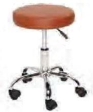 Round Stool Adjustable
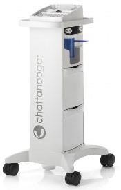 Intellect Mobile 2 Cart 5 Bins with Vacuum Set
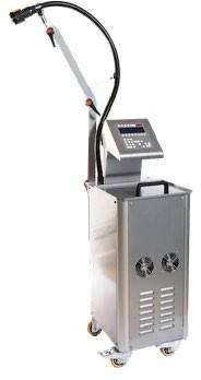 Cryo Ultrasound - Fisiocomputer
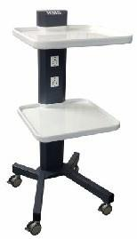 Equipment Trolley
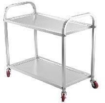 Utility Trolley - Stainless Steel
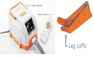 Air Compression Therapy Unit
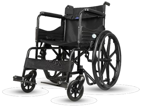 Wheel Chair - Markon
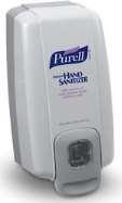 HAND SANITISER WITH GEL –PUREL
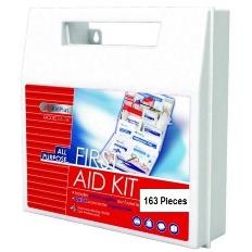 FIRST AID KIT –AIDPLUS
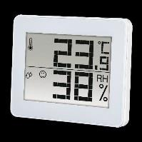 Hygrometer Intertronic
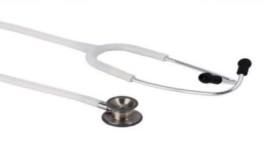 DUPLEX 2.0 RIESTER GERMANY
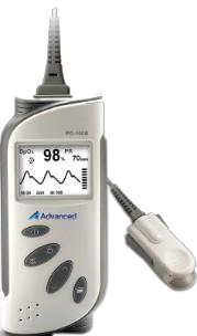 PULSE OXIMETER ADVANCED
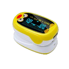 Pulse Oximeter Pediatric
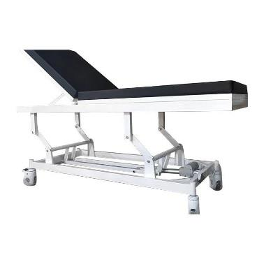 Electric Examination Couch
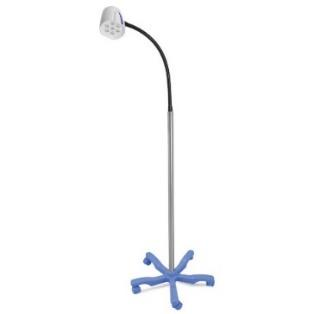 LED Examination Light
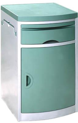 MEDICAL CART- BESIDE CABINET-SIMPLE
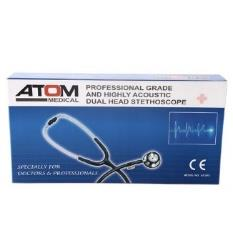 STETHESCOPE-ATOM -GERMANY
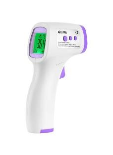 Infrared Thermometer –AIQURA
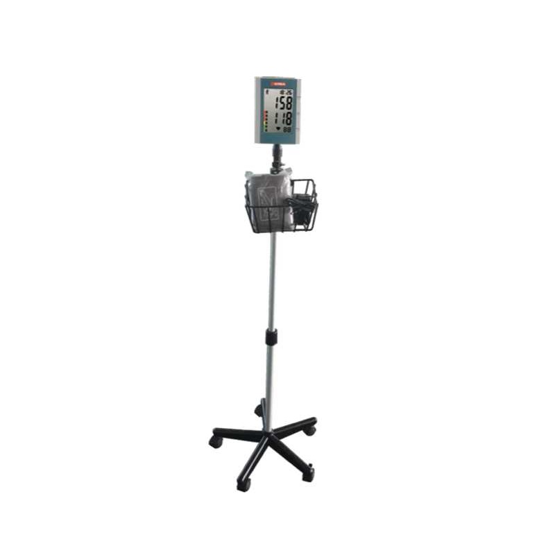 Digital BP Monitor Trolley
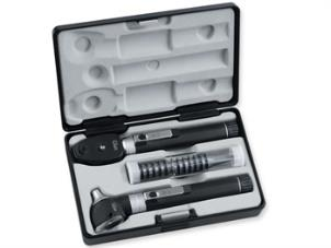 PARKER DIAGNOSTIC SET Lightweight, rigid case with one Parker otoscope 2.5 V with rechargeable handle, 3 autoclavable ear speculum (Ø 2.5, 3.5 and 4.5 mm), 2 laryngeal mirrors (size 3 and 4), bent arm throat lamp, tongue spatula, adjustable nasal speculum
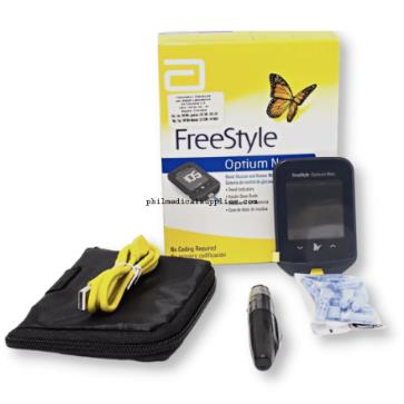 GLUCOMETER-FREESTYLE
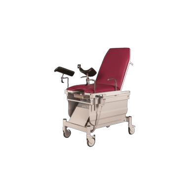 GYNAECOLOGY EXAMINATION COUCH
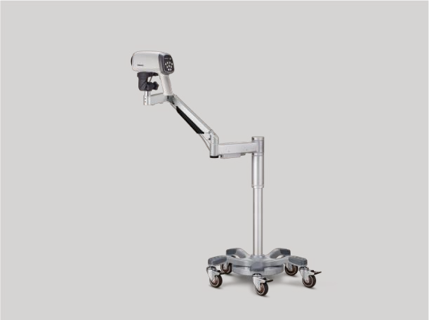 Video Colposcope With Swing Arm stand basic
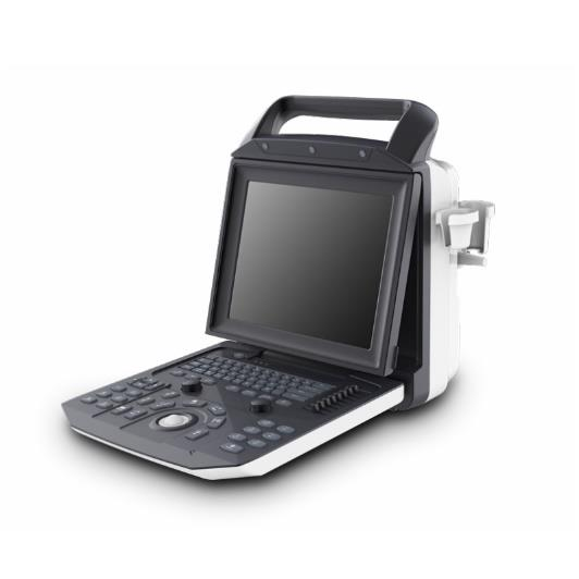 Portable Color Ultrasound System Zoncare-M5
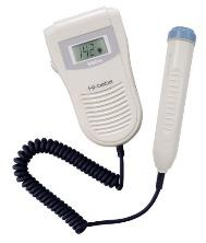 FETAL DOPPLER POCKET SIZED
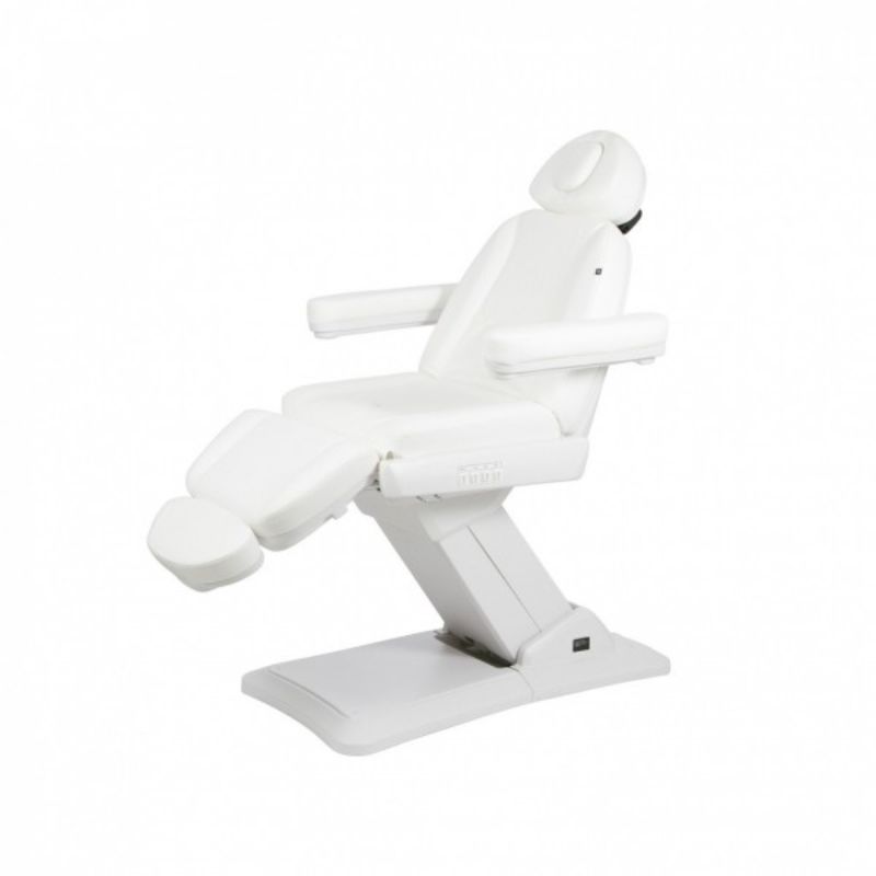 Skin Derma Chair with Four motor
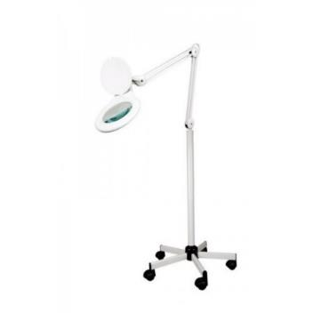 MAGNIFYING LAMP WITH TROLLEY
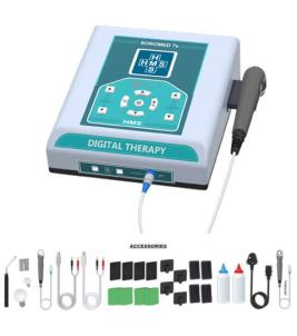 Interferential And Ultrasound Combo Machine
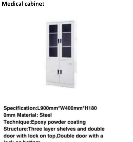 Medical Cabinet
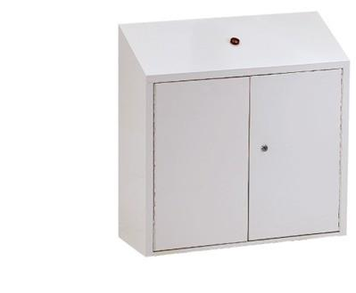 Drug Cabinet
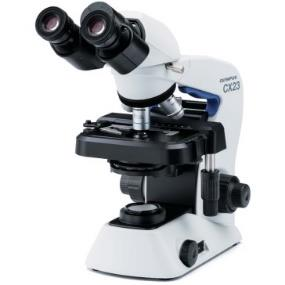 Microscope CX23 LED Olympus
 Fridge Thermometer , SP-E-16 , CHINA
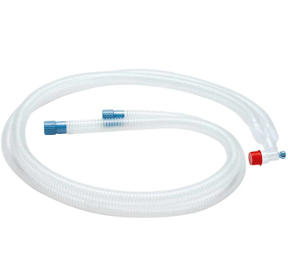 Breathing circuit Vent Set, disposable, basic, 1.5 m, not made with natural rubber latex
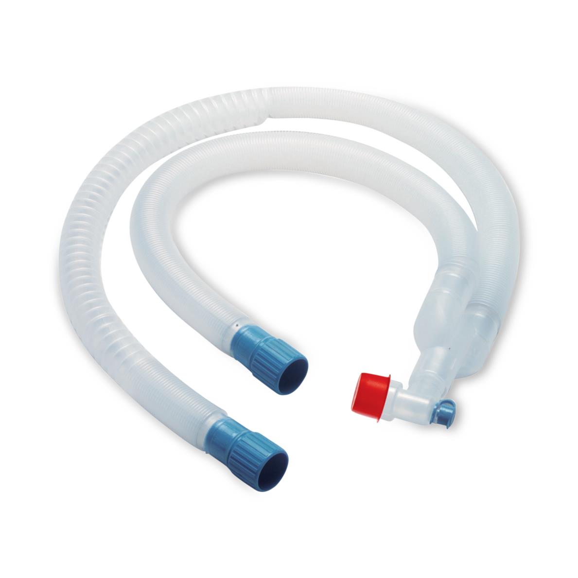 Breathing circuit VentStar", disposable, basic, 1.8 m, not made with natural rubber latex
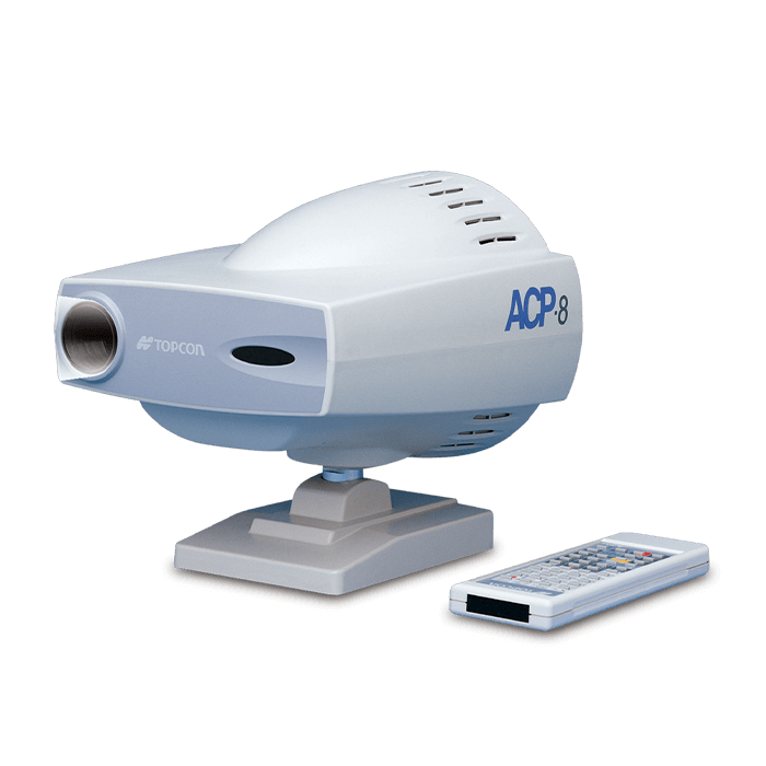 Auto chart projector ACP-8
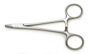 NEEDLE HOLDER
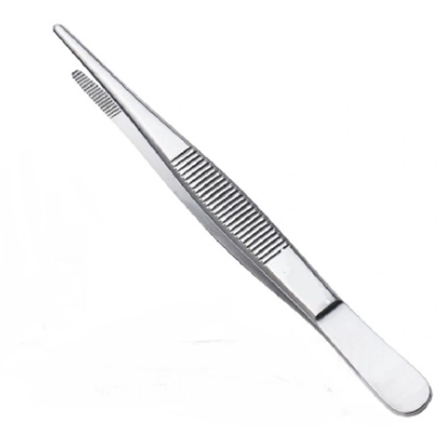 THUMB FORCEPS
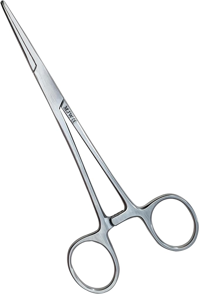 ARTERY FORCEPS
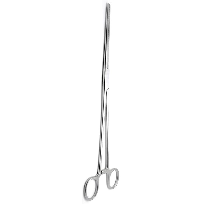 ARTERY FORCEPS LARGE
 MOSQEITO FORCEPS
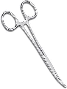 MOSQEITO FORCEPS
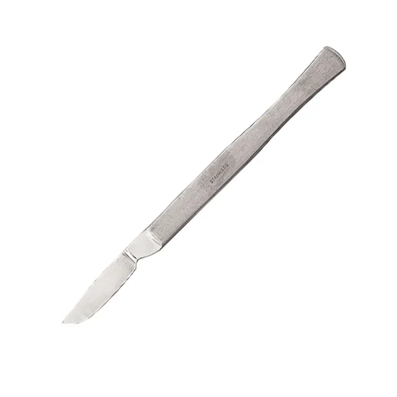 SCALPEL
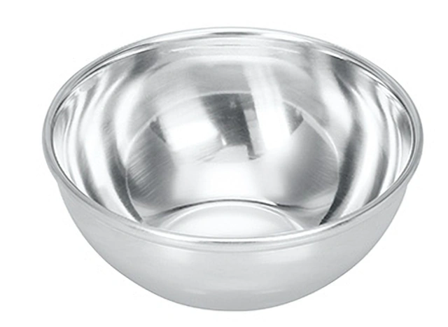 STEEL BOWEL
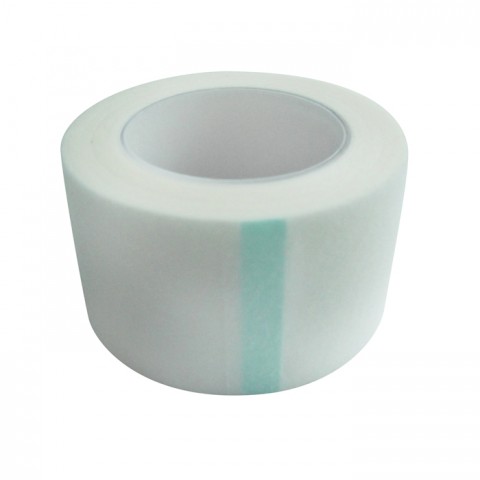 NON WOVEN (PAPER) SURGICAL TAPE 5 CM*10Y(6 ROLLS/PKT0
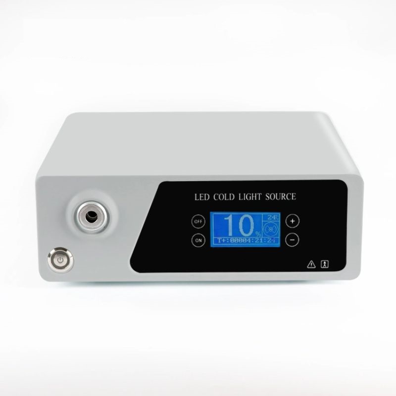 LED COLD LIGHT SOURCE
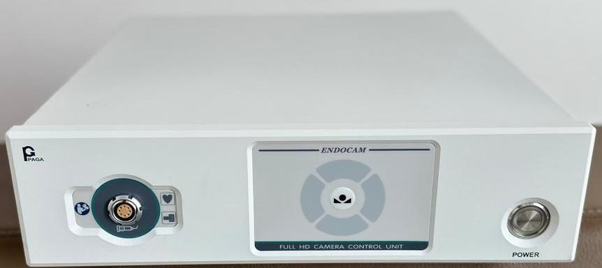 LED COLD LIGHT SOURCE
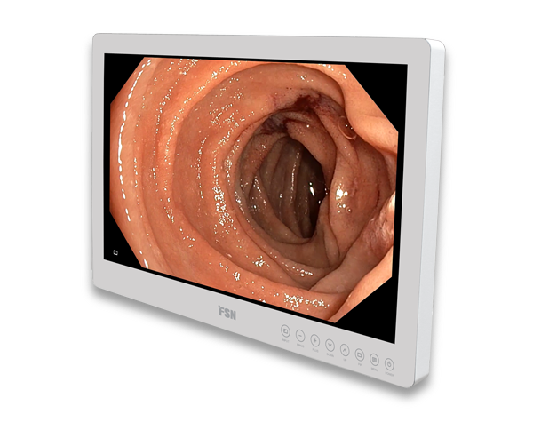 HD MEDICAL MONITOR
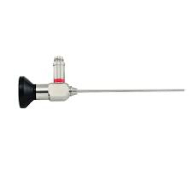 Sino scope 0°-2.7mm-175mm
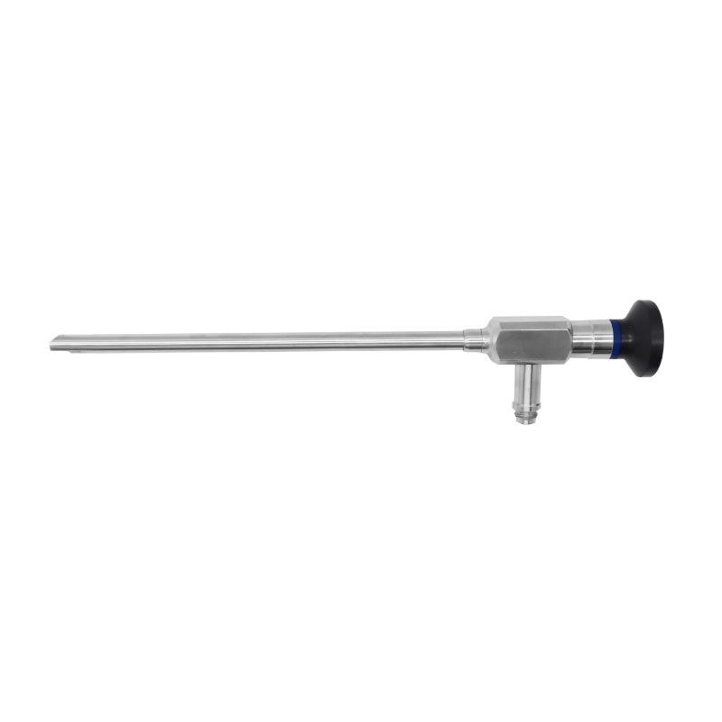 Laryngoscope 70°-6mm-184mm
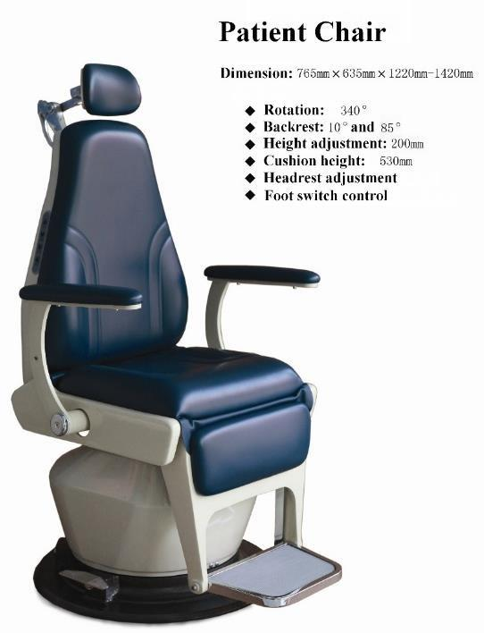 LUXURY PATIENT CHAIR
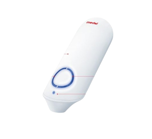 INSECT BITE HEALER
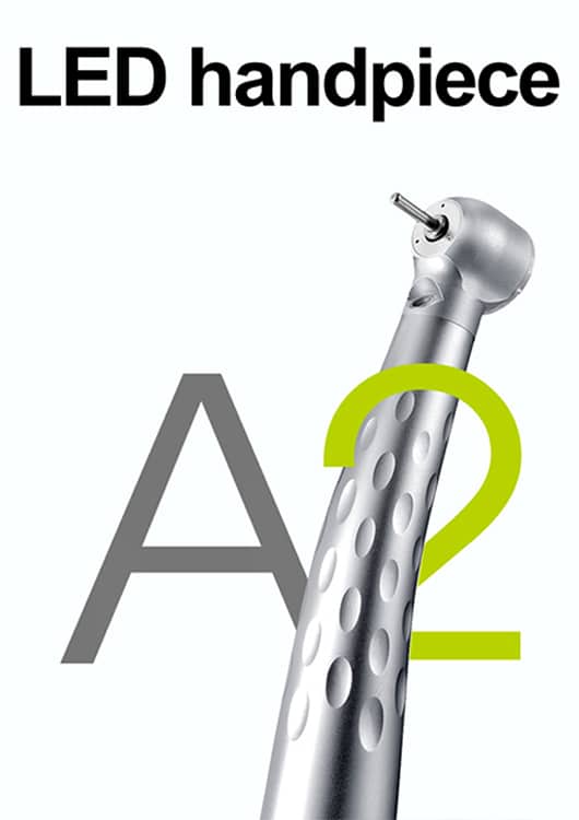 Highspeed Handpiece A2 APPLE
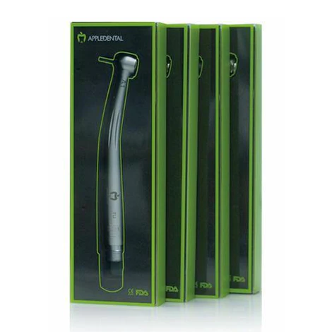 High speed Handpiece A1
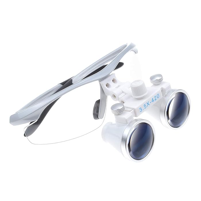 Dental Power 3.5X Binocular Loupes 420mm Working Distance Glasses
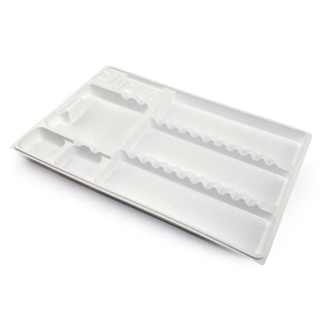 Mono Tray
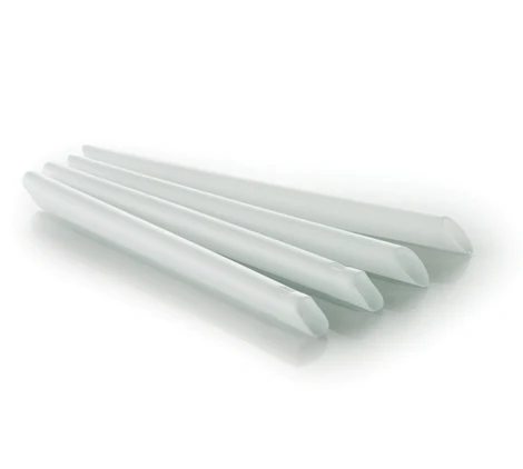 High Volume Suction Tip
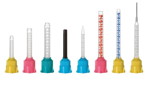 Mixing Tips
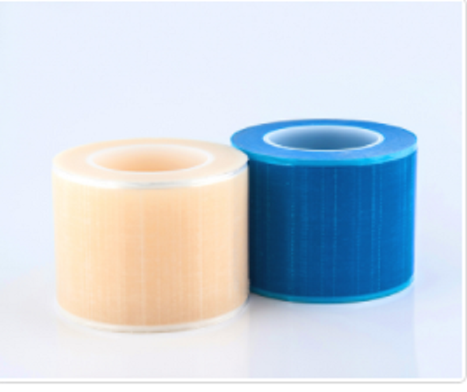 Dental Barrier Film
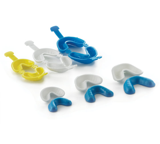 Disposable Fluoride Trays
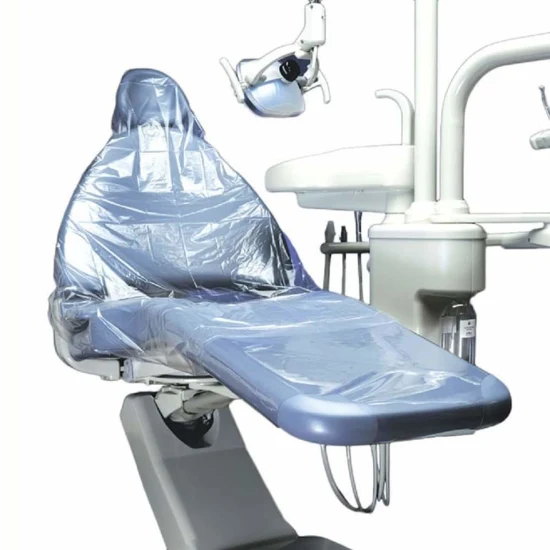 Dental Chair Cover Sleeve Full Chair Cover 125 PCS
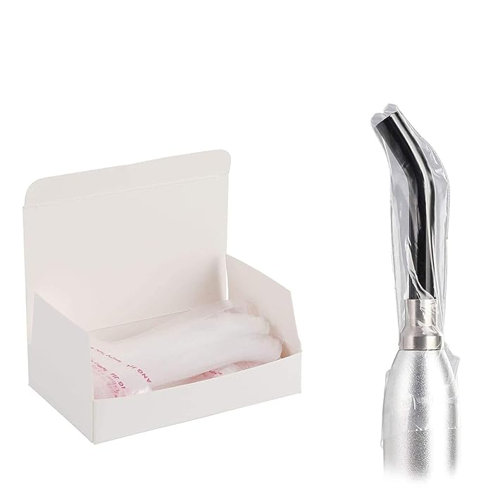 Disposable LED Light Guide Sleeves 200PCS, Implant Protective Cure Covers for Curing Tips - Plastic Optic & Transparent Sheet
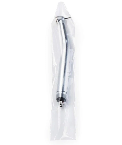 Handpiece Sleeves 500 PCS
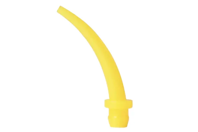 Intraoral Tips
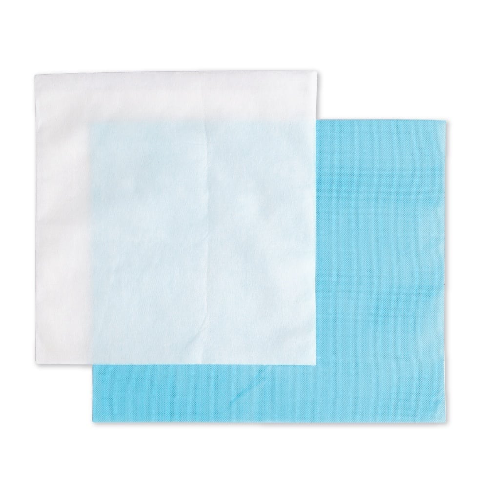 Disposable Non-Woven Headrest Covers 500 PCS
 Disposable Cups 500 PCS
 Polishing Strips Universal Kit 50 Pieces
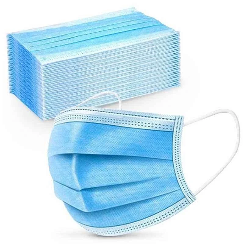 10 Pcs Disposable Surgical Face Mask Box
 Prophy Powder
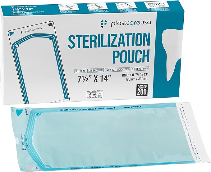 Self-Sterilization POUCH
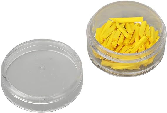 Dental Wedges
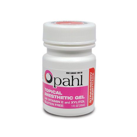 Dharma Topical Anesthetic Gel 30ml
 Pola Office Bulk Kit
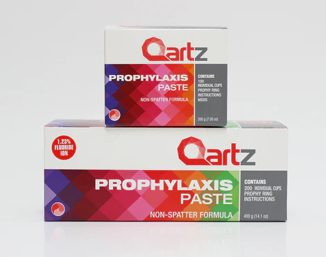 Prophy Paste
 PROPHY PASTE CHERRY WITH FLUORIDE
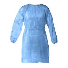 Disposable GOWNS 100 PCS
.jpg) Phosphate fluoride gel-Ionite-APF Gels
Phosphate fluoride gel-Ionite-APF Gels
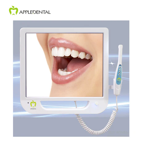 Intra Oral Camera
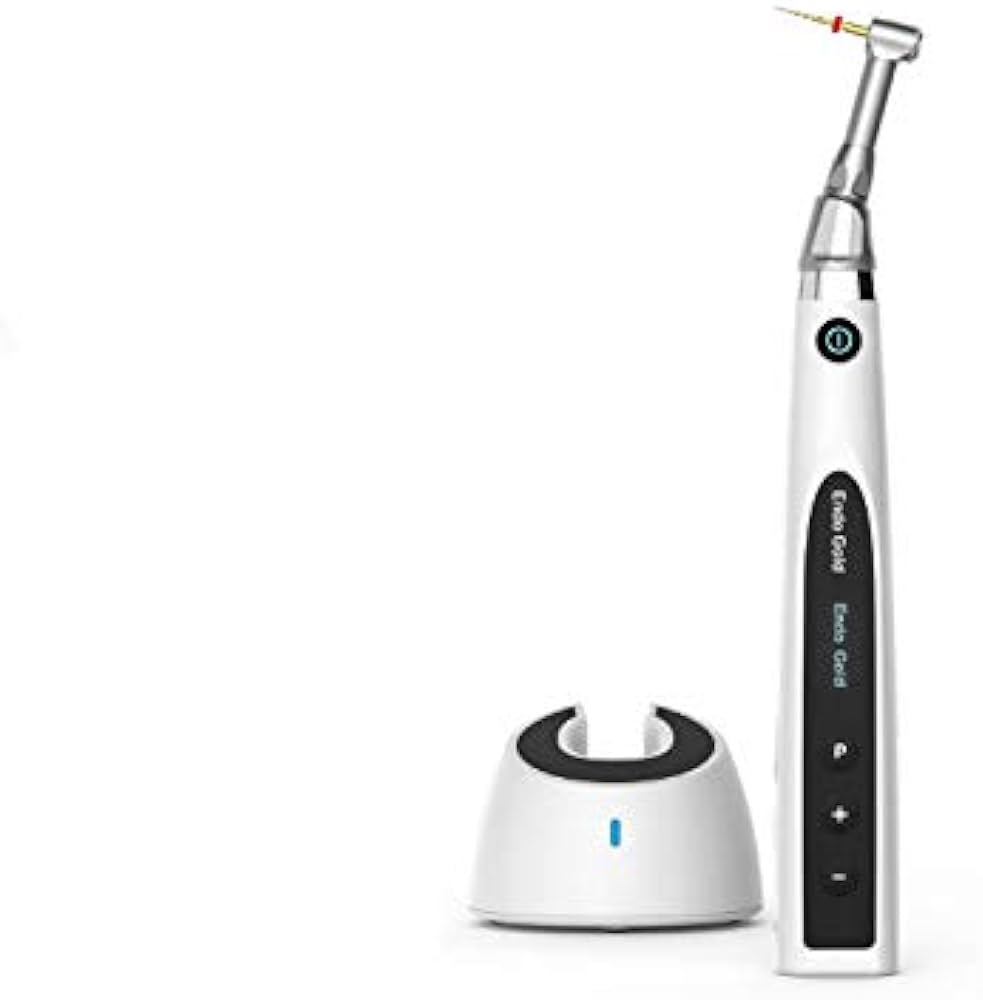 WOODPECKER ENDOMOTOR ENDOGOLD
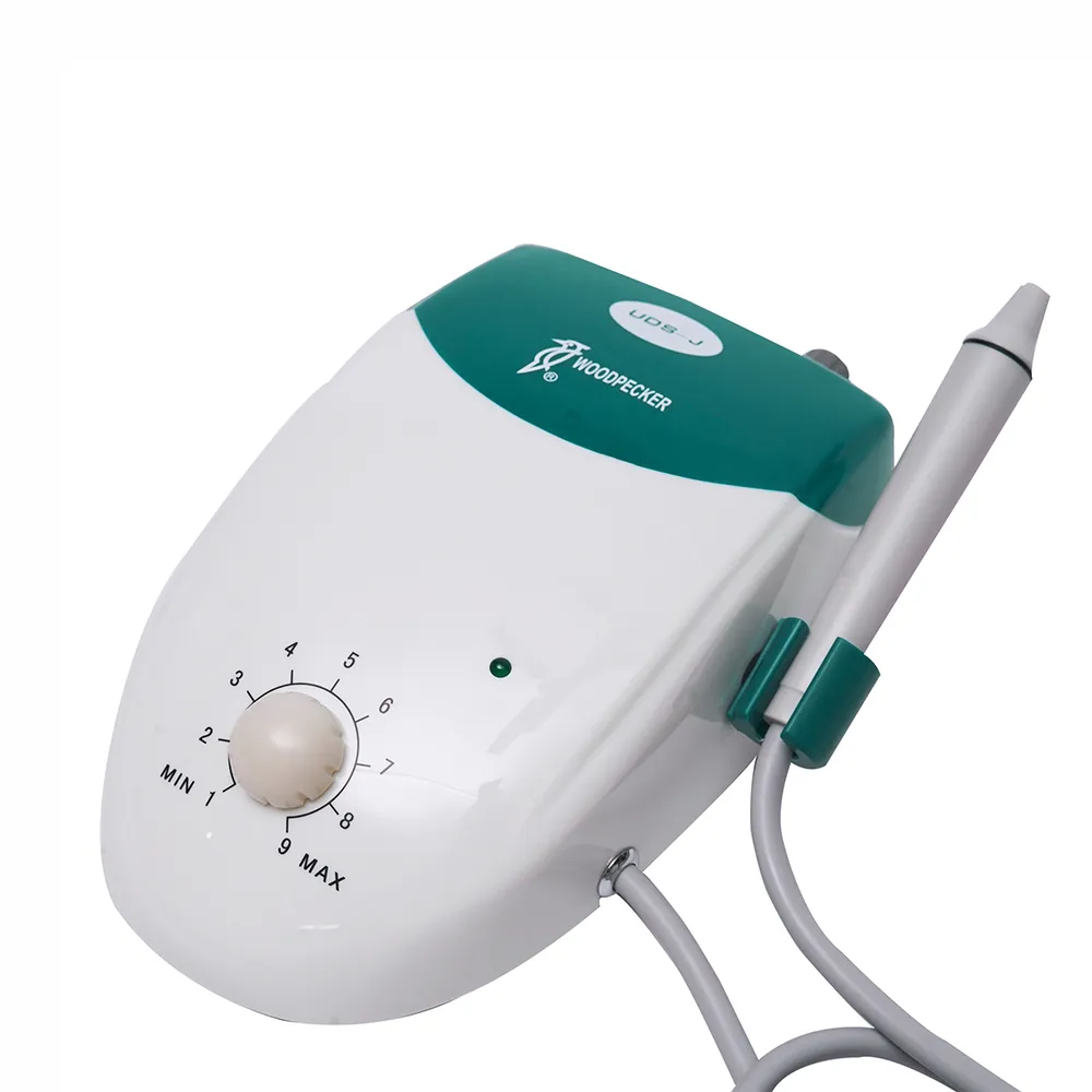 Woodpecker UDS-J Ultrasonic Scaler
.jpg) MicroMotor Marathon
MicroMotor Marathon
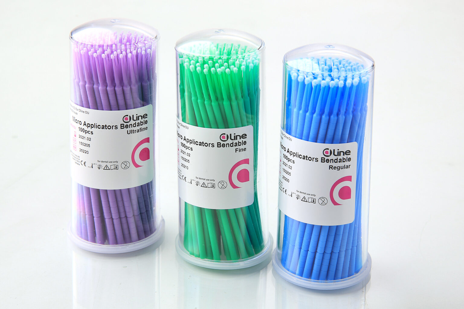 Micro Applicators 100 PCS
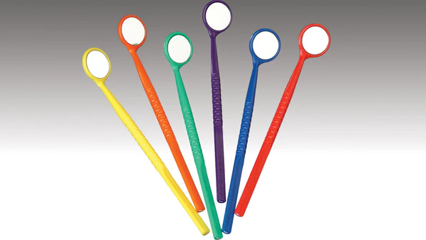 Disposable Mirrors with Handles
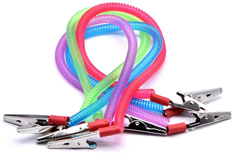 BIB Holders Coiled
 Sterilization Pouch 90*260mm 500 PCS
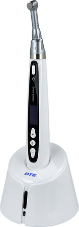 Endo-Smart Endomotor Woodpecker
 Riva Luting Plus: Modified Ionomer Powder / Liquid
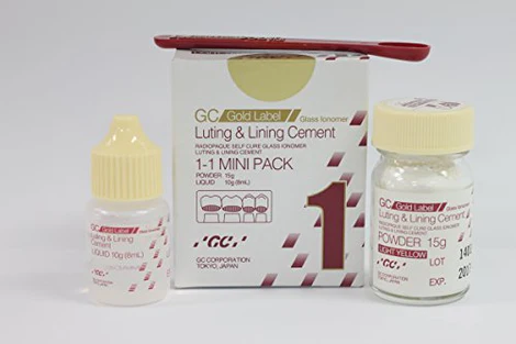 Fuji 1 Powder/ Liquid
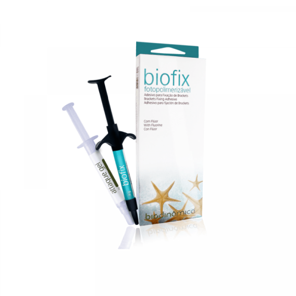 BIOFIX Adhesive brackets (4gr) - BIODINAMICA
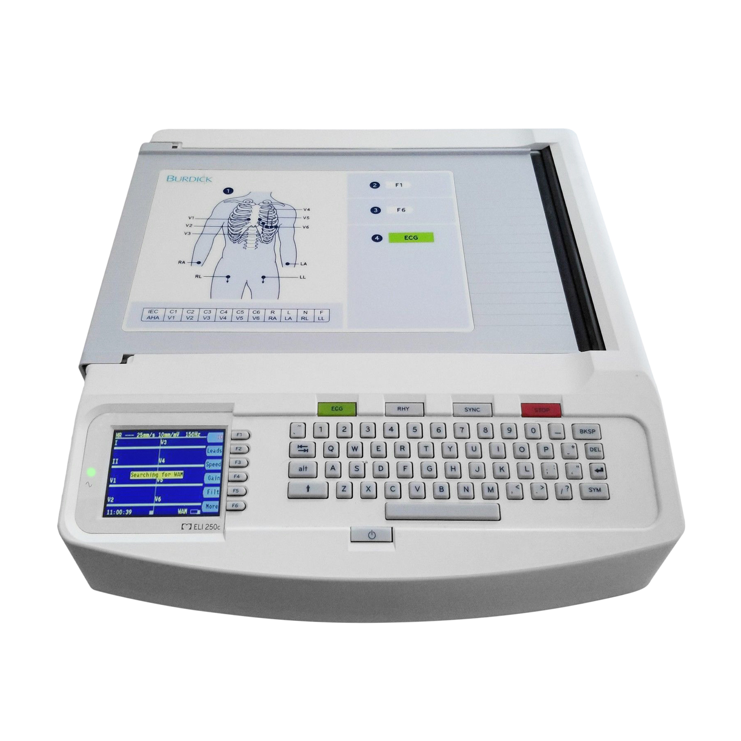 Burdick / Mortara ELI 250c EKG Machine (REFURB)
.jpg) For Sale GE HEALTHCARE Logiq C5 Ultrasound
For Sale GE HEALTHCARE Logiq C5 Ultrasound
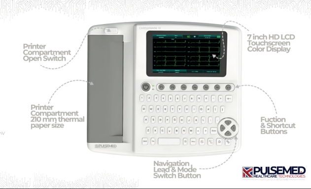 Pulsemed UK Cardiosense 12
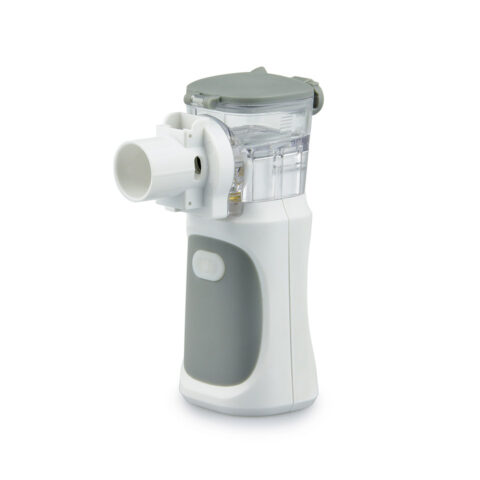 Mesh Nebuliser
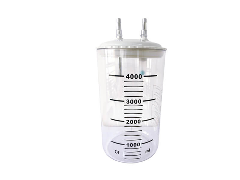 Suction Jar
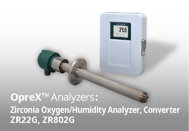 Zirconia Oxygen/Humidity Analyzer
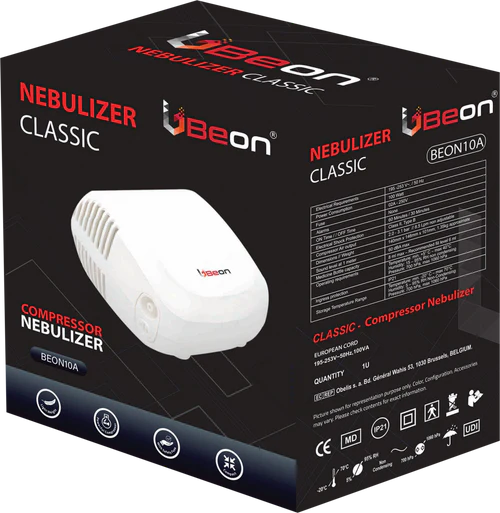 NEBULIZER BEON
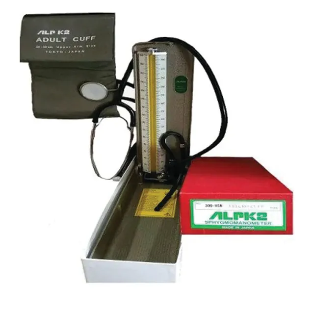 ALPK2
ALPK2
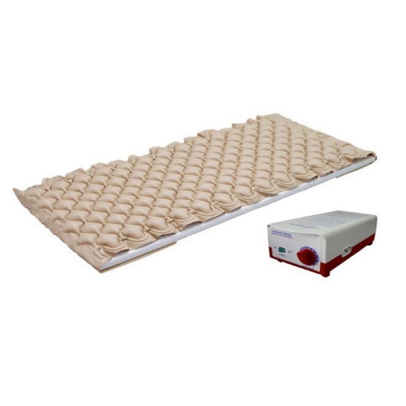 Certeza Air Mattress For Bedsores with Pump
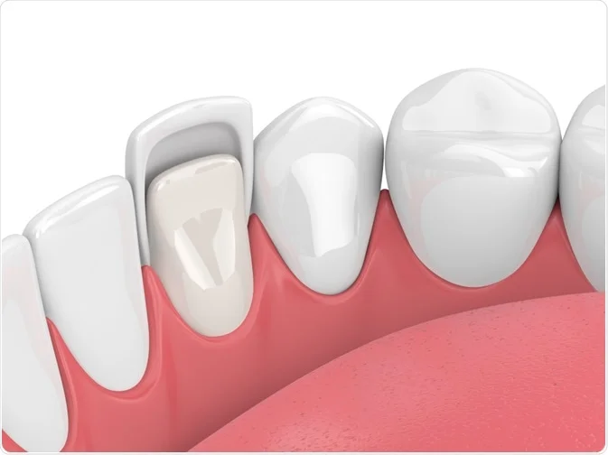 Veneers
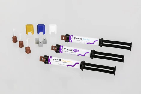 Dental composite Core It
 ECG GEL
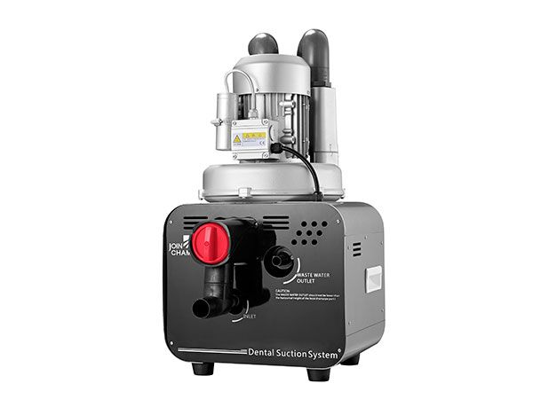 Dental Vacuum Suction System
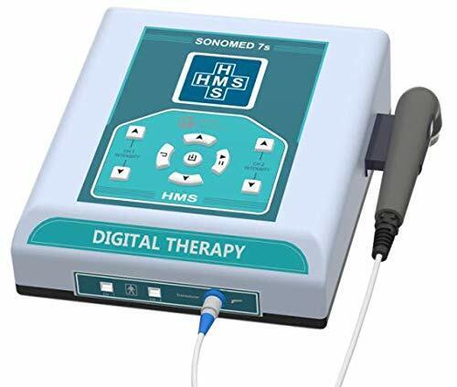 Interferential Electrotherapy Ultrasound therapy 1/3 Mhz Combination Therapy Ui
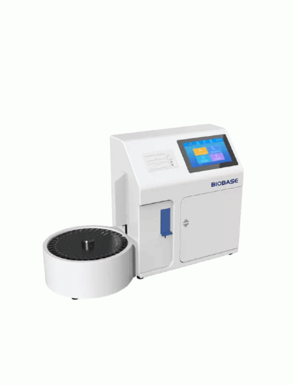 Electrolyte Analyzer
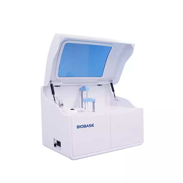 Auto Chemistry Analyzer
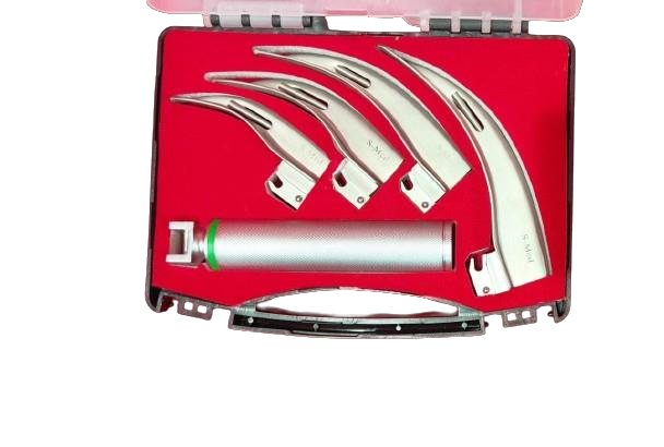 LARYGOSCOPE
.jpg) PEN LIGHT
PEN LIGHT
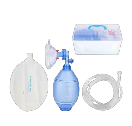 AMBUBAG ADULT
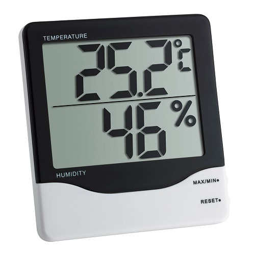 THERMO HYGROMETER
.jpg) MINISPIR
MINISPIR
.jpg) PORTABLE WASH BASIN
PORTABLE WASH BASIN
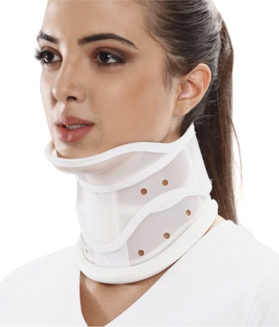 Cervical Collar Hard With Chin
.jpg) DRY BATH INCUBATOR DIGITAL
DRY BATH INCUBATOR DIGITAL
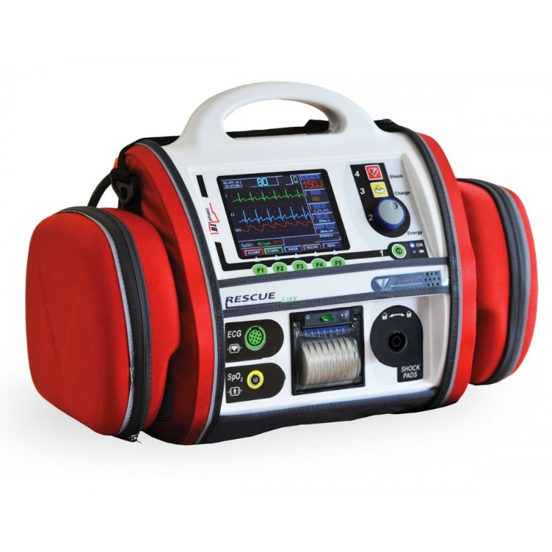 Progetti – Defibrillator
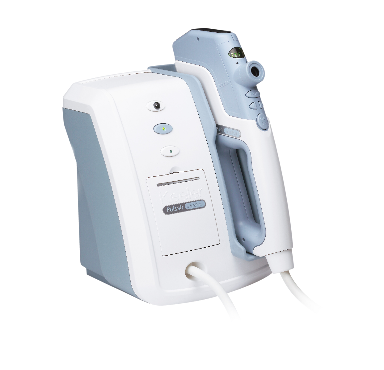 KEELER NON CONTACT TONOMETER
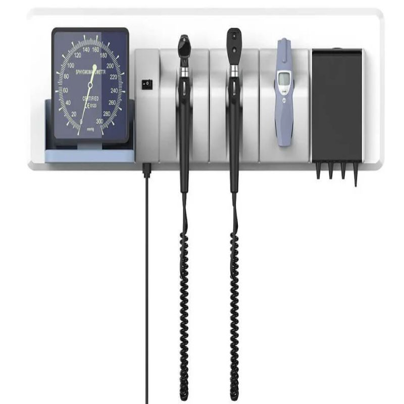 Wall-Mounted ENT Diagnostic Set
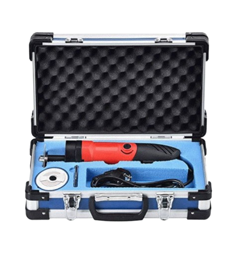 Electric Plaster Cutter
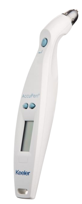 Keeler AccuPen Handheld applanation tonometer
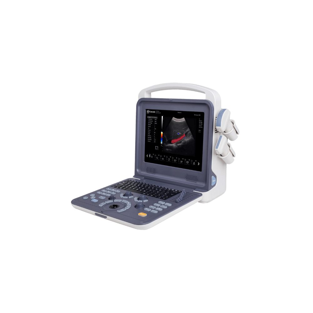 Portable Ultrasound System (Color ) – Cansonic K0
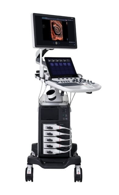 SONOSCAPE ULTRASOUND MACHINE P50
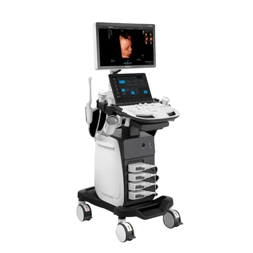 SONOSCAPE ULTRASOUND MACHINE P11 ELITE
.png) SONOSCAPE ULTRASOUND MACHINE P15
SONOSCAPE ULTRASOUND MACHINE P15
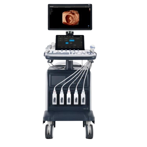 SONOSCAPE ULTRASOUND MACHINE S60
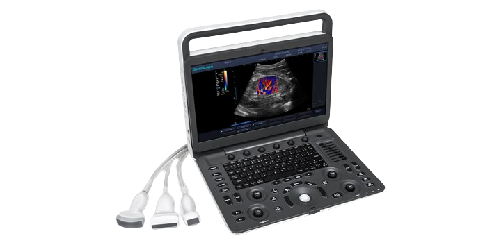 SONOSCAPE PORTABLE ULTRASOUND MACHINE E3
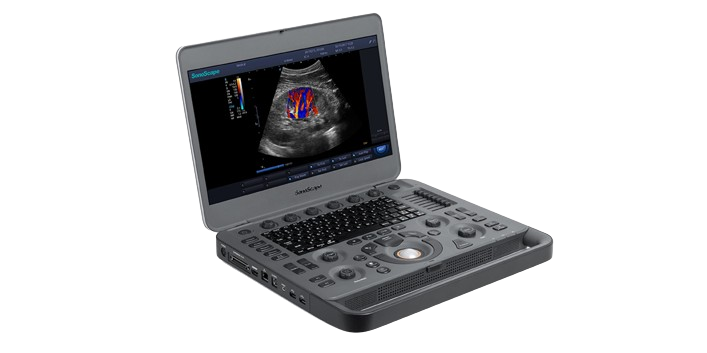 SONOSCAPE PORTABLE ULTRASOUND MACHINE X3
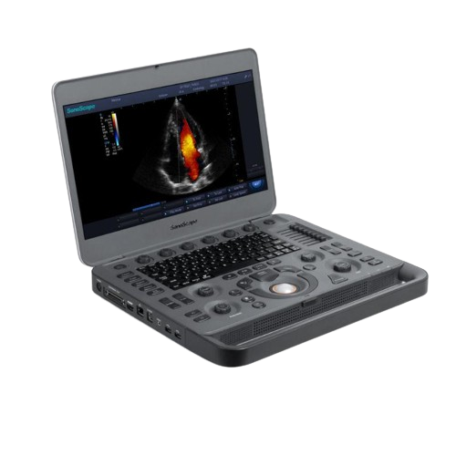 SONOSCAPE PORTABLE ULTRASOUND MACHINE X5
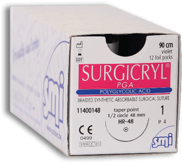 SMI Surgicryl Polyglycolic Acid Violet Sutures
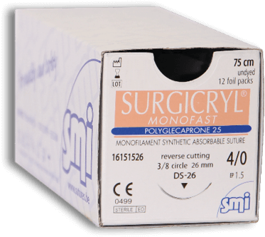 SMI Surgicryl Monofast Polyglecaprone 25 Violet Suture
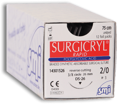 SMI Surgicryl Rapid Polyglycolic Acid Undyed Suture
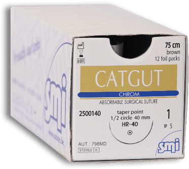 SMI Catgut Chrom Brown Suture
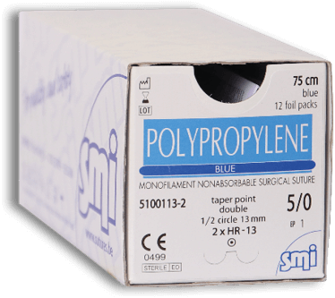 SMI Polypropylene Blue Suture
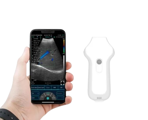 ECOLESS WIRELESS CARDIAC ULTRASOUND MACHINE
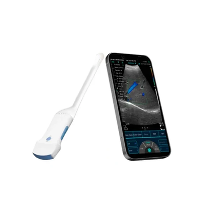 ECOLESS WIRELESS CONVEX ULTRASOUND MACHINE
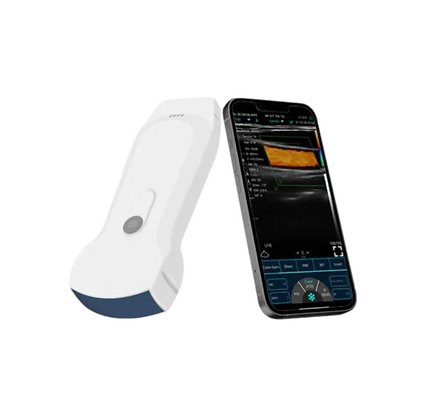 ECOLESS WIRELESS 3 IN 1 ULTRASOUND MACHINE
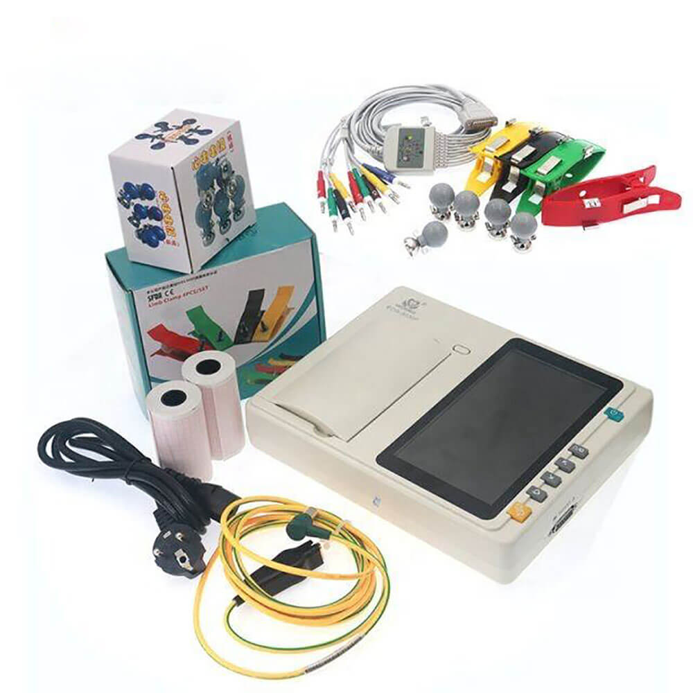 3 Channel 7 Inch Touch Screen Portable Electrocardiograph ECG Machine
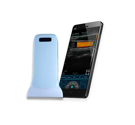 ECOLESS WIRELESS LINEAR ULTRASOUND MACHINE
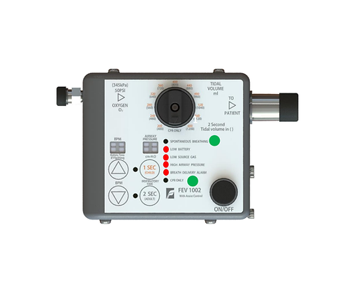 EMERGENCY VENTILATOR
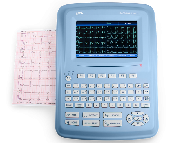 BPL ECG MACHINE 12 CHANNEL
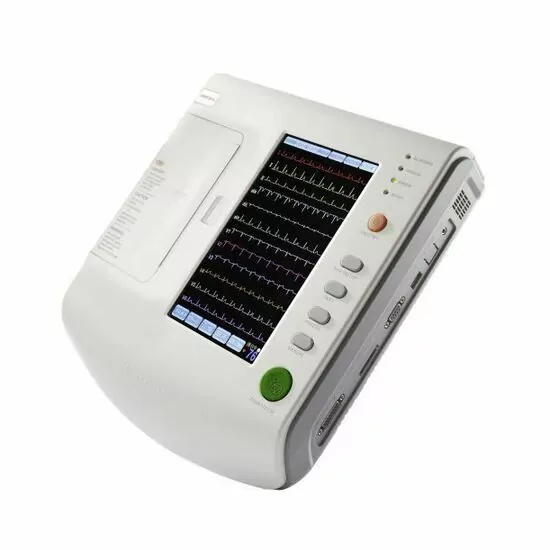 Zoncare 12 Channel ECG Machine
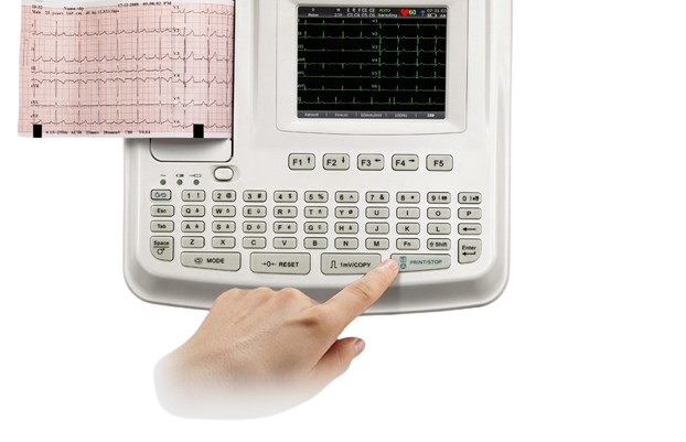 Edan ECG Machine SE-601 Series 6-Channel ECG
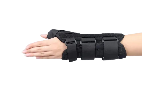 Wrist Thumb Spica Splint
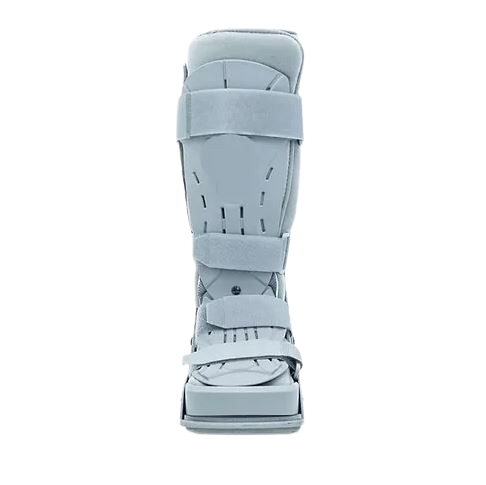 Walker Brace
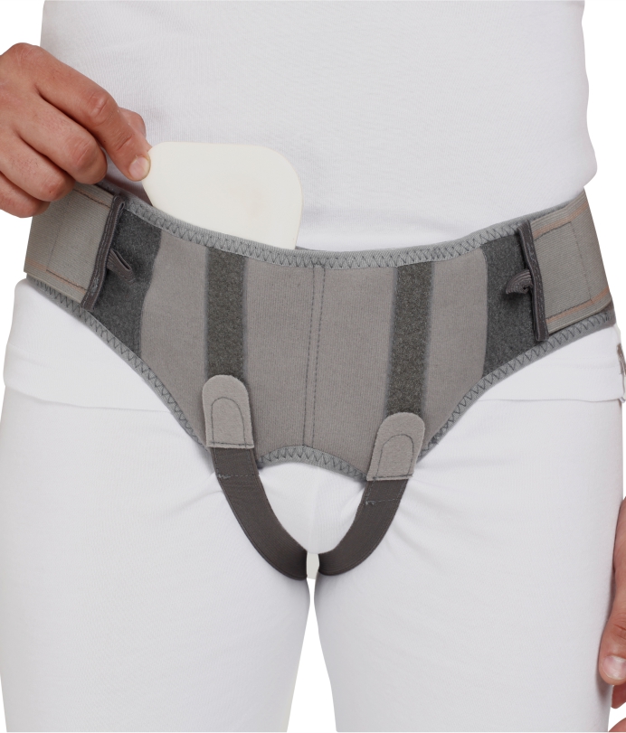 Hernia Belt
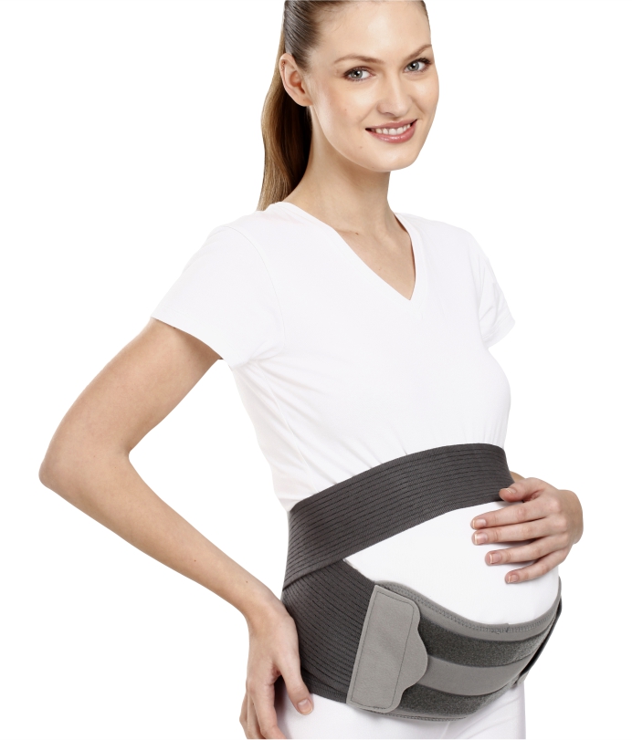 Pregnancy Back Support
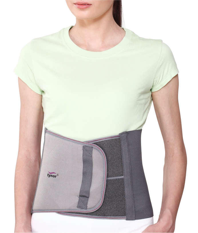 Abdominal Support
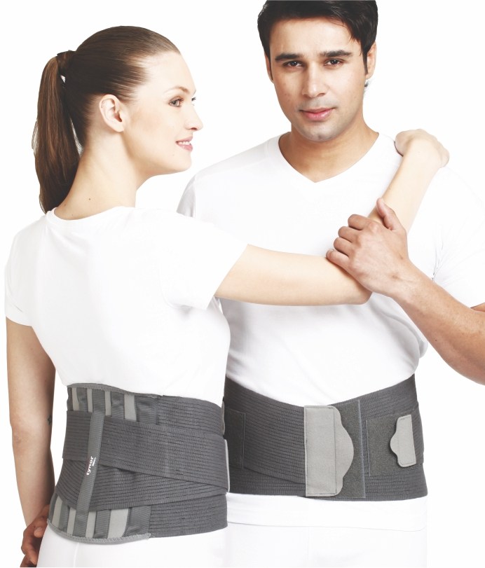 Lumbopore
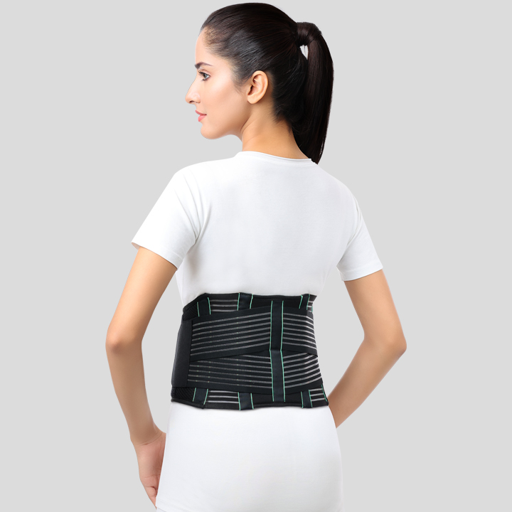 Lumbo Sacral Belt
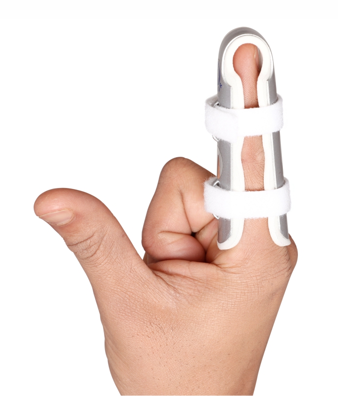 Finger Cot
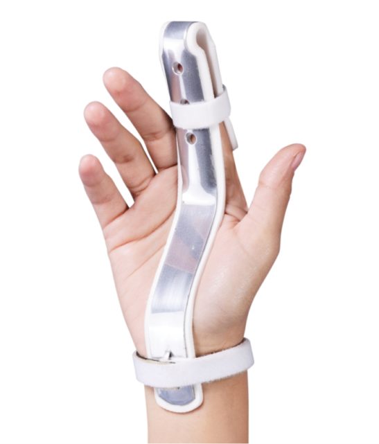 Finger Extension Splint
.jpg) Cervical Collar Soft with Support
Cervical Collar Soft with Support
 Collar Soft (Firm Density)
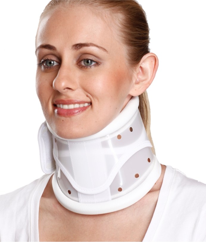 Cervical Collar (Hard Adjustable Height)
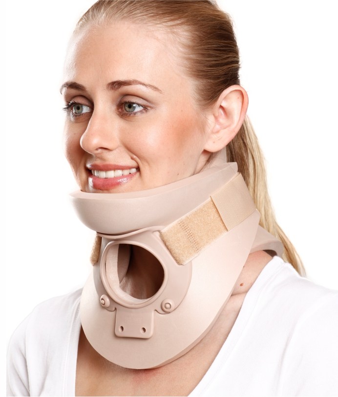 Cervical Orthosis (Philadelphia)
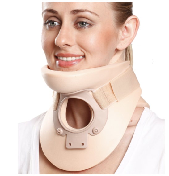 Cervical Orthosis (Philadlphia) Plastazote
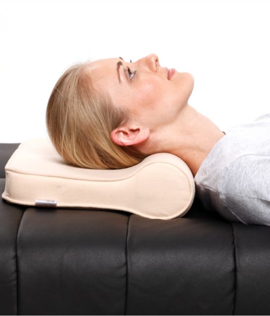 Cervical Pillow Regular
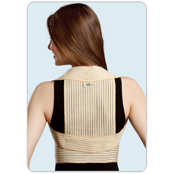 Clavicle Posture Shoulder Brace
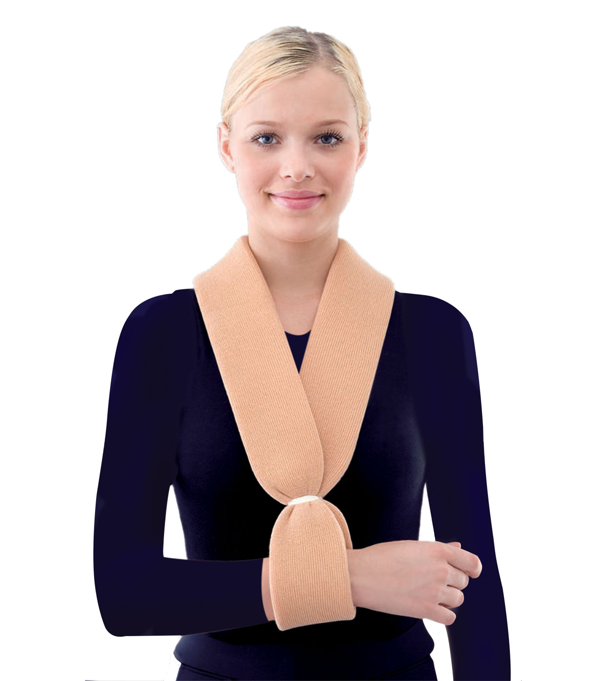 Collar & Cuff Sling
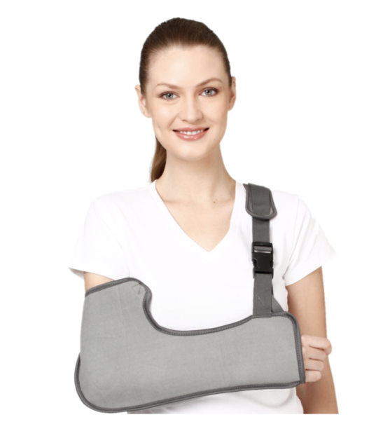 Pouch Arm Sling (Tropical)
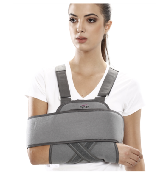 Universal Shoulder Immobiliser
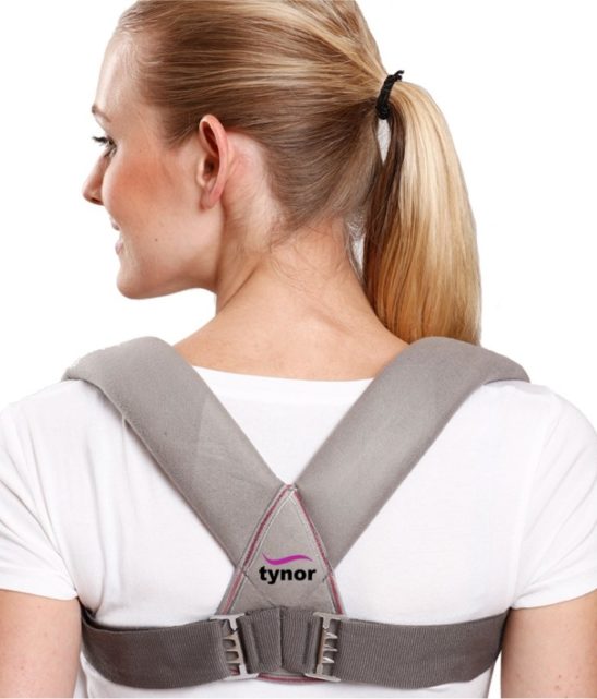 Clavicle Brace with Buckle
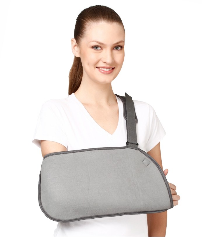 Pouch Arm Sling (Baggy)
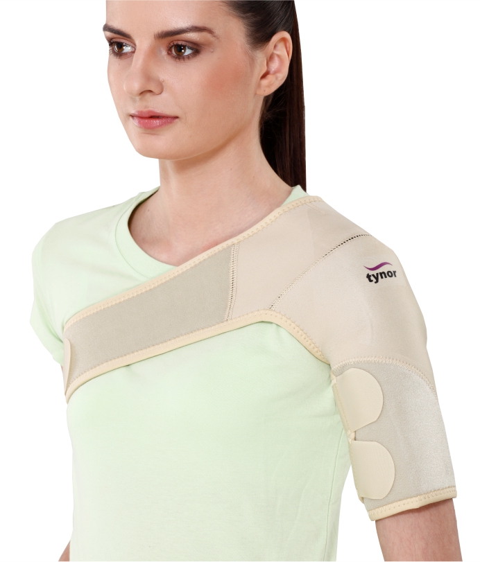 Shoulder Support (Neoprene)
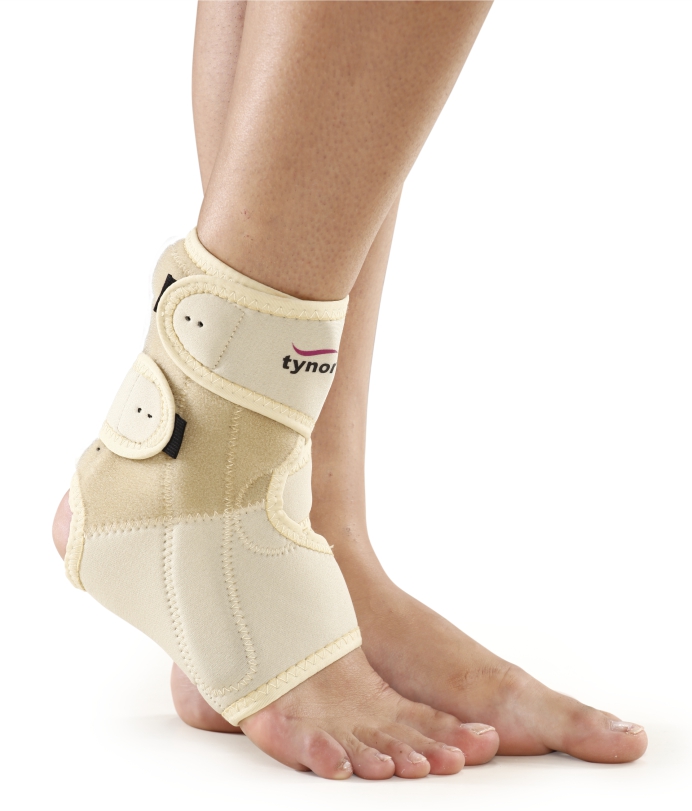 Ankle Support
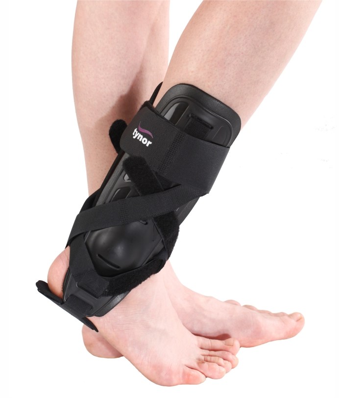 Ankle Splint
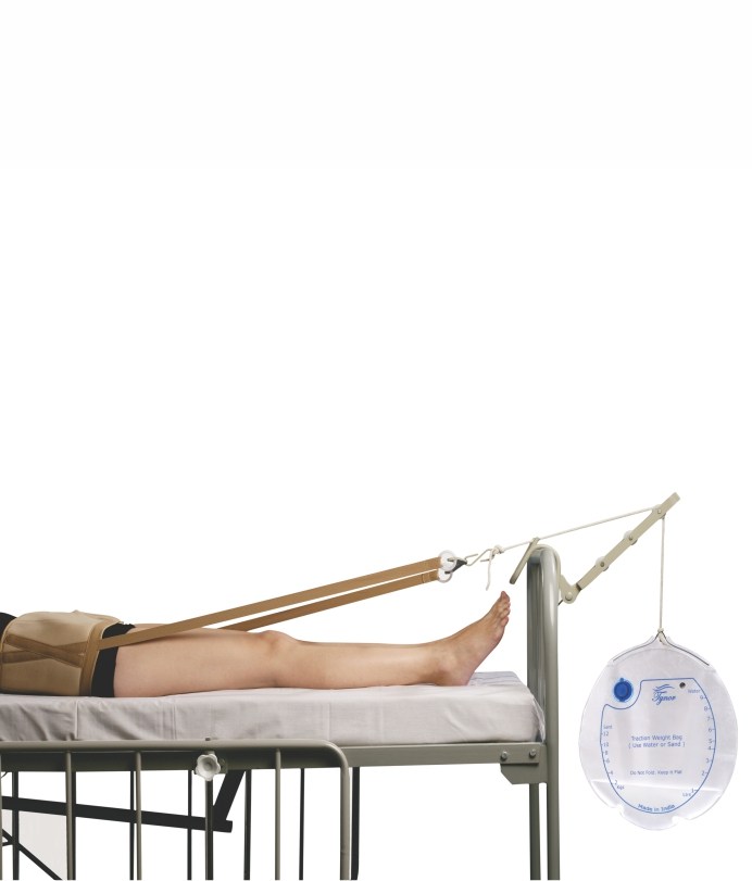 Pelvic Traction Kit
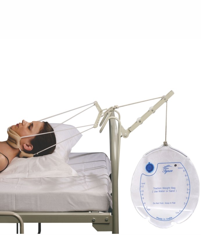 Cervical Traction Kit
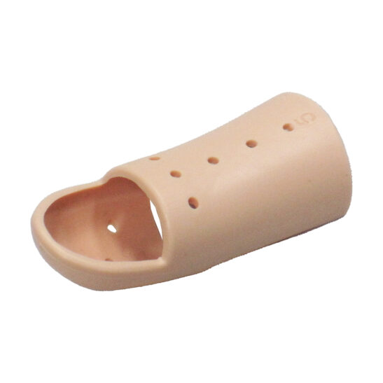 Mallet Finger Splint
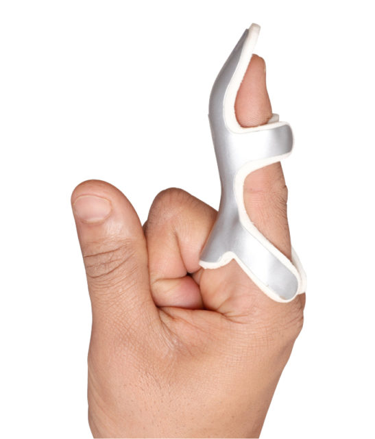 Frog Splint
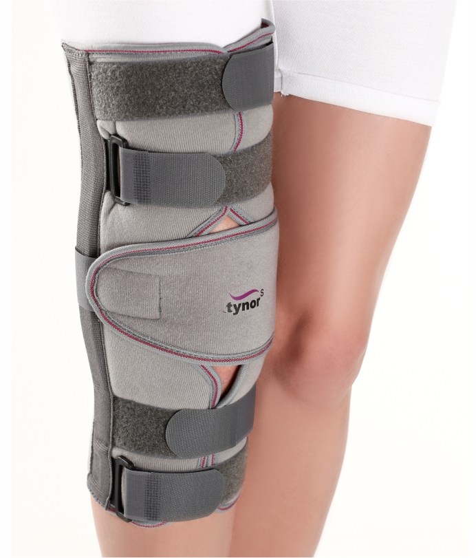 Knee Immobilizer
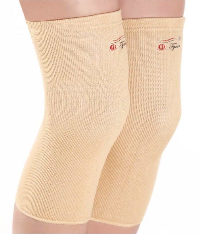 Knee Cap
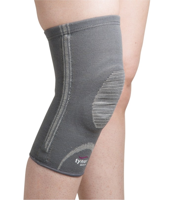 Knee Cap with Patellar Ring
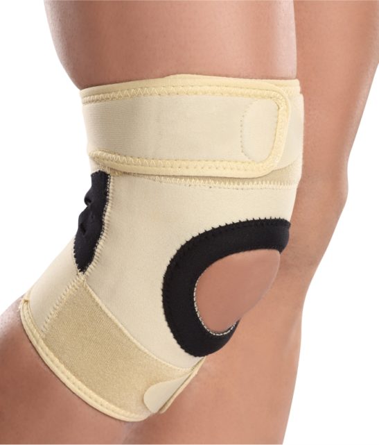 Knee Support Sportif
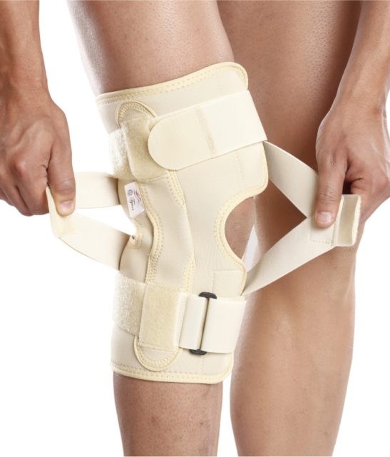 OA Knee Support
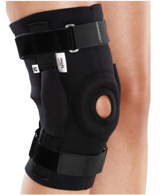 Knee Wrap Hinged
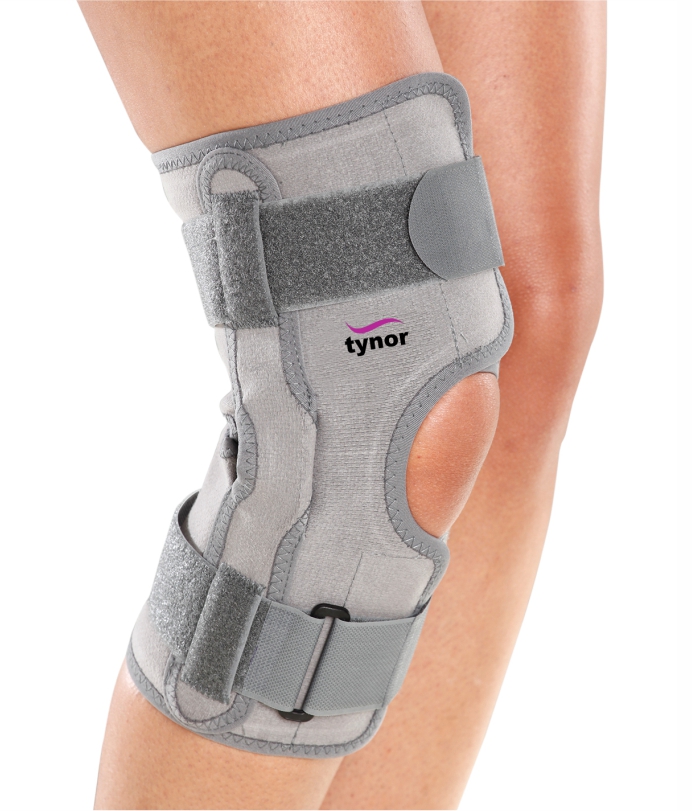 Functional Knee Support
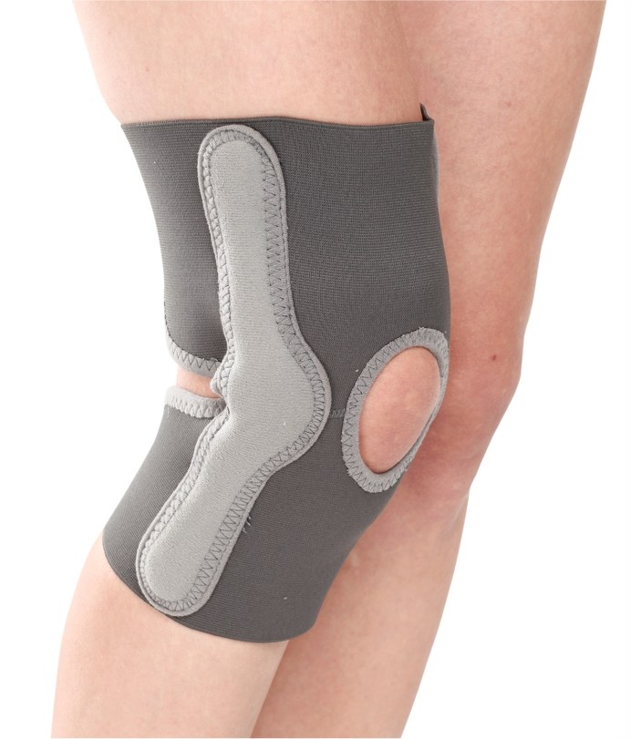 Elastic Knee Support
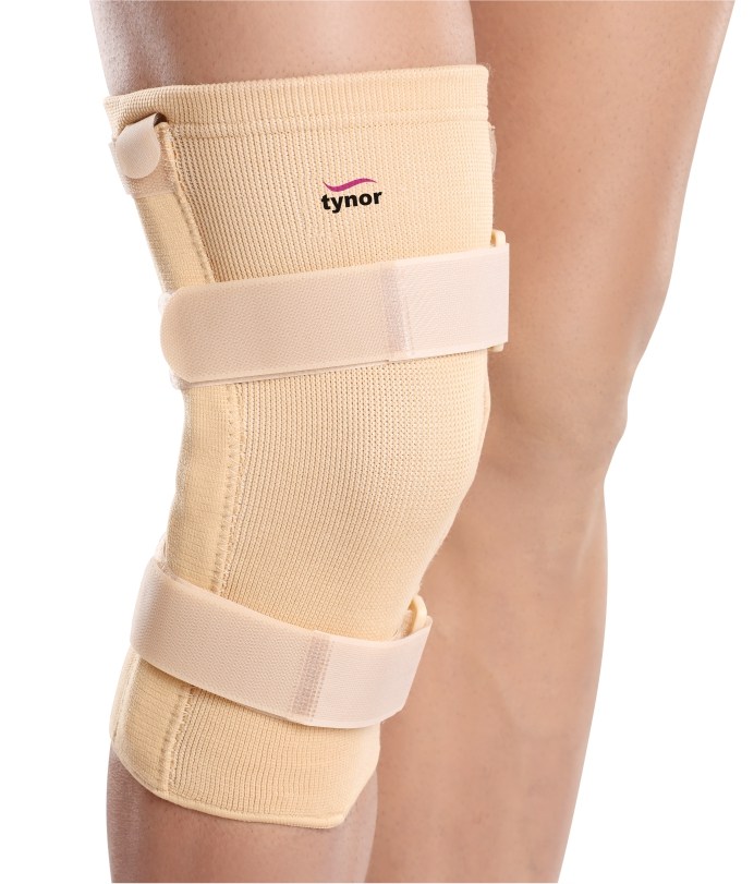 Knee Cap (with Rigid Hinge)
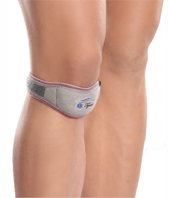 Pattelar Support
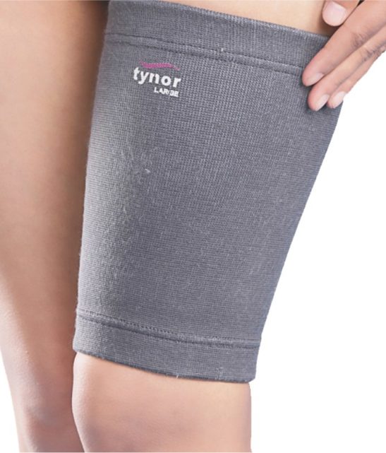 Thigh Support
.png) Electric 5 Function Hospital Bed With Mattress
Electric 5 Function Hospital Bed With Mattress
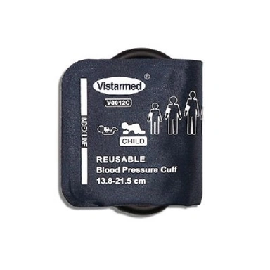 VISTARMED BP CUFF for patient monitors and sphygmomanometers - Single tube - CHILD
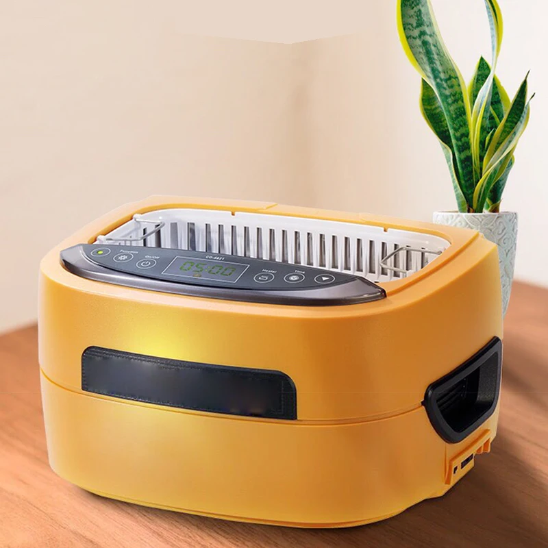 PROFESSIONAL ULTRASONIC CLEANER
 CONTEC ECG600G Digital 6 channel Electrocardiograph ECG machine EKG CE
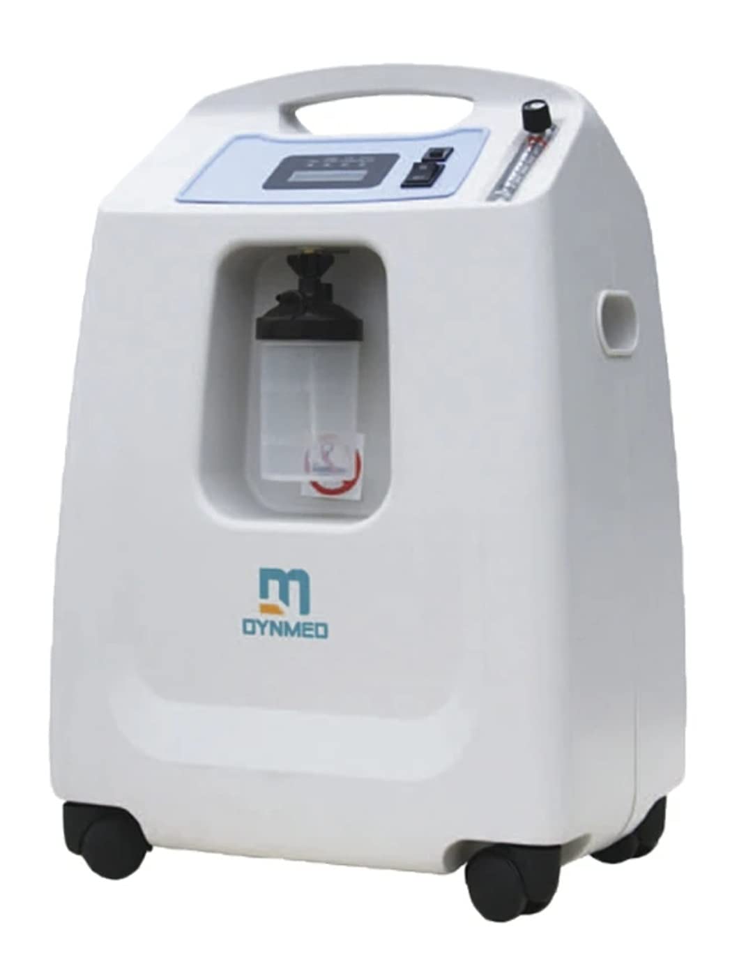 Dynmed oxygen concentrator 10 ltr
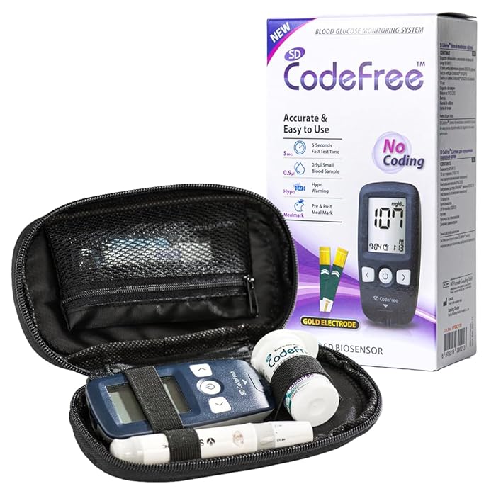 Codefree blood glucose monitor
 Medikro ® Pro Laboratory Spirometer
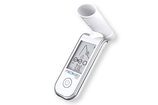 Medikro ® Duo - The 2 minute asthma and COPD screener
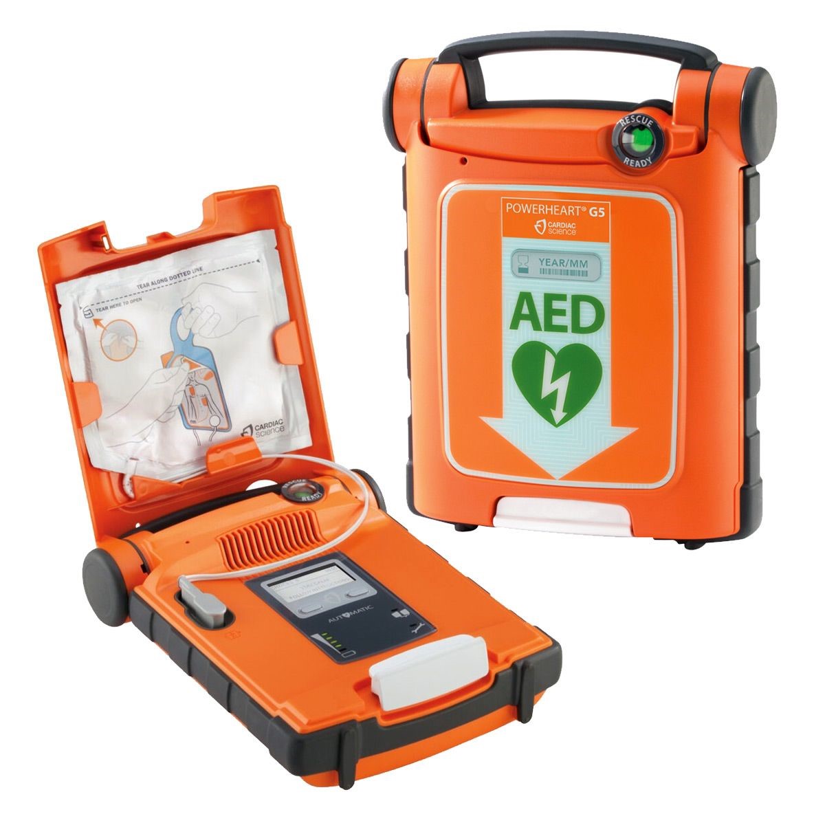 POWERHEART® G5 AED
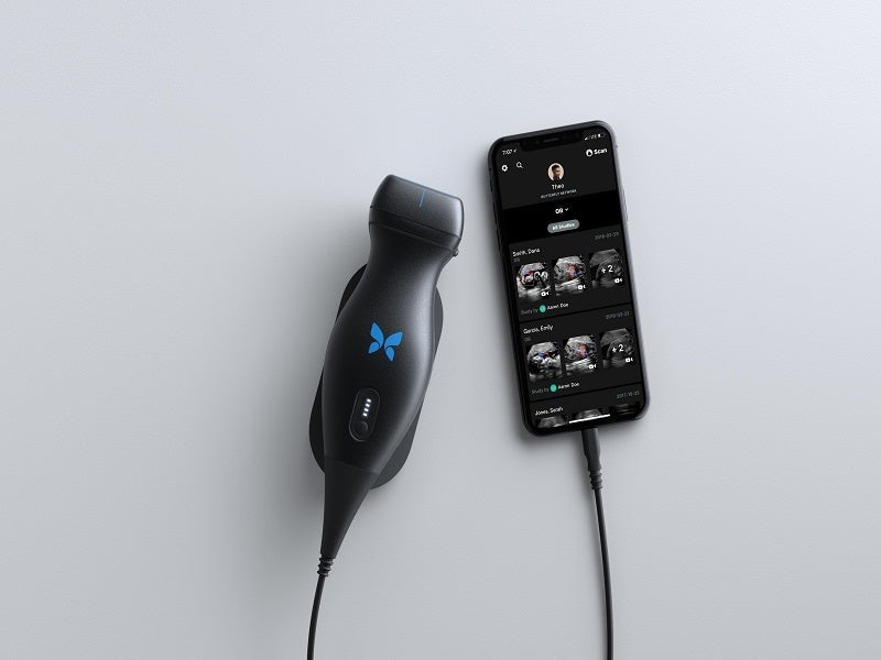 Handheld, whole-body imaging - Ultrasound
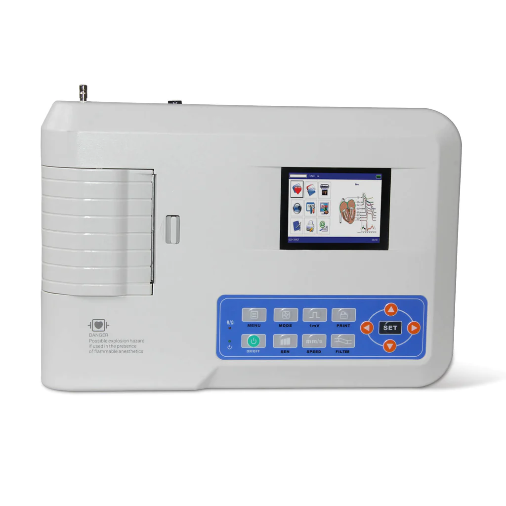 CONTEC ECG300G Electrocardiograph
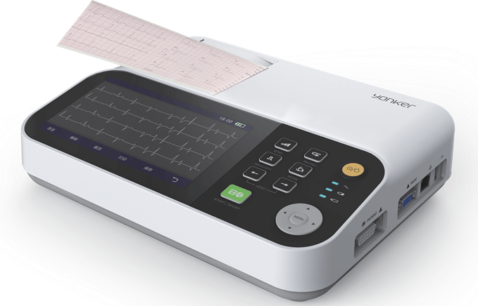 Yonker 7inch Display 3 Channel ECG Machine With Touch Screen
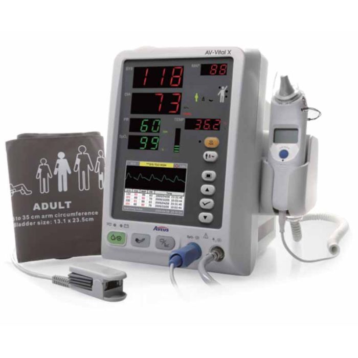 Vital Sign Monitor Aveus AV-Vital X
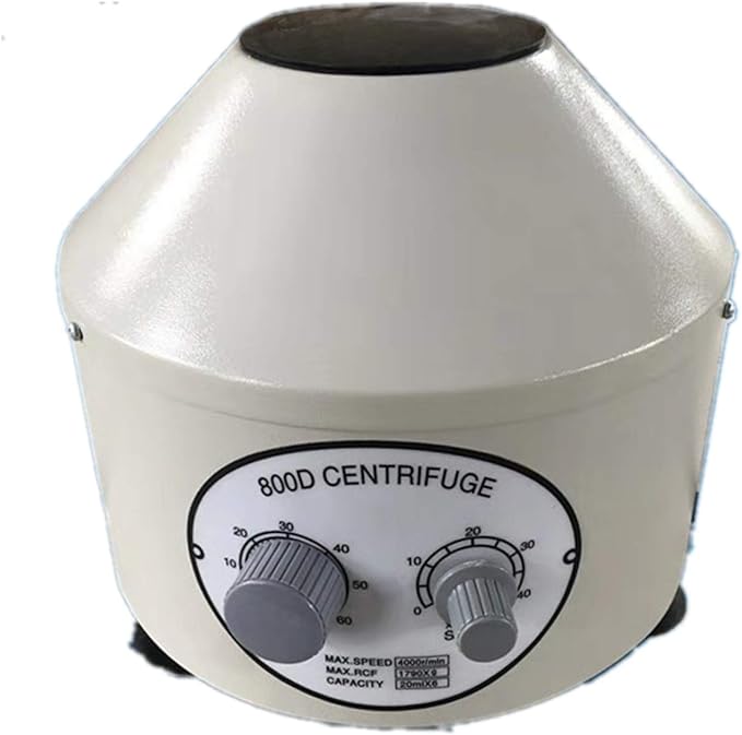 Large Capacity Desktop Clinical Lab 800D Centrifuge
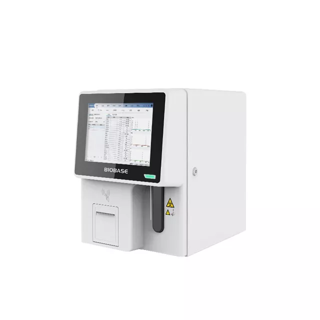 Auto Hematology Analyzer
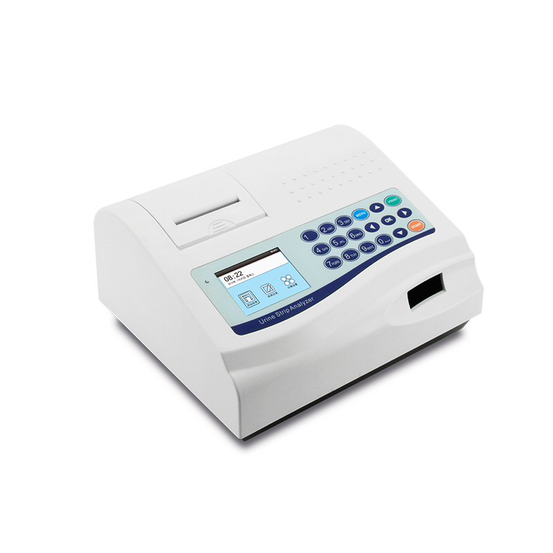 BC400 Urine Analyzer
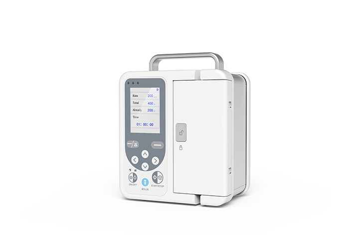 SP750 Infusion Pump
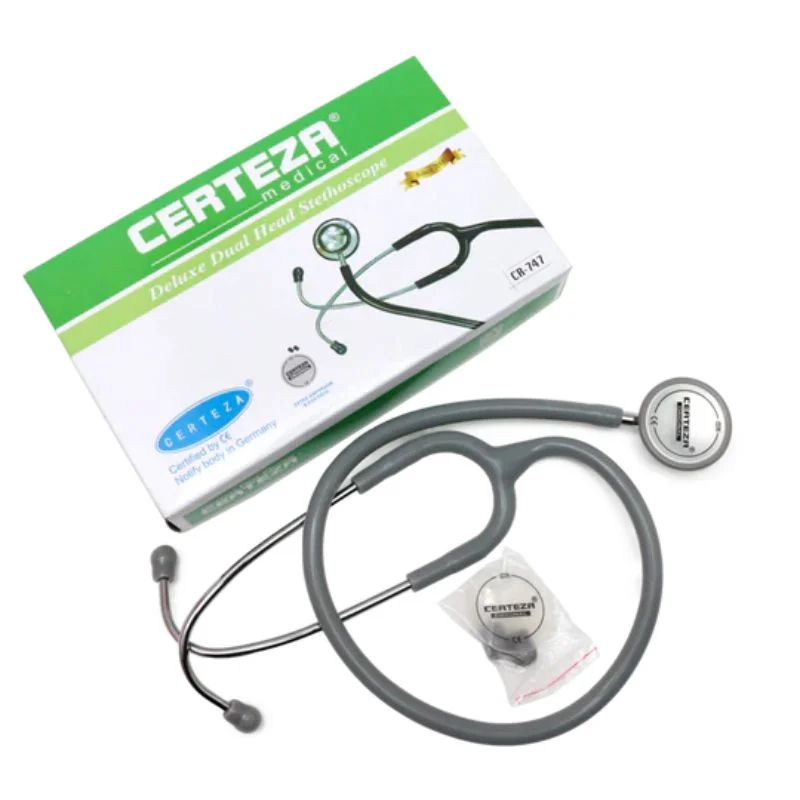 Certeza Stethoscope
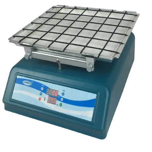 Laboratory automatic Oscillator shaker
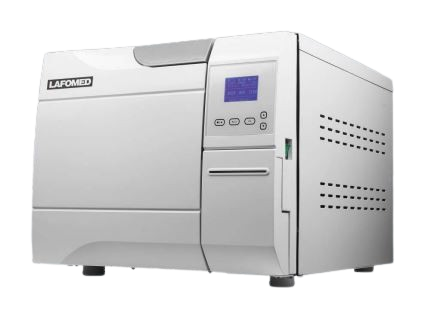 Lafomed Autoclave 23 ltr
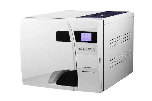 Lafomed Autoclave 18 ltr
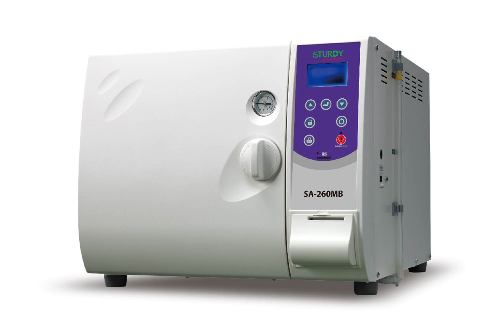 Sturdy autoclave 24 ltr
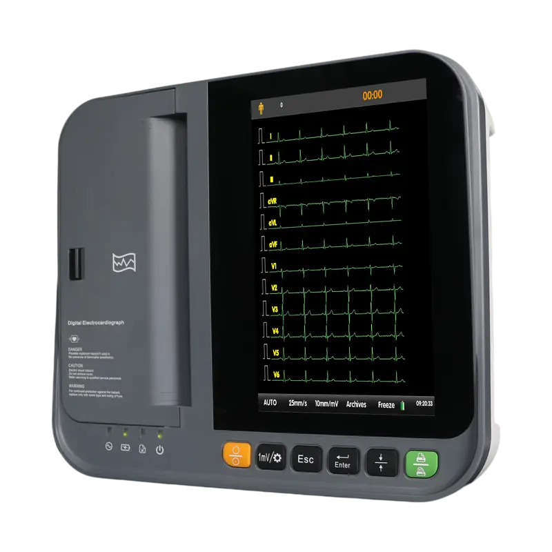 DAWEI 12Channel ECG Machine
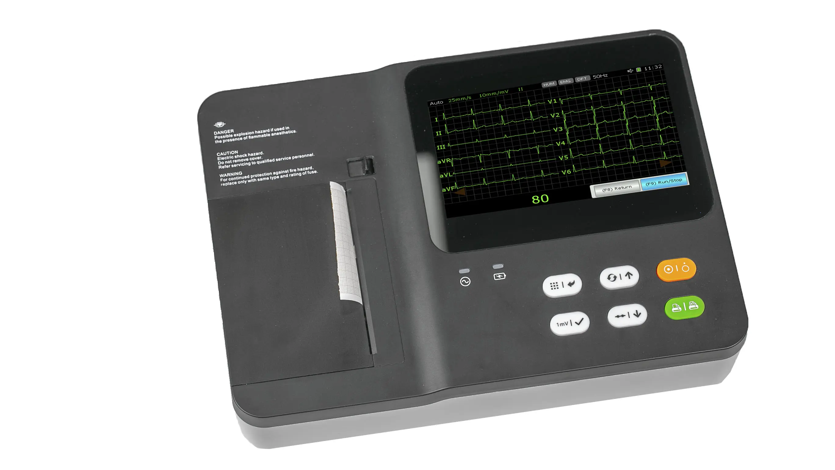 Dawei ECG Machine 3 Channel
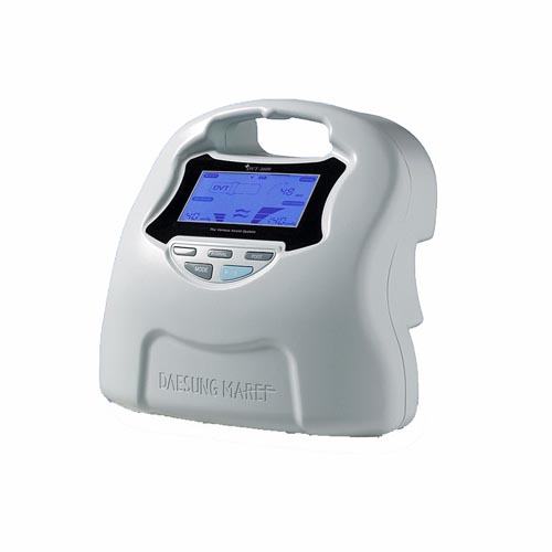 DVT 2600
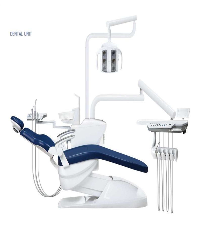 WOSON DENTALCHAIR SONZ
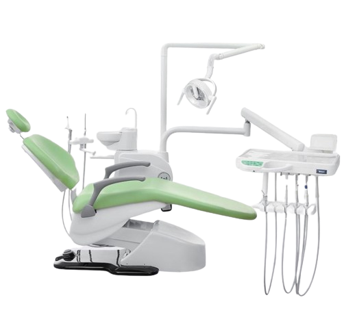 WOSON DENTAL CHAIR WODO MILLE
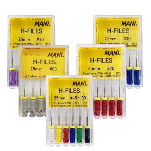 H FILES
 POLISHING BRICKS
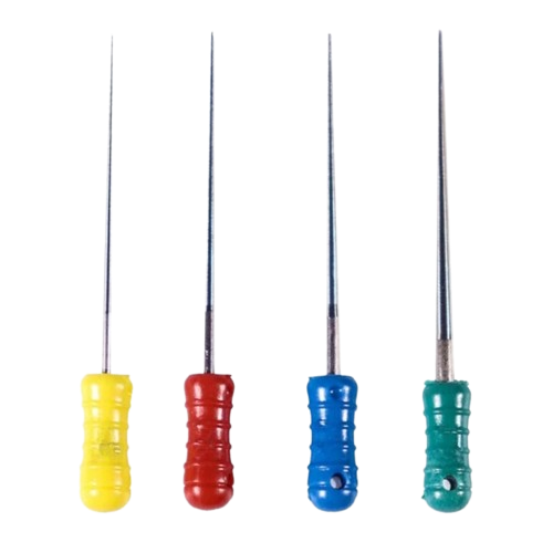 SPREADER
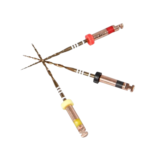 RC FILES
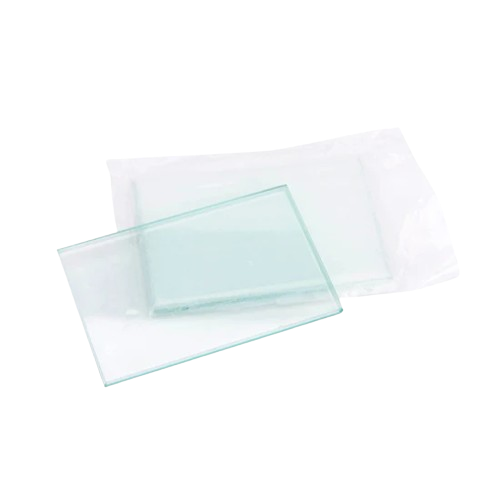 MIXING PLATE/GLASS
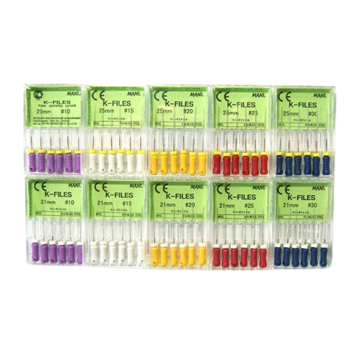 K FILES
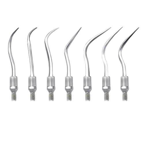 SCALER TIPS
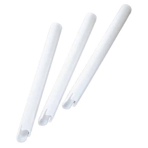 VENTED TIPS
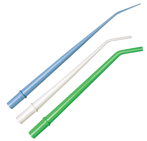 SURGICAL TIPS
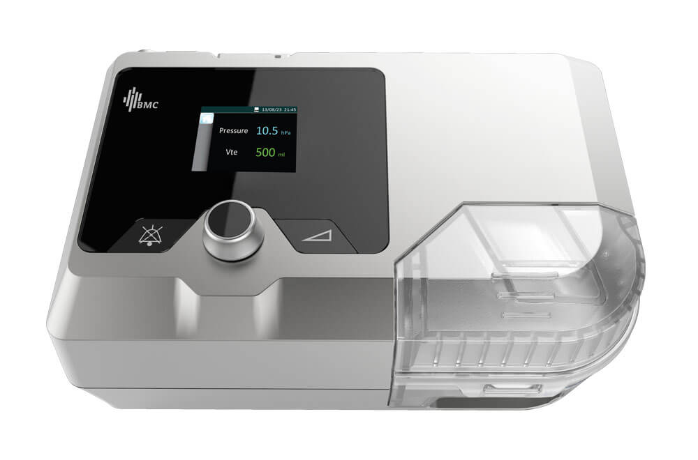 BMC G2S B25VT BiPAP ST WITH HUMIDIFIER AND MASK
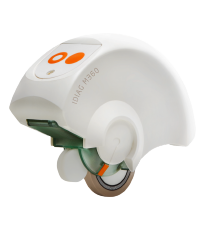 SPINAL MOUSE IDIAG M360
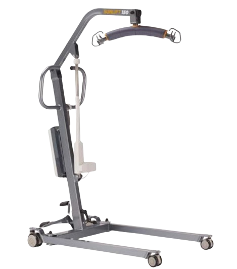 PATIENT LIFTER
.webp) HANDHELD PULSE OXIMETER WITH NEONATAL PROBE
HANDHELD PULSE OXIMETER WITH NEONATAL PROBE
.webp) HANDHELD PULSE OXIMETER WITH PAEDIATRIC PROBE
HANDHELD PULSE OXIMETER WITH PAEDIATRIC PROBE
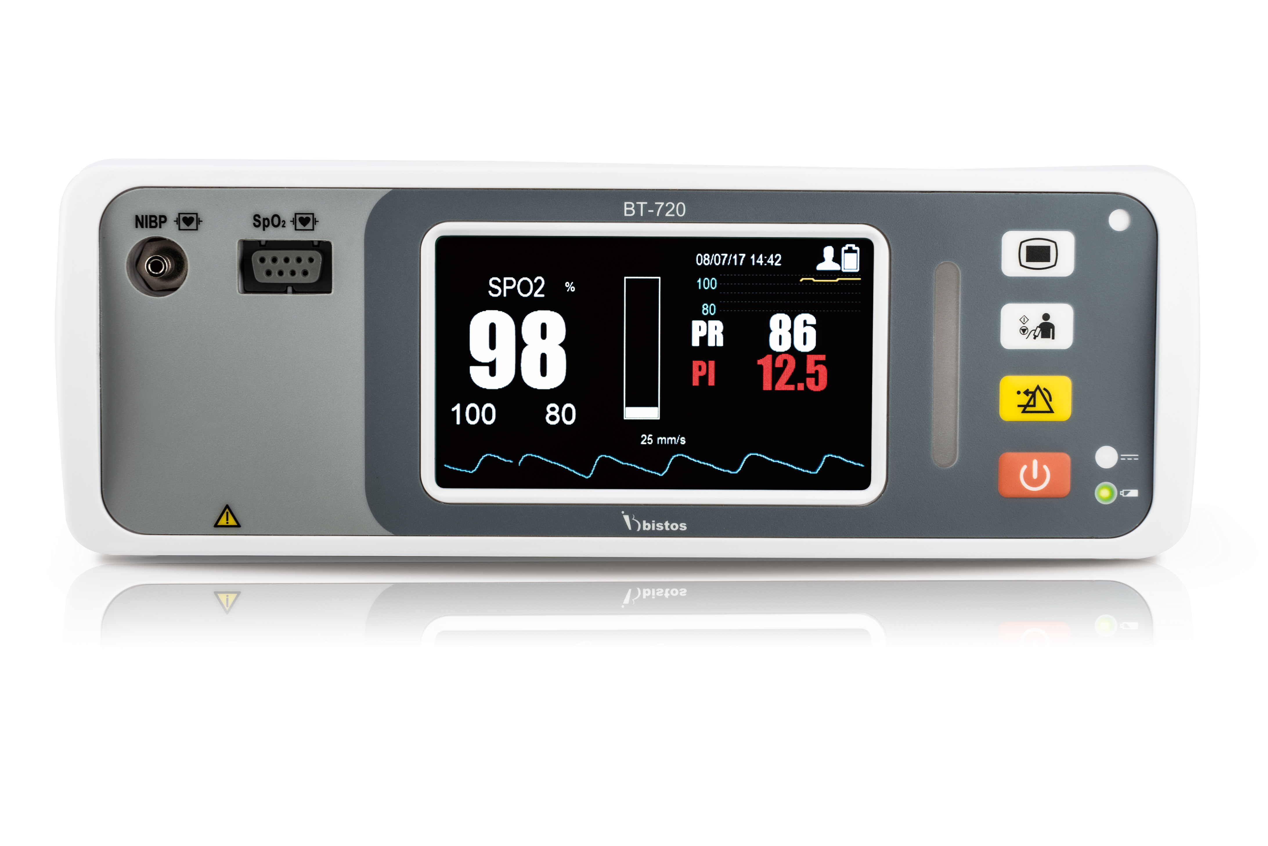 BISTOS BT-720 VITAL SIGN MONITOR
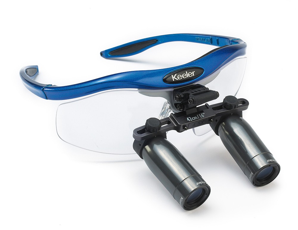 PRISMATIC LOUPES 3.5X MAGNIFICATION
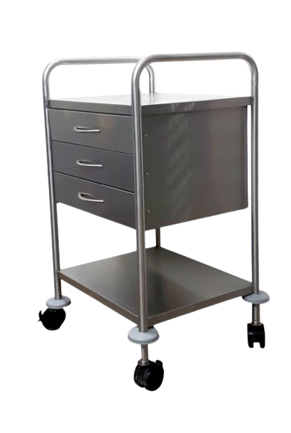 DRESSING TROLLEY 3 DRAWER 1 SHELVE PIPE WITH SIDE RAIL
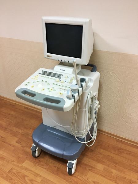 SSI-5000 Sonoscape ultrasound machine Used
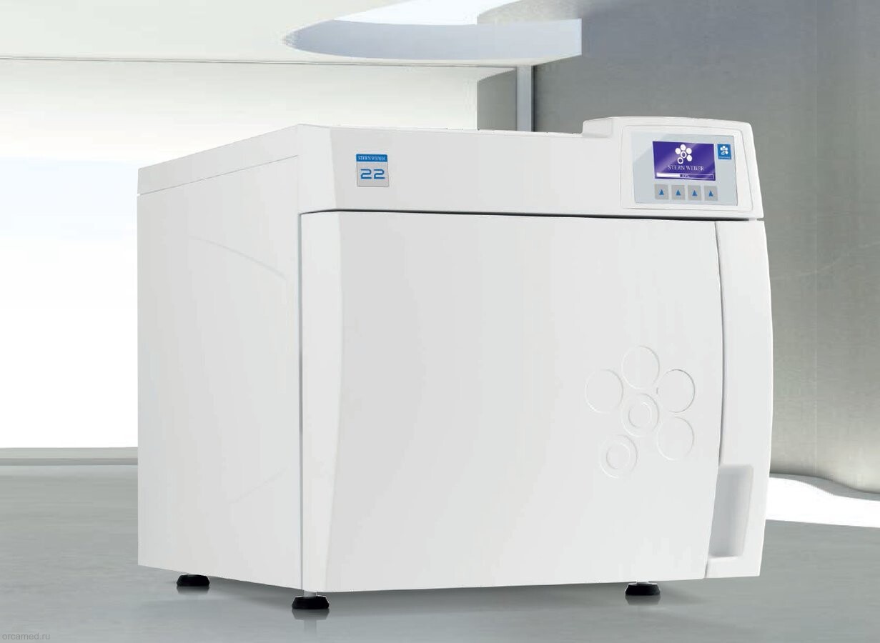 STERN WEBBER AUTOCLAVE 22 LTR USED
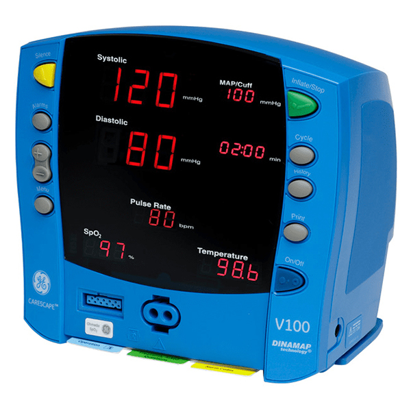 GE Vital Signs Monitor Carescape V100 Dinamap Refurbished
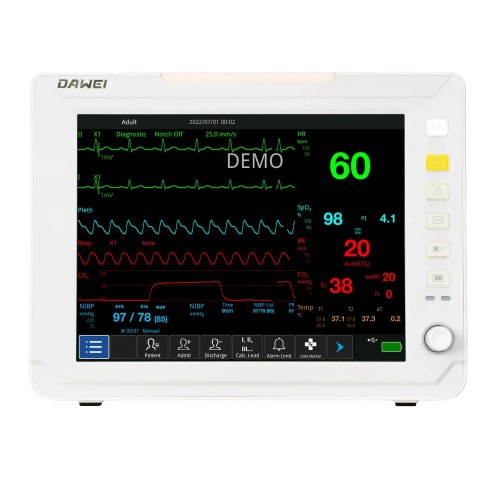 Dawei HM10 multi-parameter patient monitor
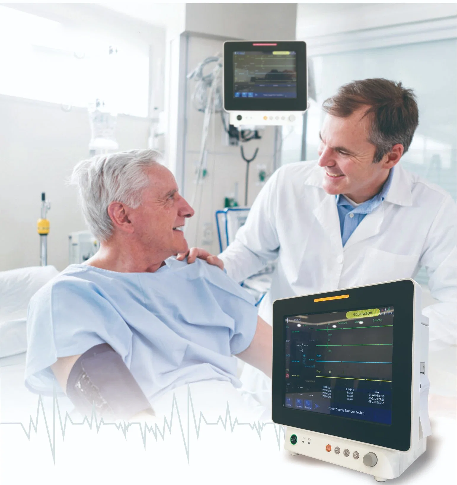 Superstarmed SP J12 multi-parameter patient monitor
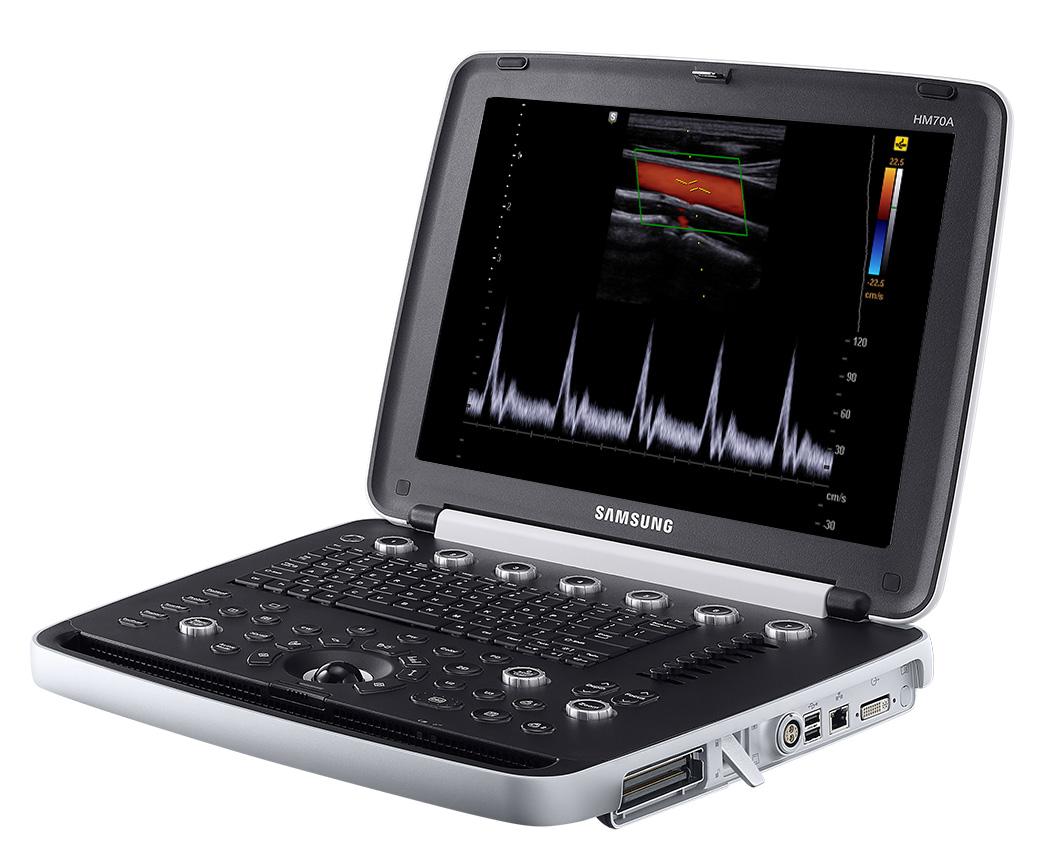 Samsung Ultrasound HM70A
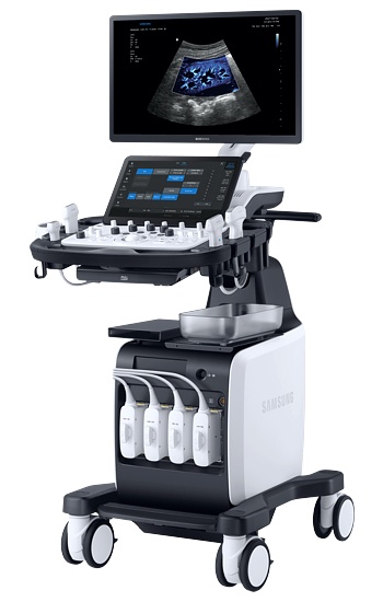 SAMSUNG V8 HIGH END ULTRASOUND SYSTEM
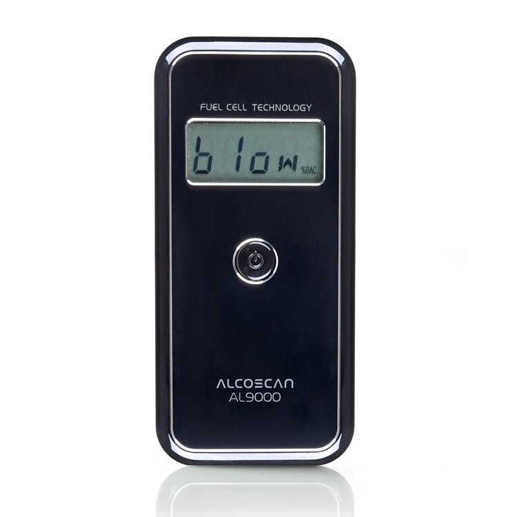 Alcoscan AL9000 Fuel Cell Breathalyzer
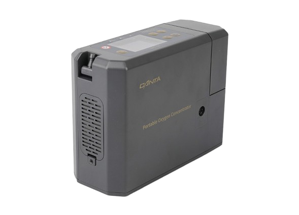 Canta Portable Oxygen Concentrator 1-5L
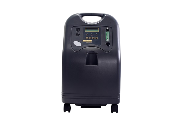 Oxygen Concentrator 5L CANTA
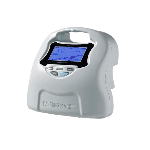 DS MAREF DVT 2600
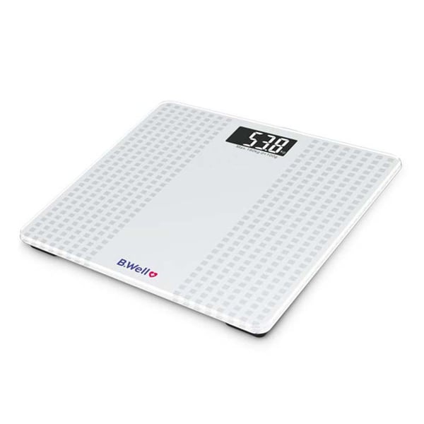 B.well Pro-166 Electronic Personal Weighing Scale
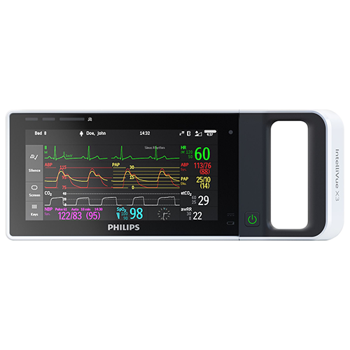 Philips IntelliVue X3 Multiparameter Monitor
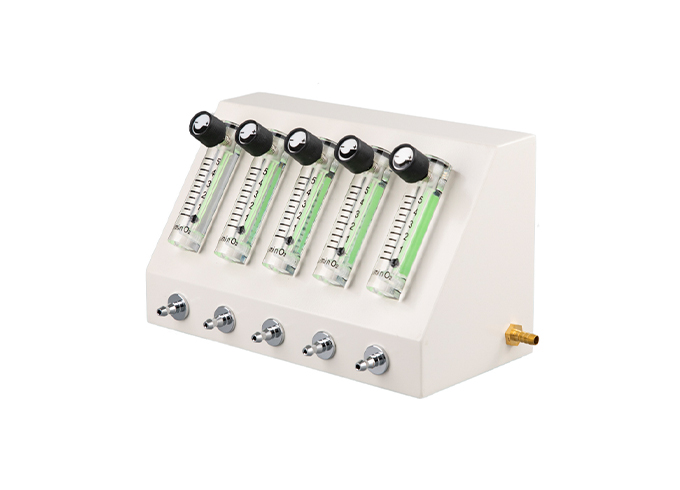 Flow Splitter For Oxygen Concentrator
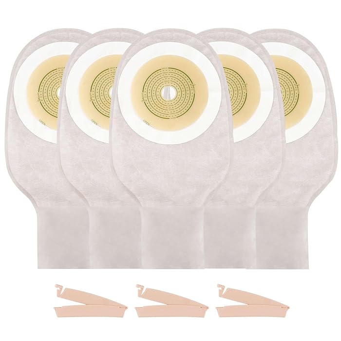 Colostomy Bags, 30 pcs in a Box
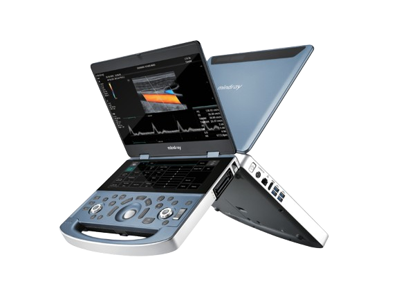 Mindray Ultrasound System MX7
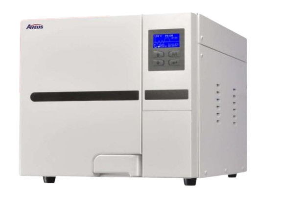 Aveus Table Top Autoclave 23ltr
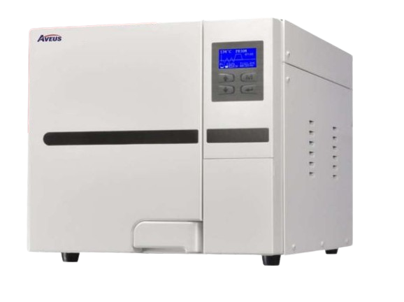 Aveus Table Top Autoclave 18ltr
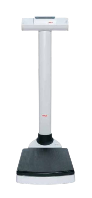 Weighing seca 703
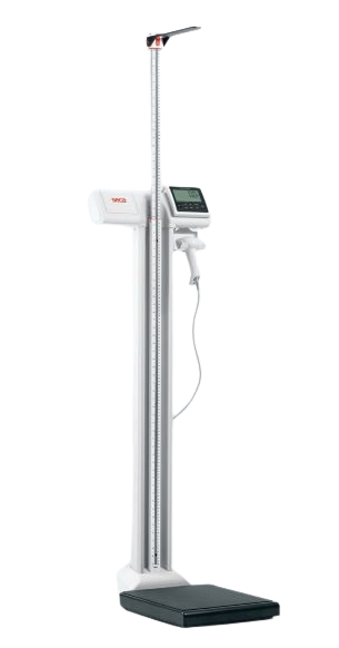 Seca Adult Weight Scale With Height Measuring Rod
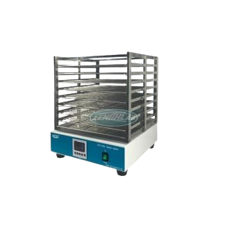 PLATELET AGITATOR
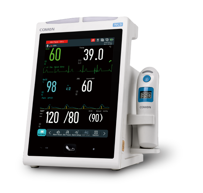 Comen – NC5 Vital Signs Monitor
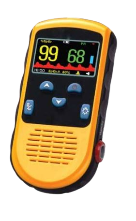 PC-66B HAND HELD PULSE OXIMETER WITH ADULT PROBE
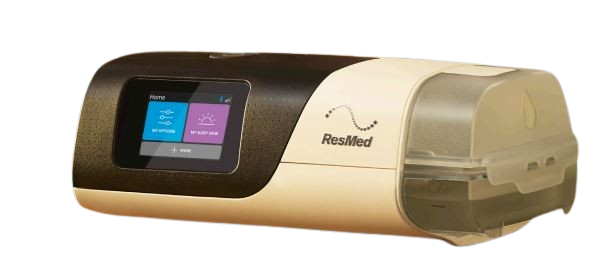 RESMED AirSense 11 CPAP
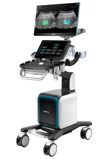 Consona N9 Mindray
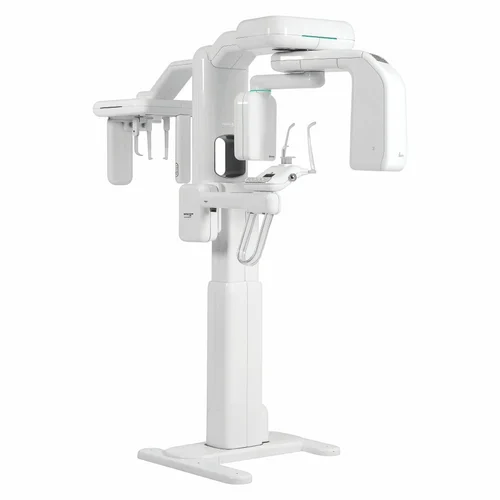 Dental X ray - Genoray - Papaya 3D Plus
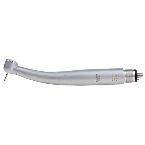 HIGH SPEED TURBINE HAND PIECE - MODEL : H 36
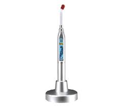 LED Curing light
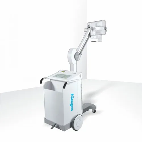 Allegers MARS 15 KW Mobile X-Ray machine
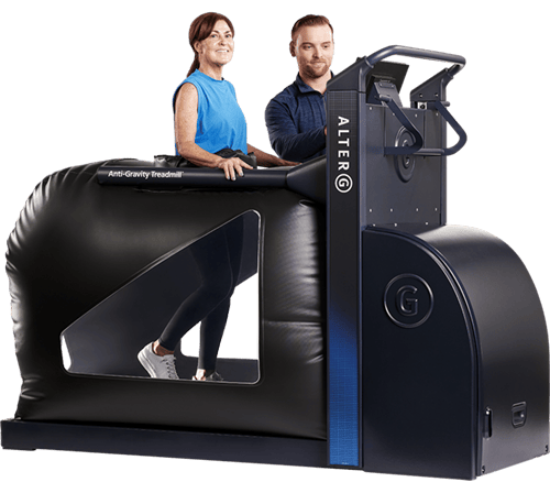 AlterG VIA Anti-Gravity Treadmill
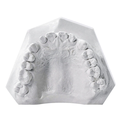 DENTITALIYA DIE STONE IV
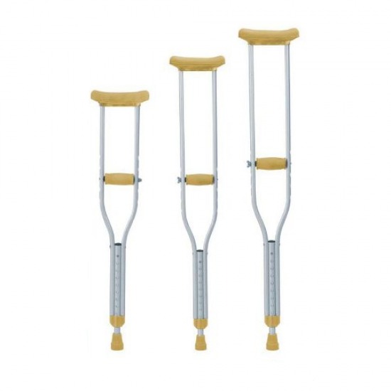 Aluminium crutches underarm M,L,S
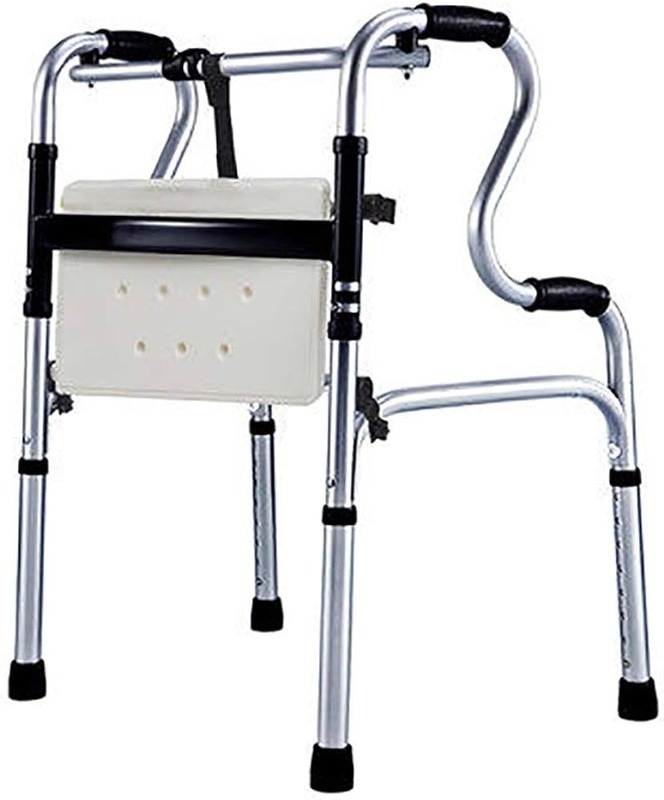 Double bend aluminium walker with chair
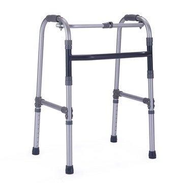 Single bend aluminium walker
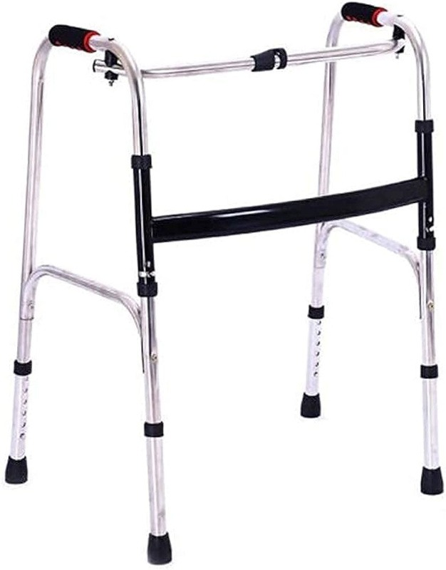 Single bend stainless stell walker
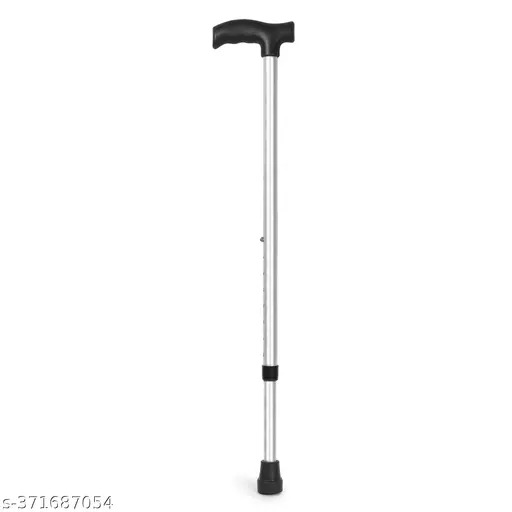 Aluminium walking stick
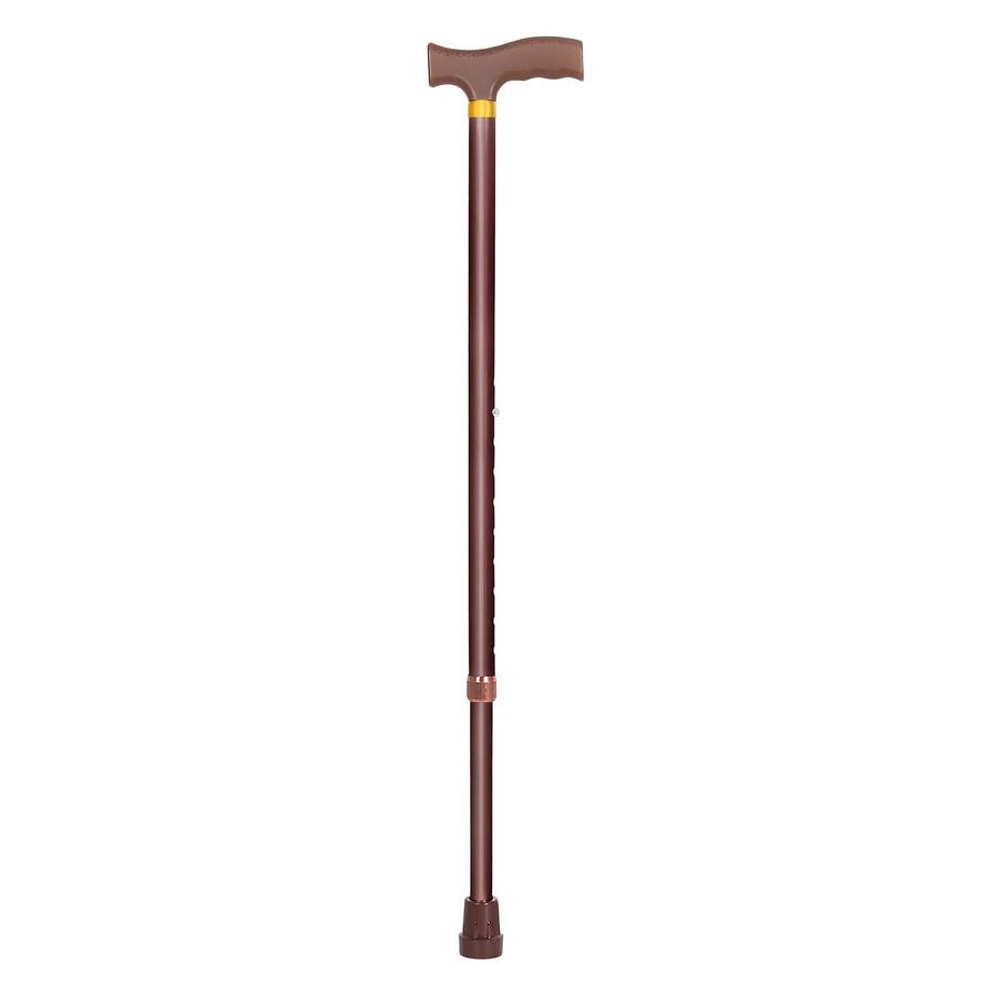 Copper color aluminium walking stick
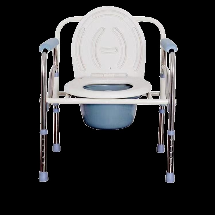 Stainless steel chair with toilet
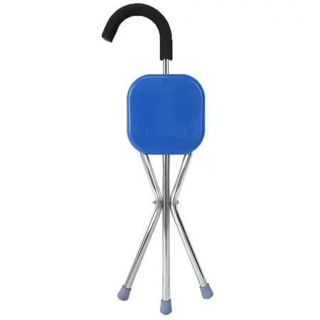 Walking stick with seat
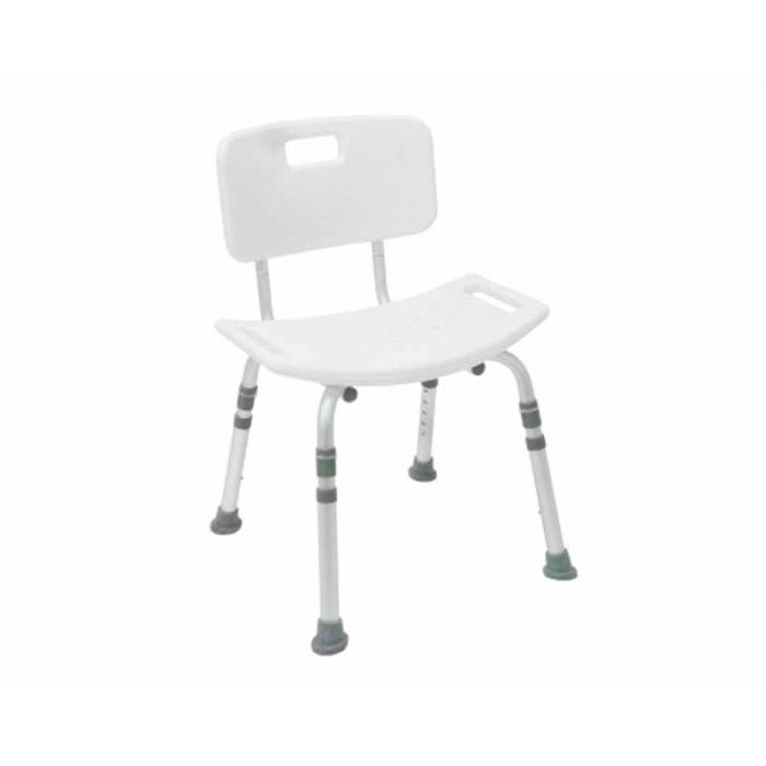 Shower chair with back support
.jpeg) Shower chair
Shower chair
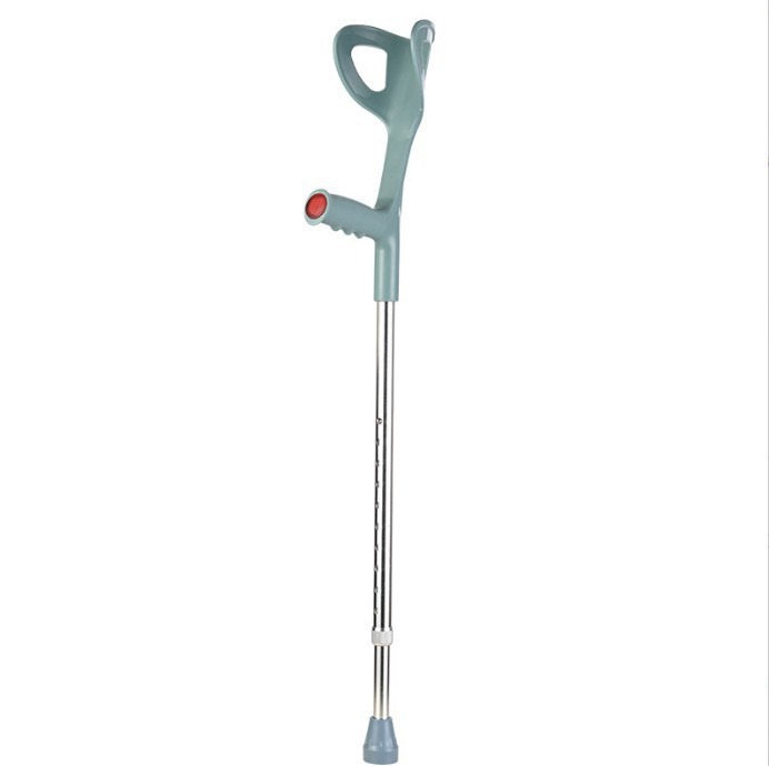 Half circle crutches forearm
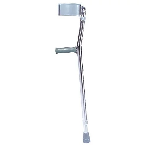 All-inclusive elbow crutch
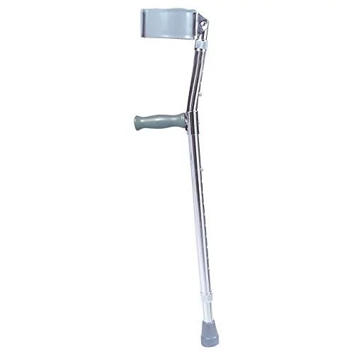 Stainless steel walking stick
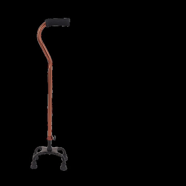 Bronze curved handle four-legs waiking stick
.jpeg) BABY WEIGHING SCALE
BABY WEIGHING SCALE
.jpeg) Comen – 12 Channel ECG Machine, CM1200
Comen – 12 Channel ECG Machine, CM1200
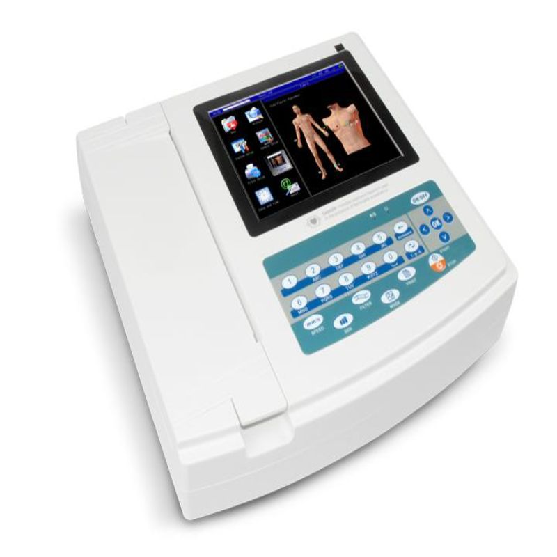 ECG 1200G – Contec
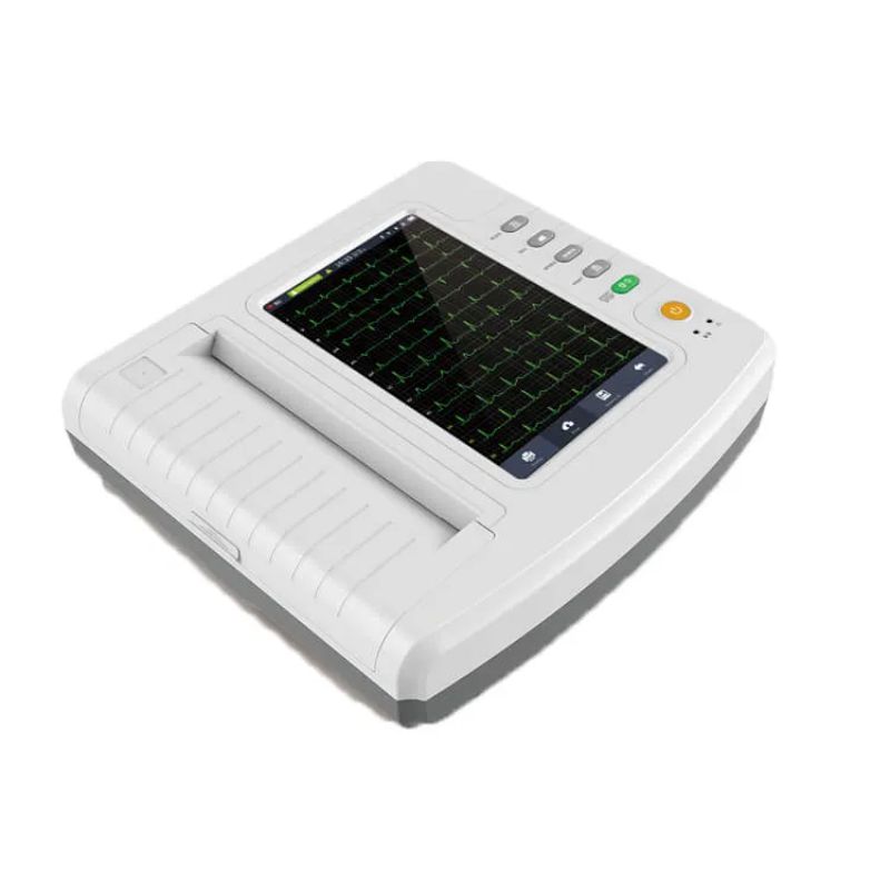 ECG 1212G – Contec
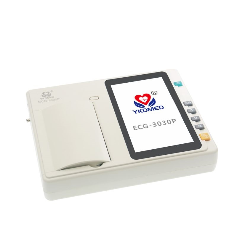 YKD MED ECG 3 CHANNEL
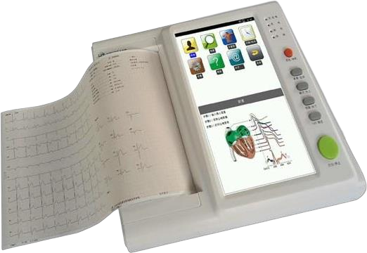 ECG 12 CHANNEL 3A CANADA
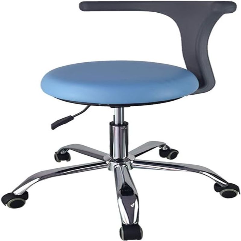 STOOL WITH BACKREST
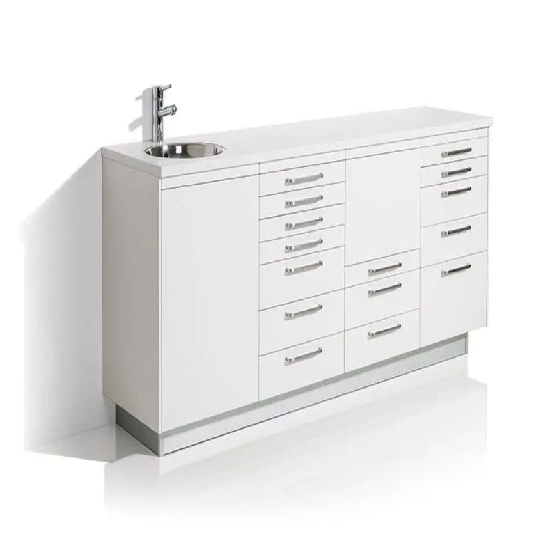 DENTAL CABINET
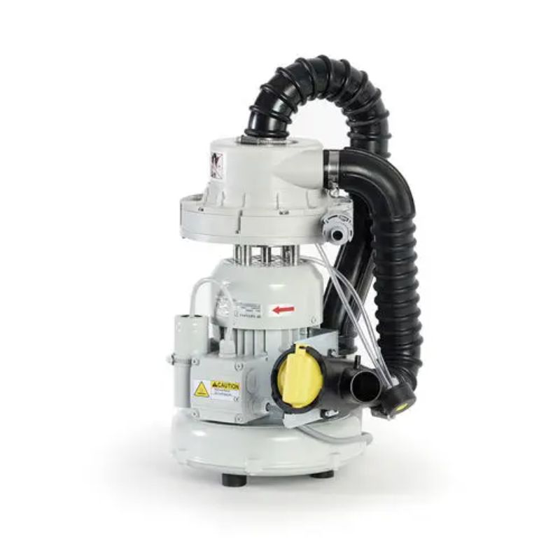 DENTAL SUCTION- YARA DENT
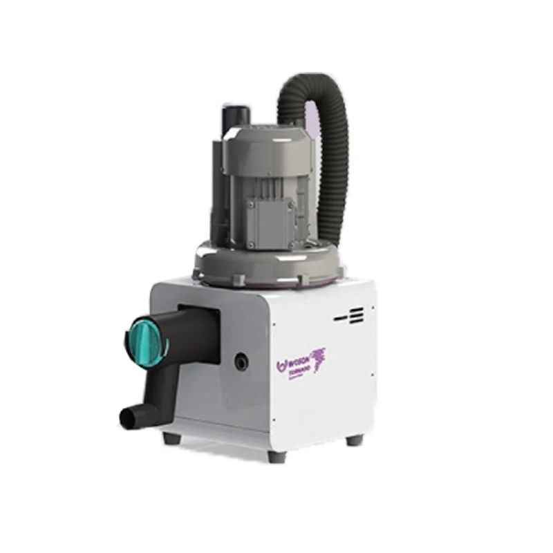 DENTAL SUCTION- WOSON
 STORM OILLESS COMPRESSOR- WOSON
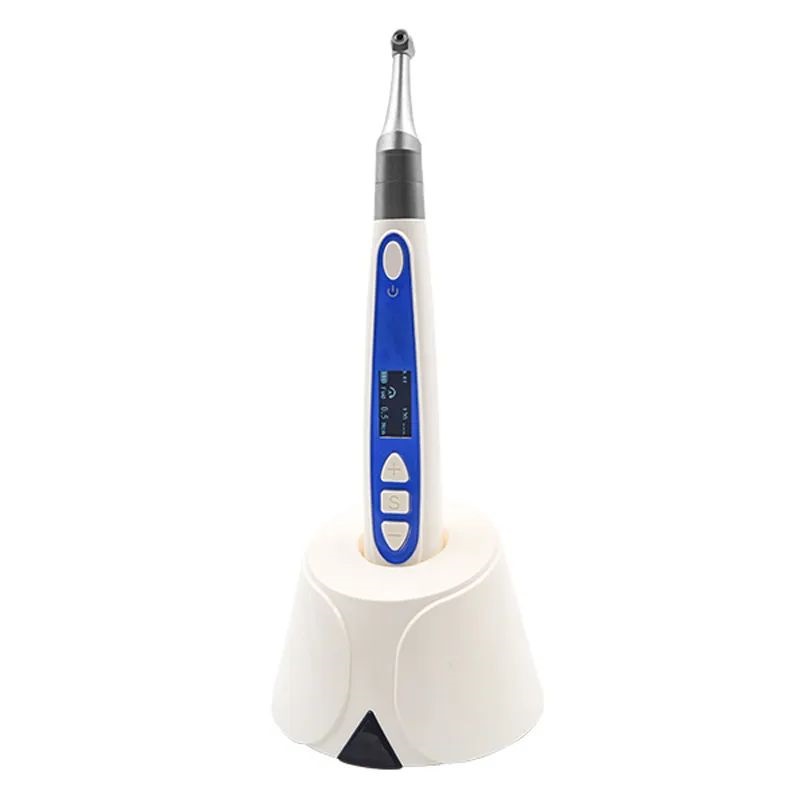
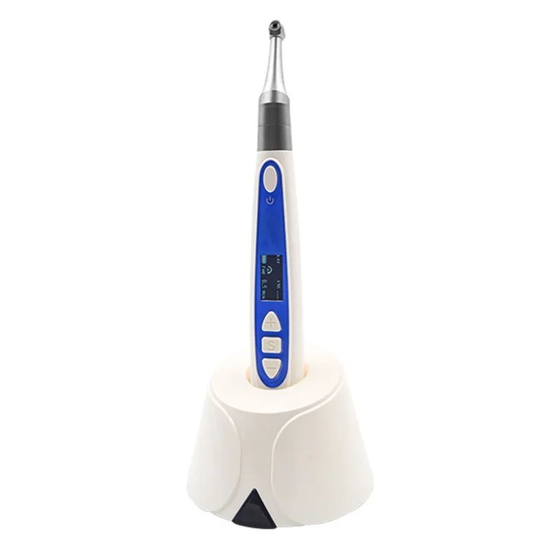 WIRELESS ENDOMOTOR WITH APEX LOCATOR- BMEDM 5 2 IN 1
 AMALGAMATOR- BM-L009
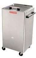 Chattanooga Hydrocollator 8M SS-2
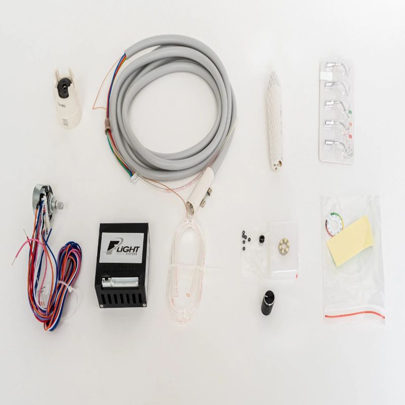 Built-in Ultrasonic Scaler without LED – TM-3HKB
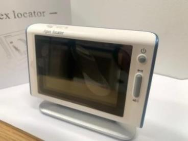 APEX LOCATOR- BM- AL 2
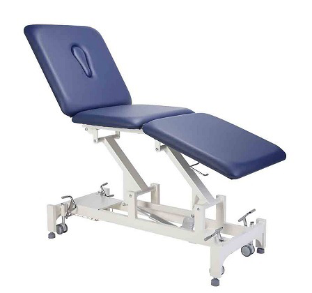 Three Section Couch Electrica
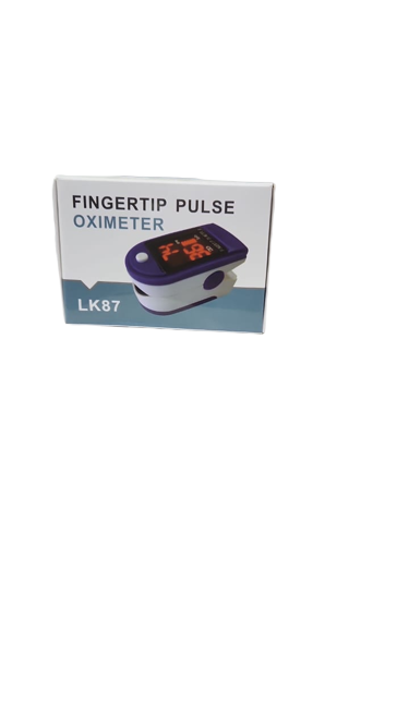 PULSE OXIMETER
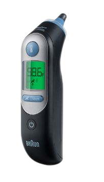 BRAUN THERMOSCAN 7
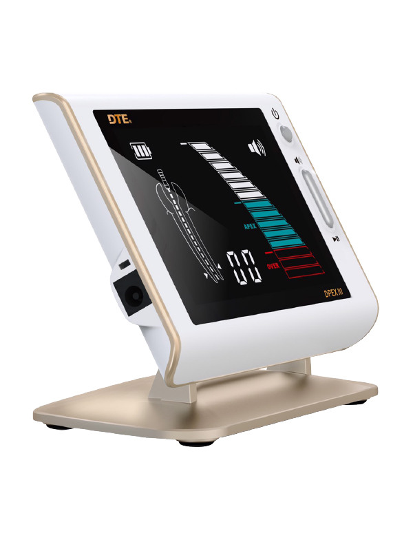 APEX LOCATOR
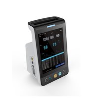 VITAL SIGN MONITOR
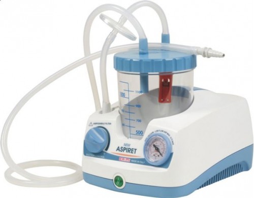 CAMI NEW ASPIRET SUCTION MACHINE
.jpg) Astringent Retraction Paste
Astringent Retraction Paste
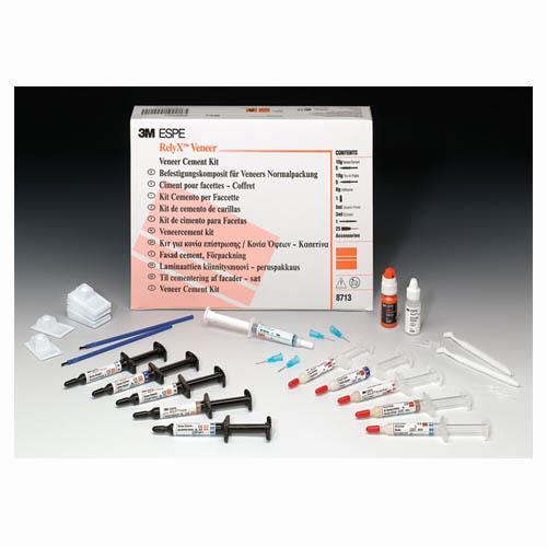 RelyX Veneer Introductory Kit
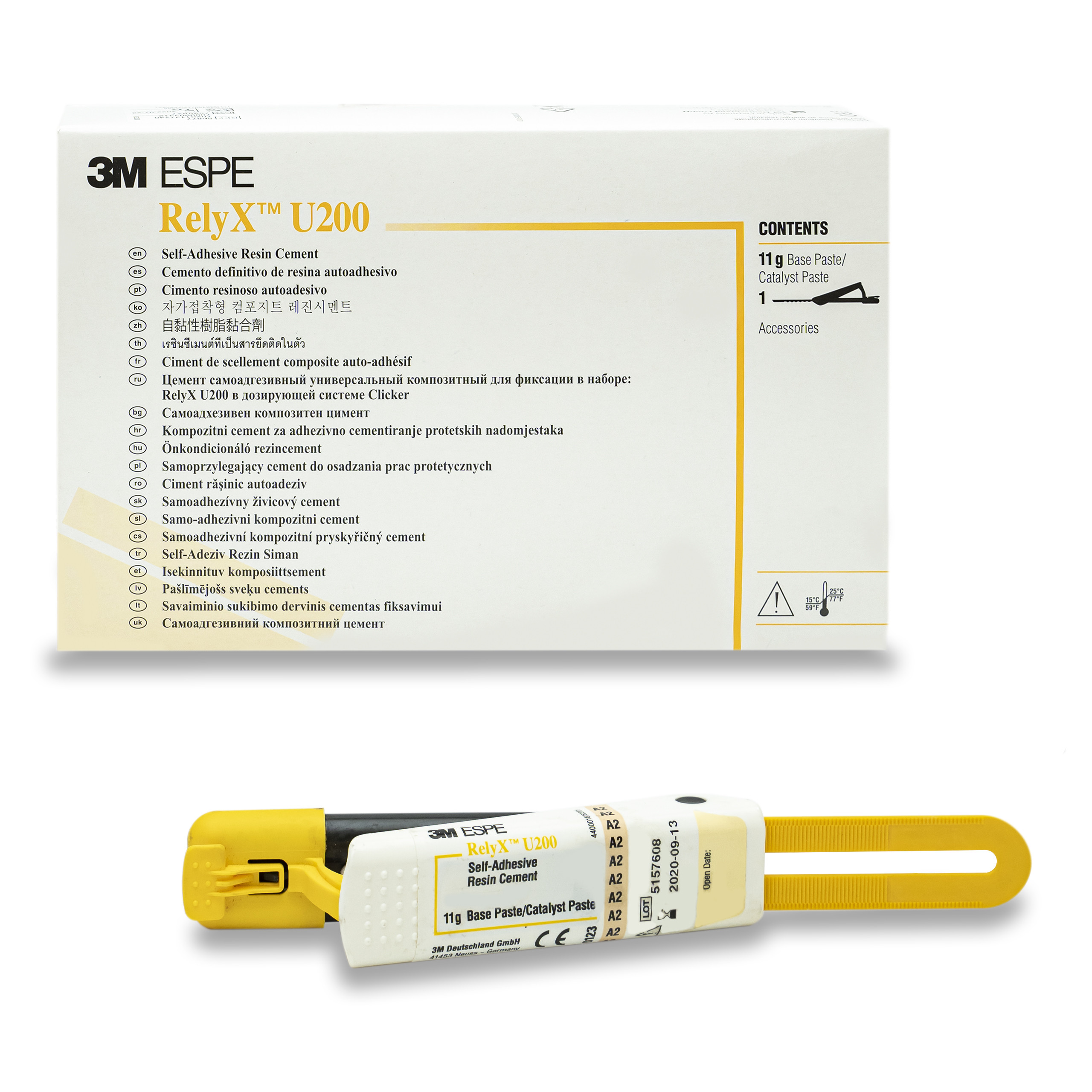 3M ESPE RelyX U200
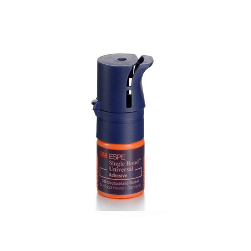 3M ESPE Single Bond Universal Adhesive
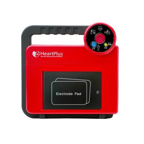 Heart Plus AED
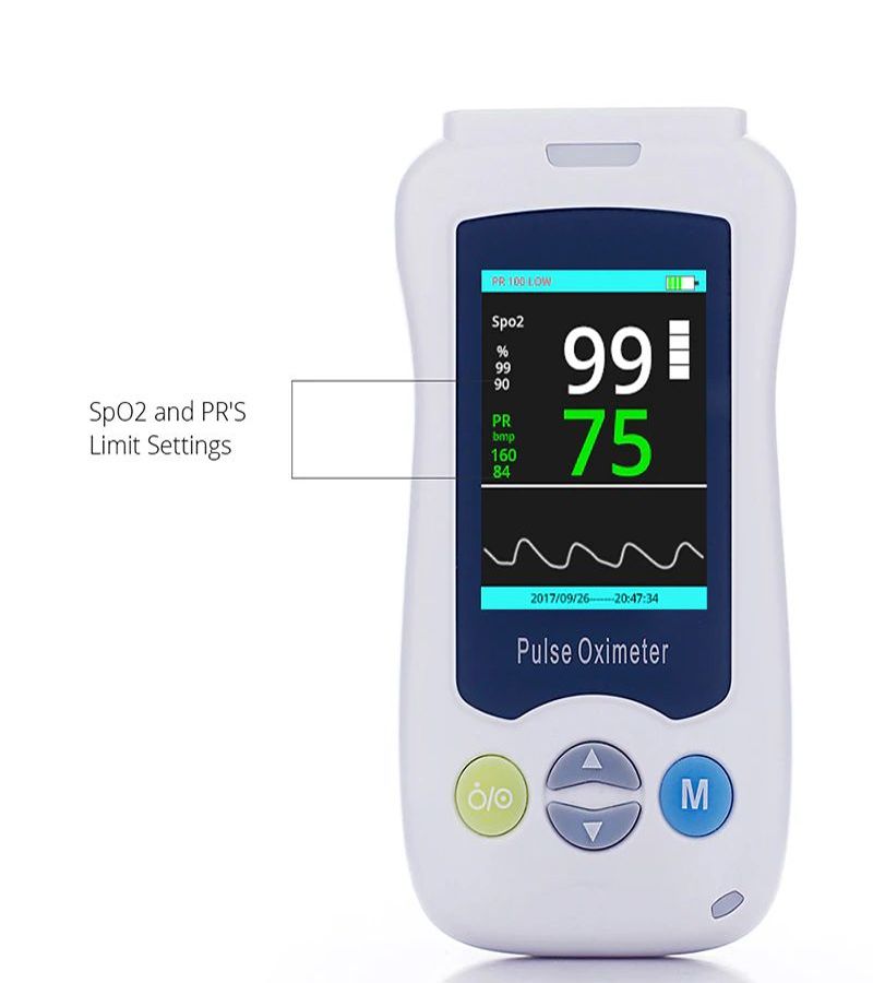 HANDHELD PULSE OXIMETER
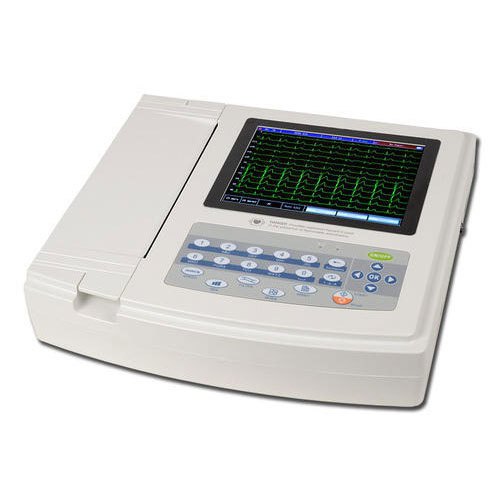 CONTEC ECG MACHINE
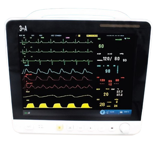 3A PATIENT MONITOR DUBAI
 PPM DUBAI
.jpeg) DENTAL CHAIR BORNDENT
DENTAL CHAIR BORNDENT
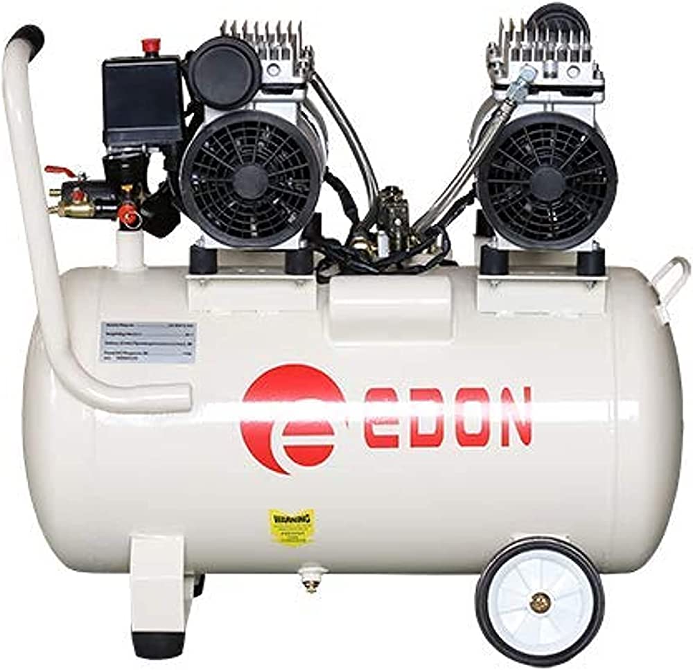 DENTAL COMPRESSOR
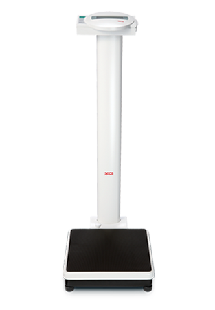 SECA WEIGHING SCALE
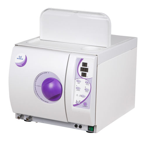 WOSON AUTOCLAVE 18L
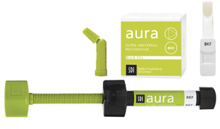 SDI Aura Bulk Fill Composite
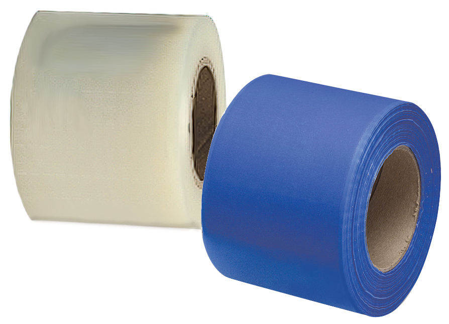 BARIER FILIM
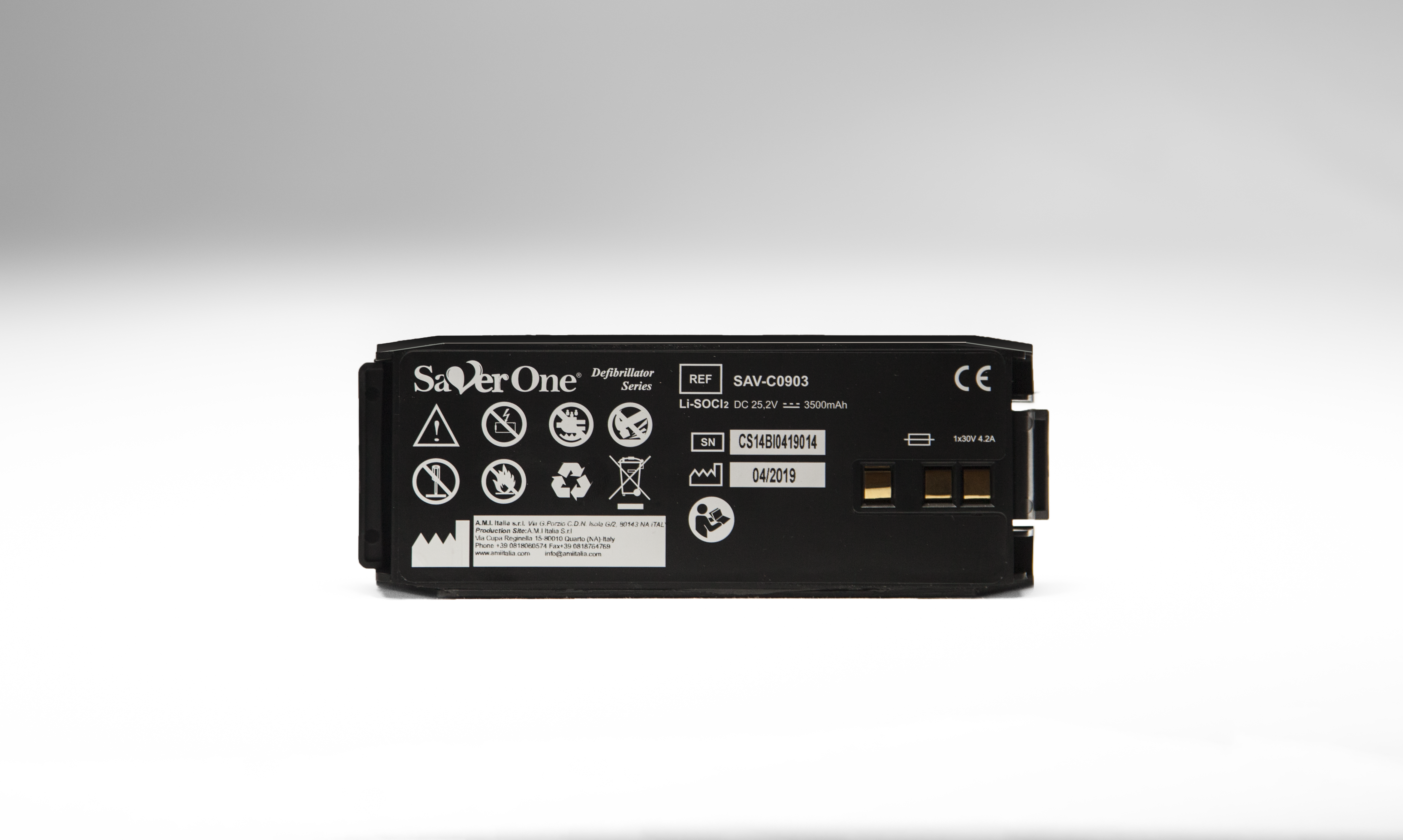 AMI ITALIA SAVER ONE BATTERY
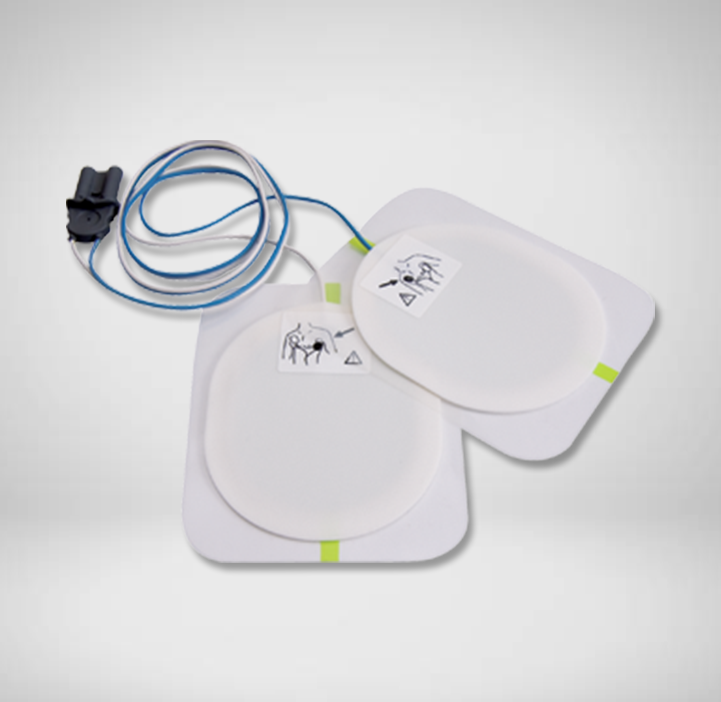 AMI ITALIA SAVER ONE AED PAD ADULT
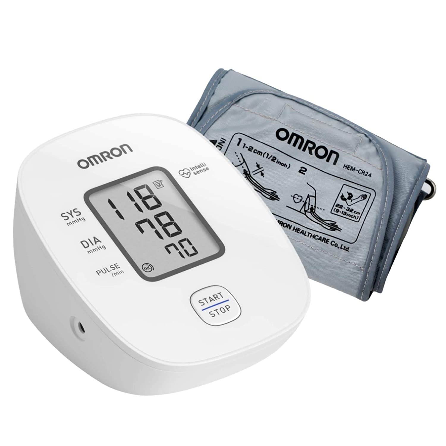 OMRON M1 BASIC
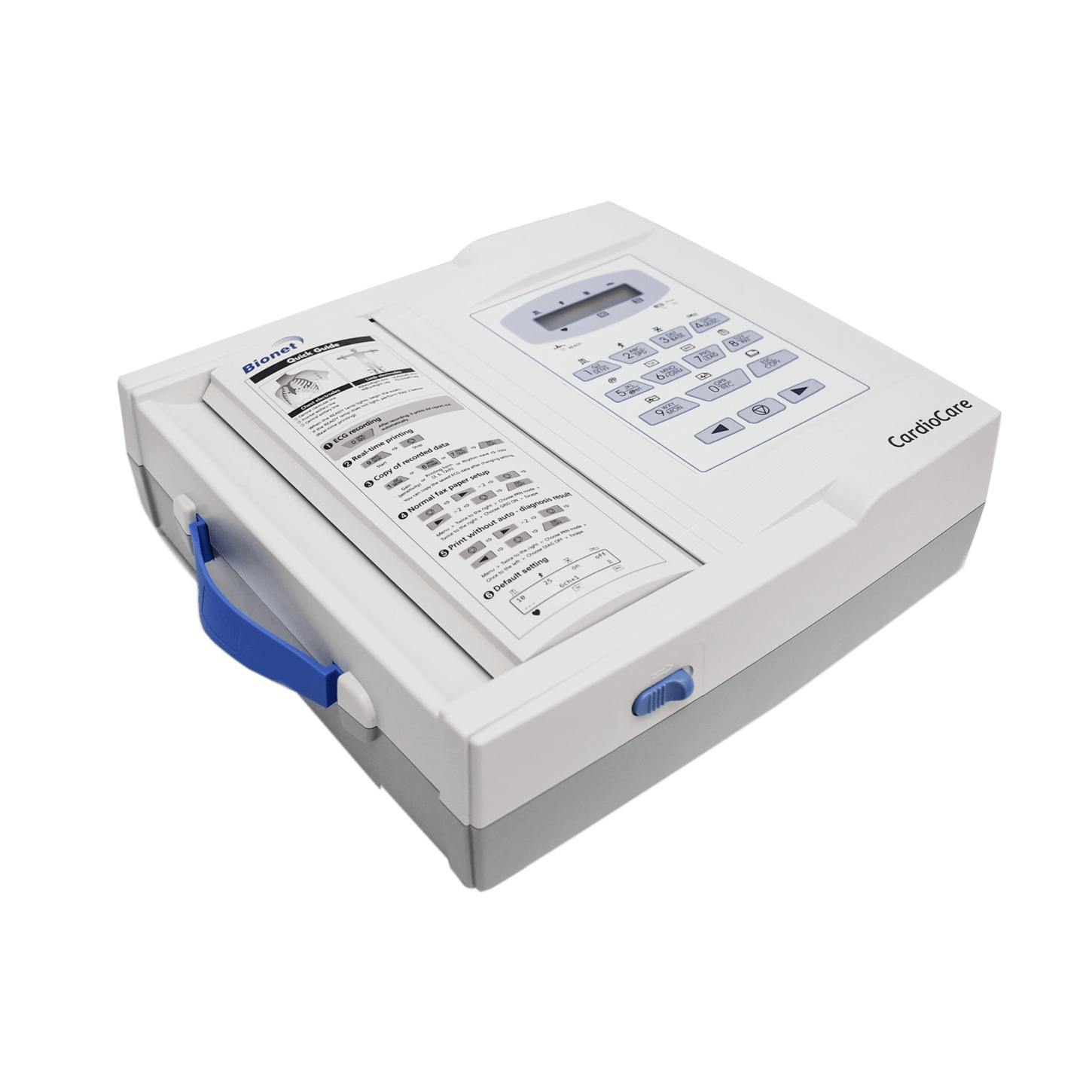 CardioCare 2000 ECG
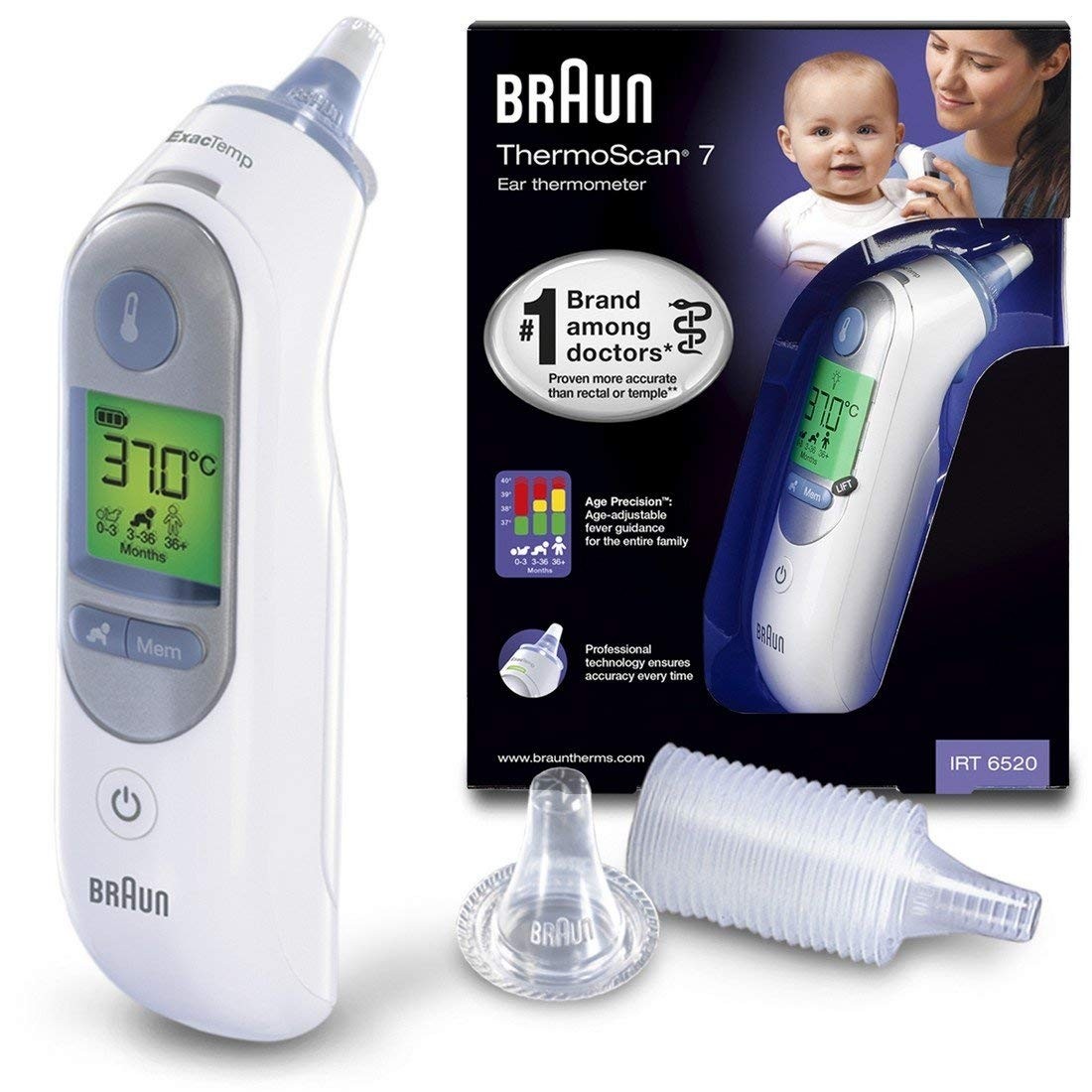 BRAUN Thermoscan 7 With Age Precision Multi Functional Ear
 VivaChek Ino Blood Glucose Test Strips 50 Tests
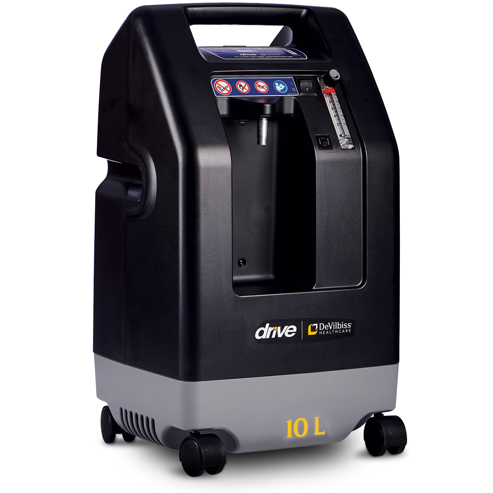 Drive DeVilbiss 10 Litre oxygen Concentrator
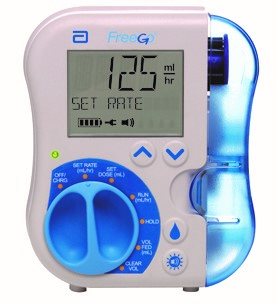 Abbott FreeGo feeding pump
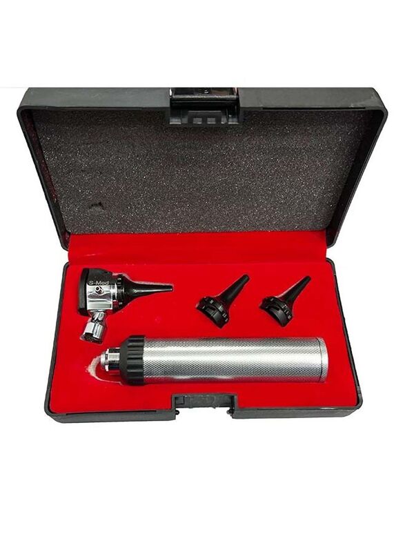 Otoscope Magnification Diagnostic Ear Scope
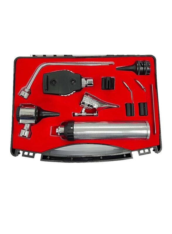 ENT Diagnostic Set
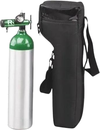 2.5L Portable aluminium light weight oxygen cylinder full set
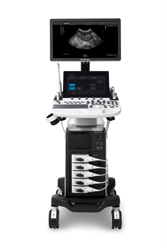 SONOSCAPE P40 Elite Ultrasound system
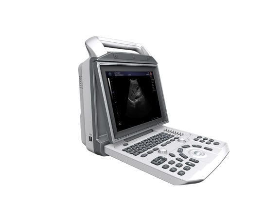 Portable ultrasound system zoncare-i50
.png) Sonoscape P10 Ultrasound
Sonoscape P10 Ultrasound
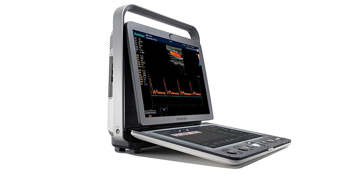 Sonoscape S9Ultrasound Colour Doppler
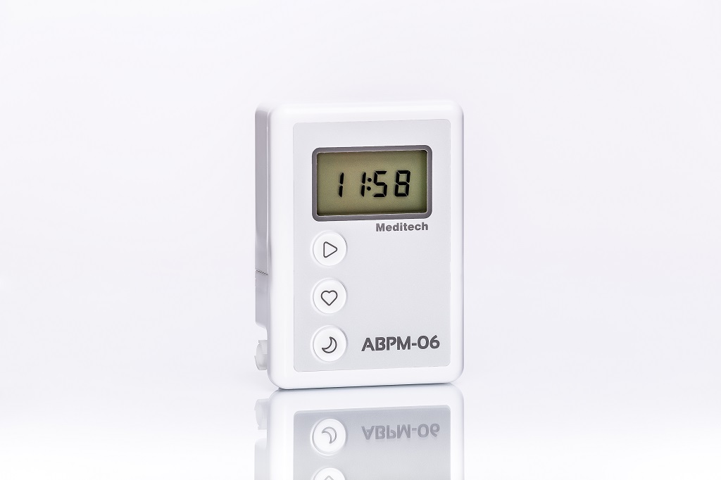 Meditech ABPM-06
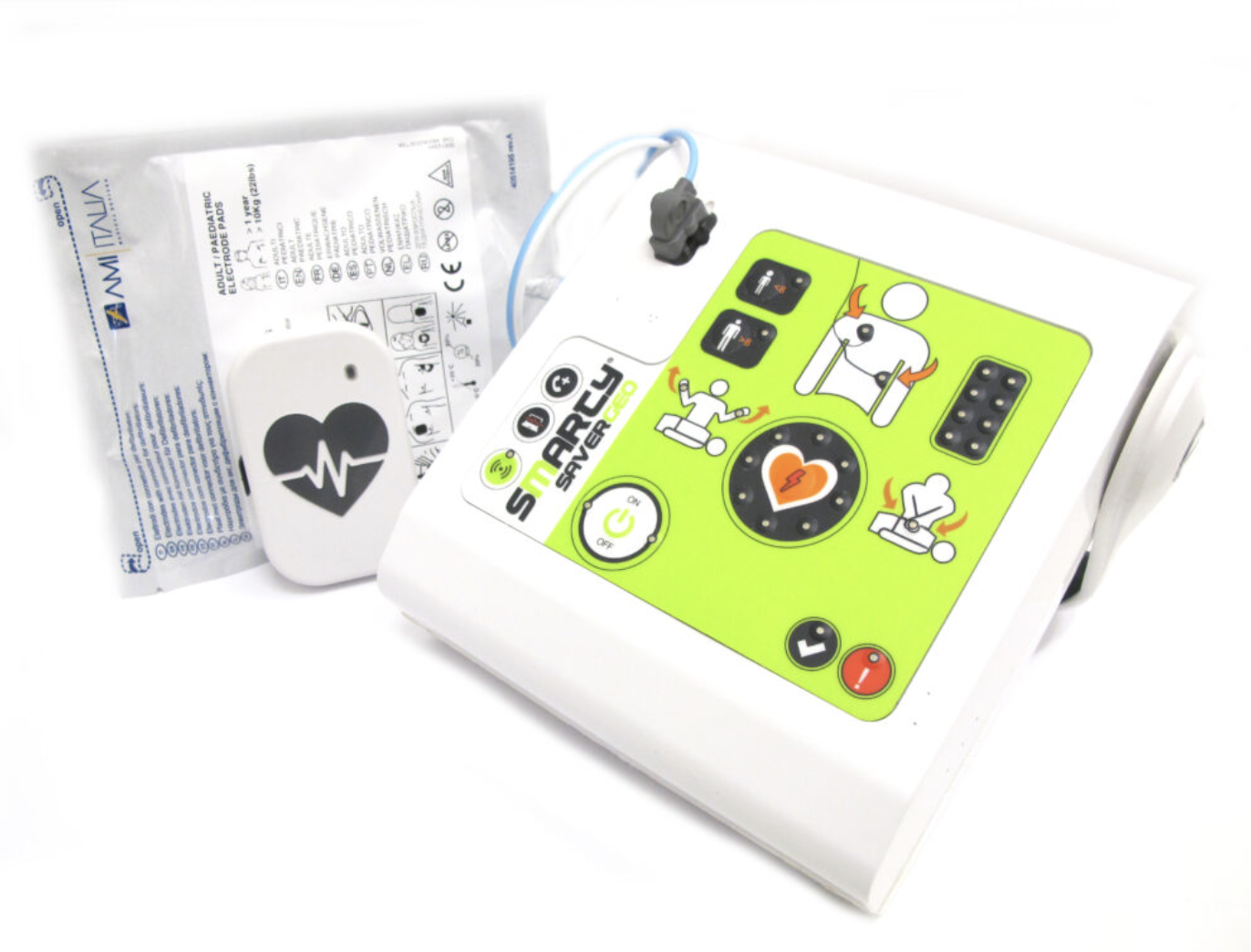 SMARTY SAVER AMI ITALIA
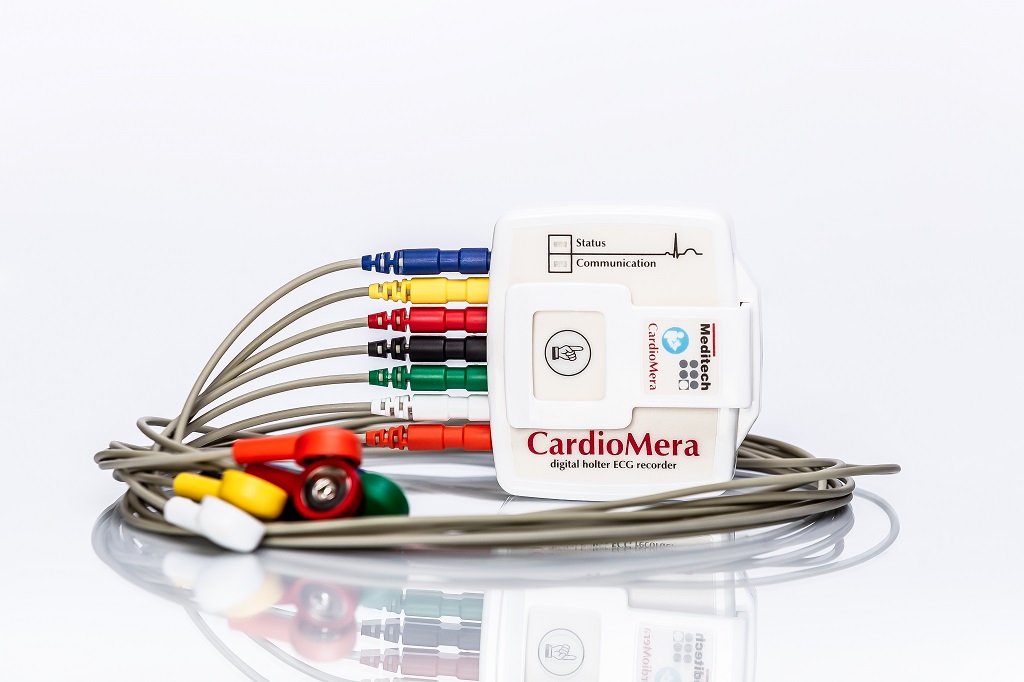 MEDITECH CARDIOMERA ECG Holter monitor
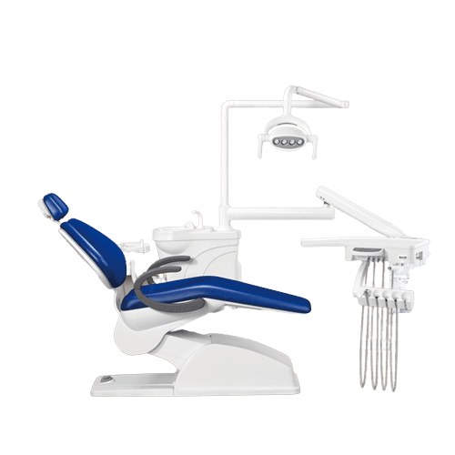 DENTAL CHAIR DENFLY
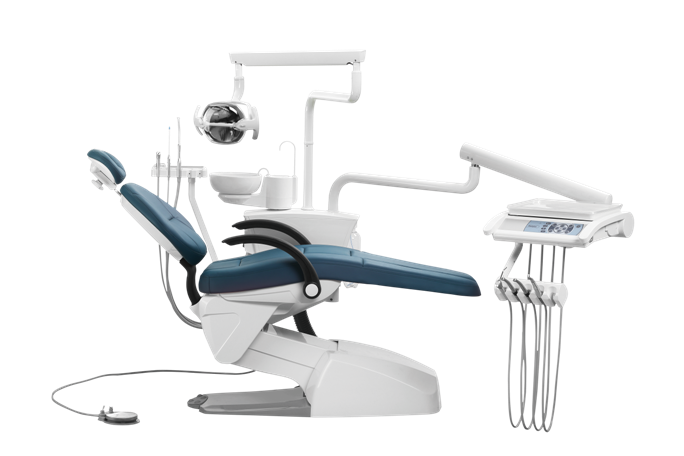 DENTAL CHAIR RUNDEER
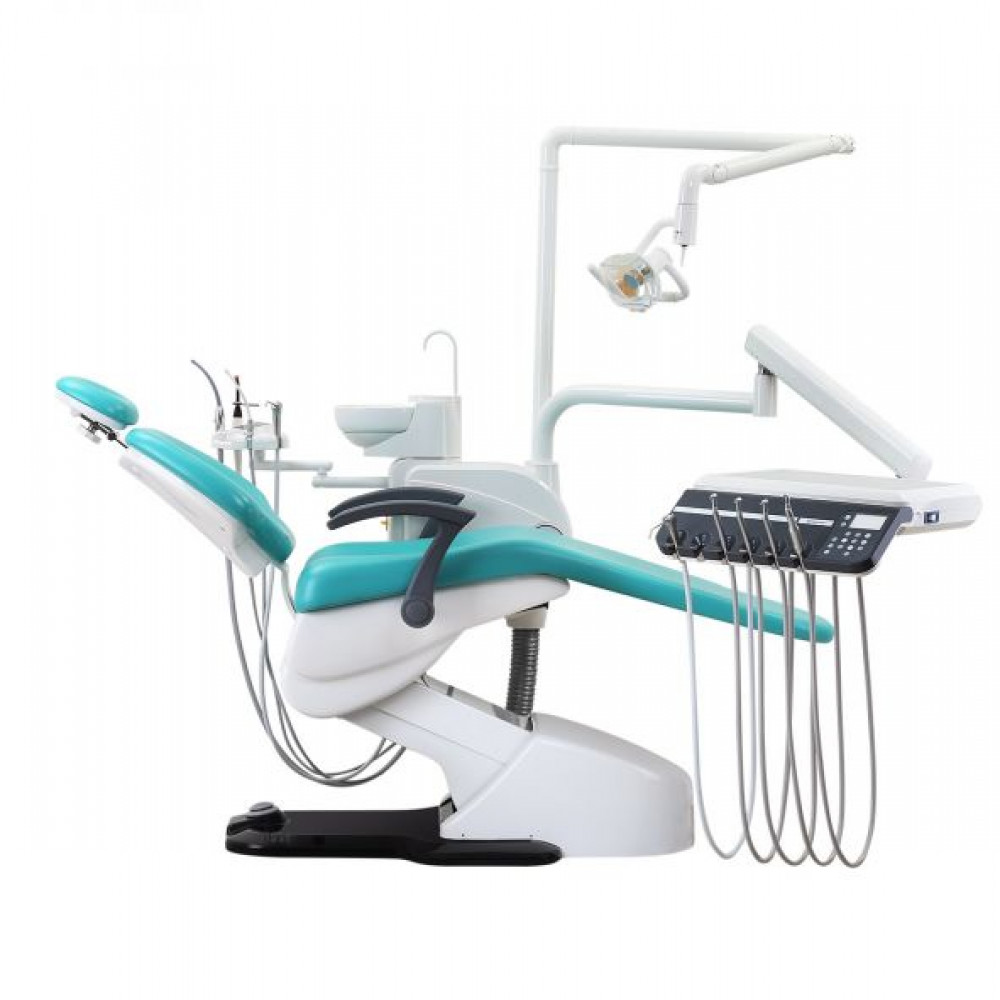 DENTAL CHAIR WOSON
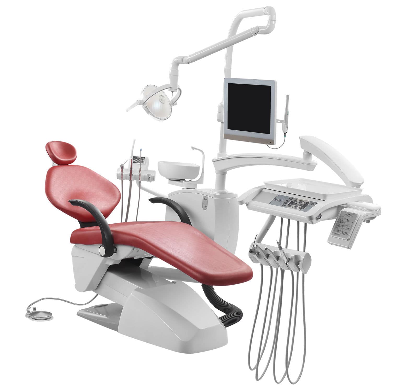 DENTAL CHAIR RUNNEYS
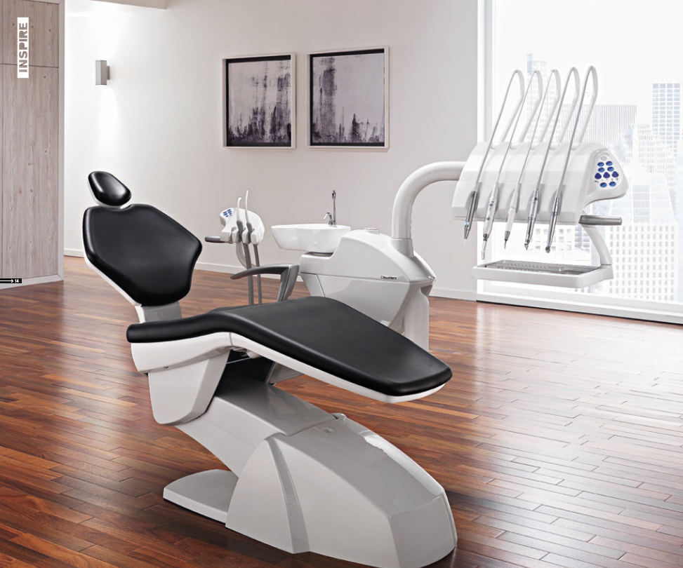 DENTAL CHAIR SWIDENT
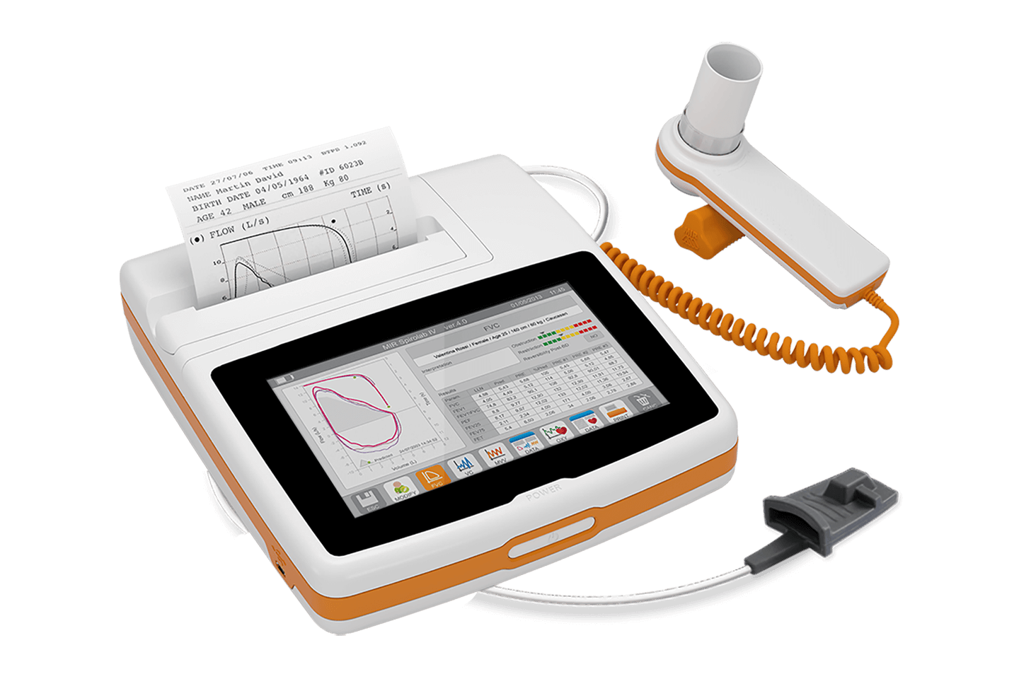 SPIROMETER SPIROLAB
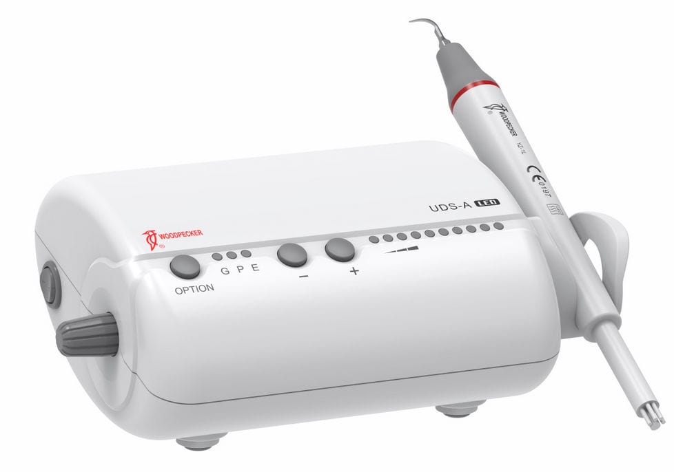 Dental Scaler Ultrasonic Woodpecker UDS-A
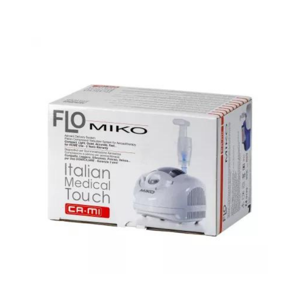 NEBULIZER CAMI, FLO MIKO
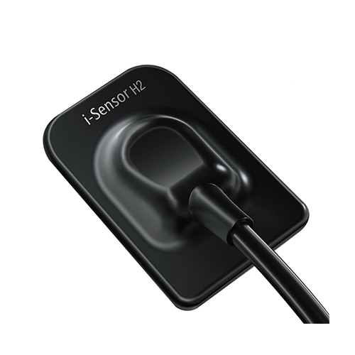 WOODPECKER RVG
.jpeg) FEEDING BAG
FEEDING BAG
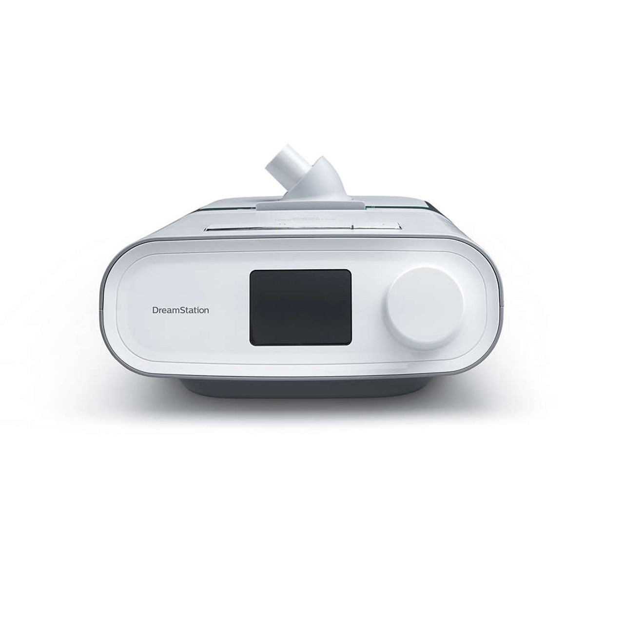 Philips Respironics DreamStation Auto CPAP
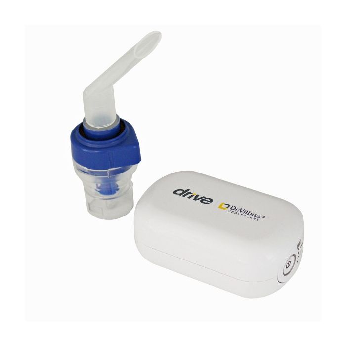 DeVilbiss AirForce Mini Compact Compressor Nebulizer
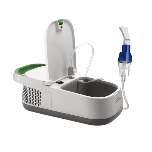 Philips Respironics Innospire Deluxe Nebuliser Compressor System
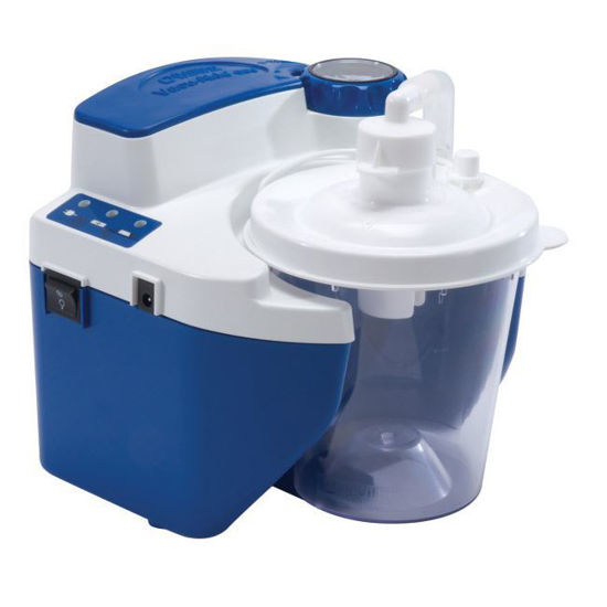 DeVilbiss Vacu Aide Suction Unit With Battery
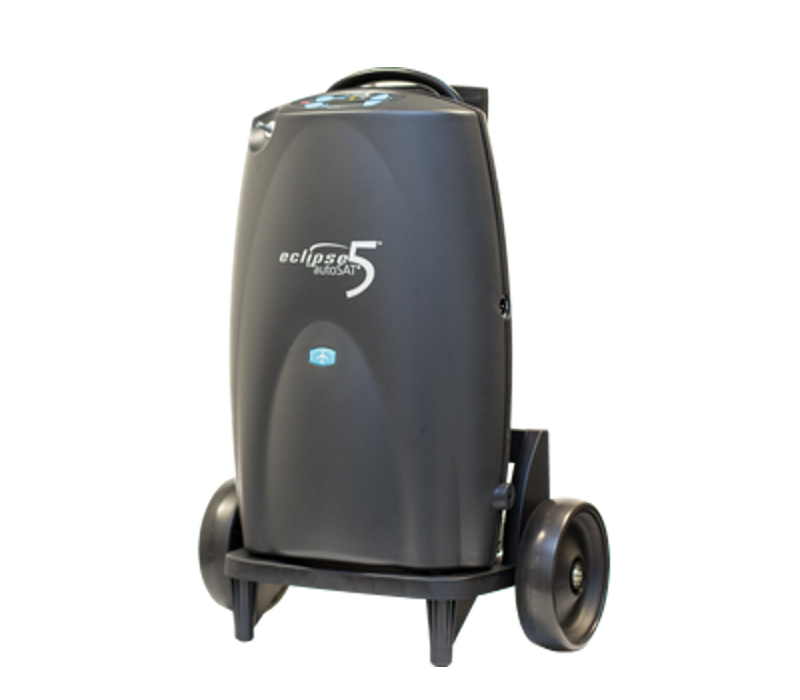 Caire Eclipse 5 PORTABLE OXYGEN CONCENTRATOR
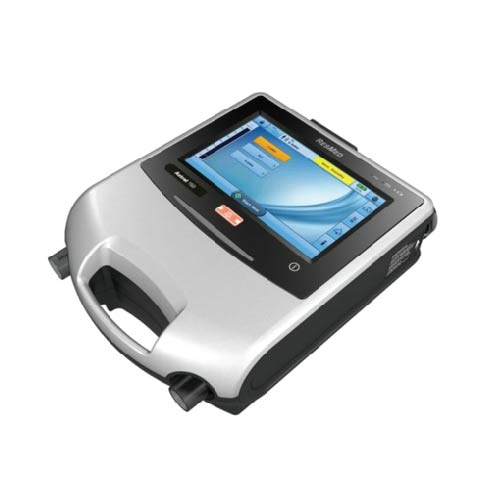 Astral 150 Portable Ventilator
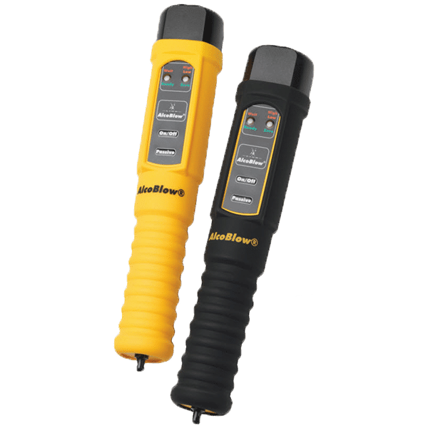 BREATH ANALYZER
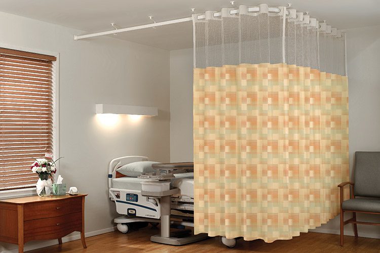 MEDICAL CURTAINS
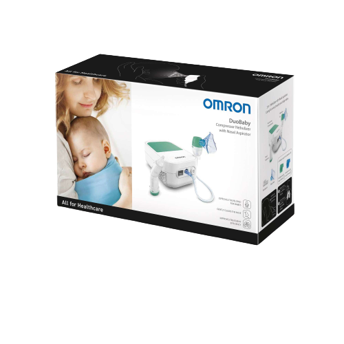 Omron Duobaby Compressor Nebulizer
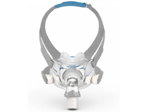 ResMed AirFit F30 Full Face CPAP
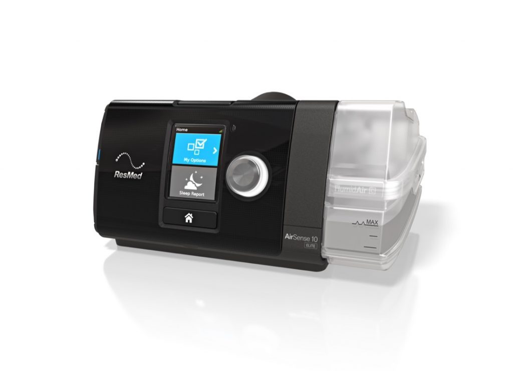 Resmed Airsense S10 Autoset CPAP
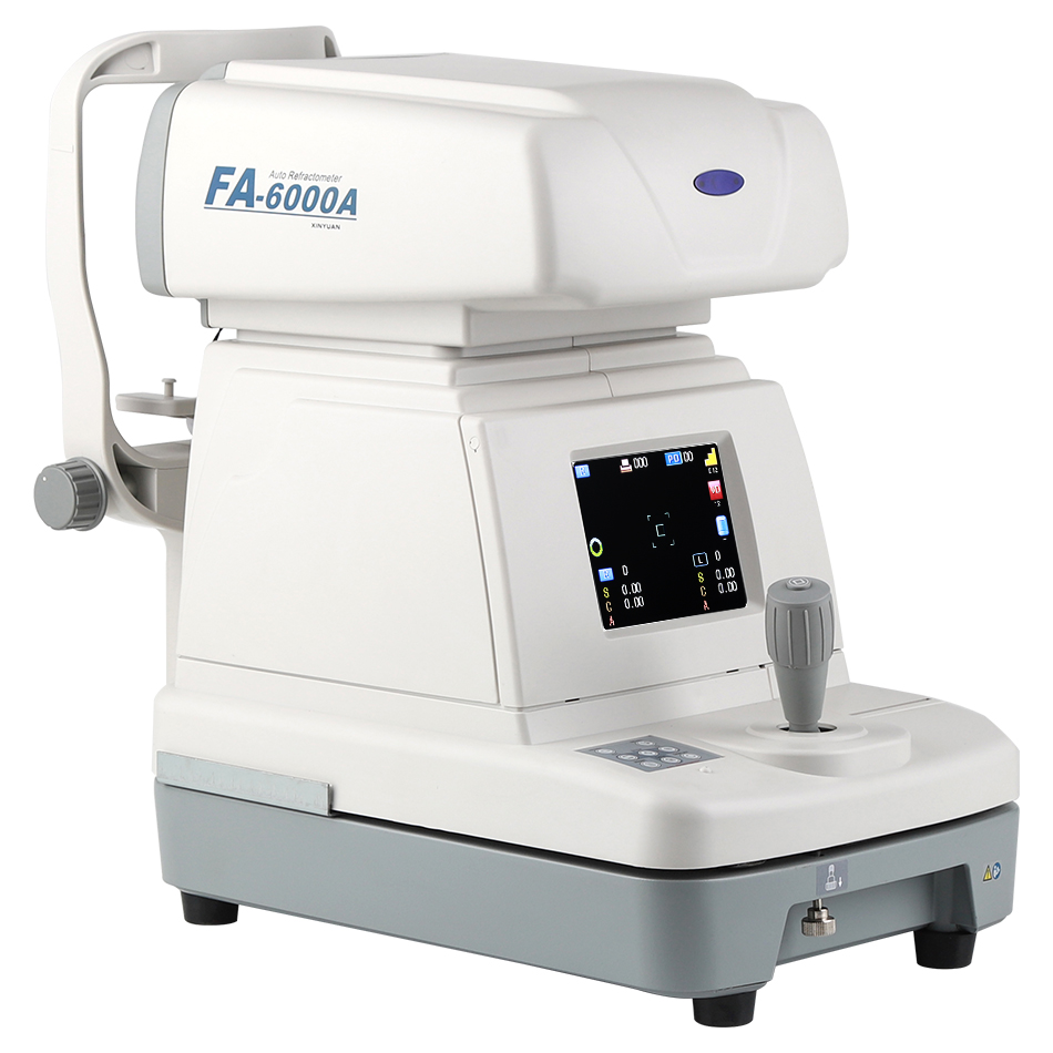 AUTO REFRACTOMETER FA 6000A
.png) Auto CPAP with Heated Humidifier and Mask
Auto CPAP with Heated Humidifier and Mask
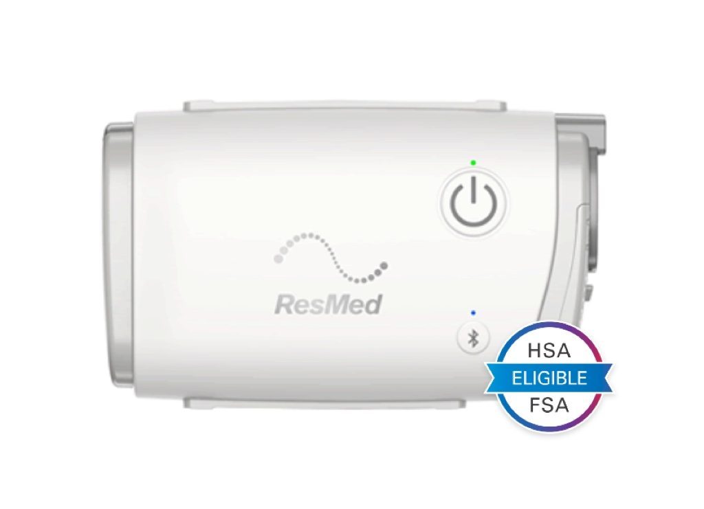 AirMini Portable CPAP
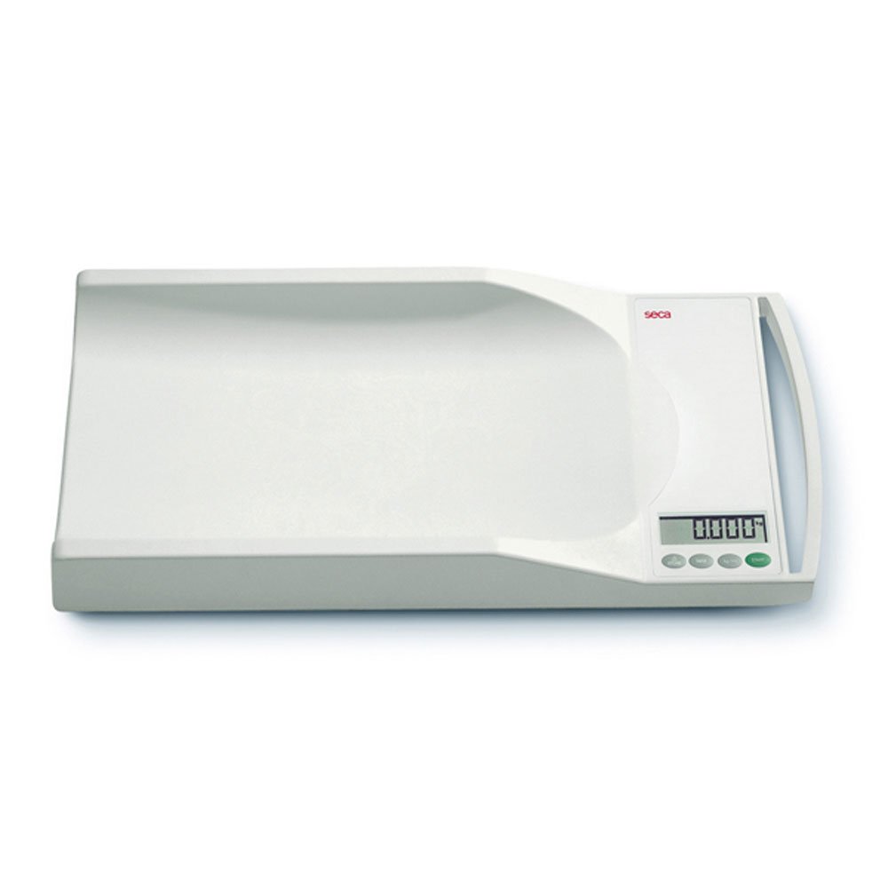 SECA BABY WEIGHING SCALE 334
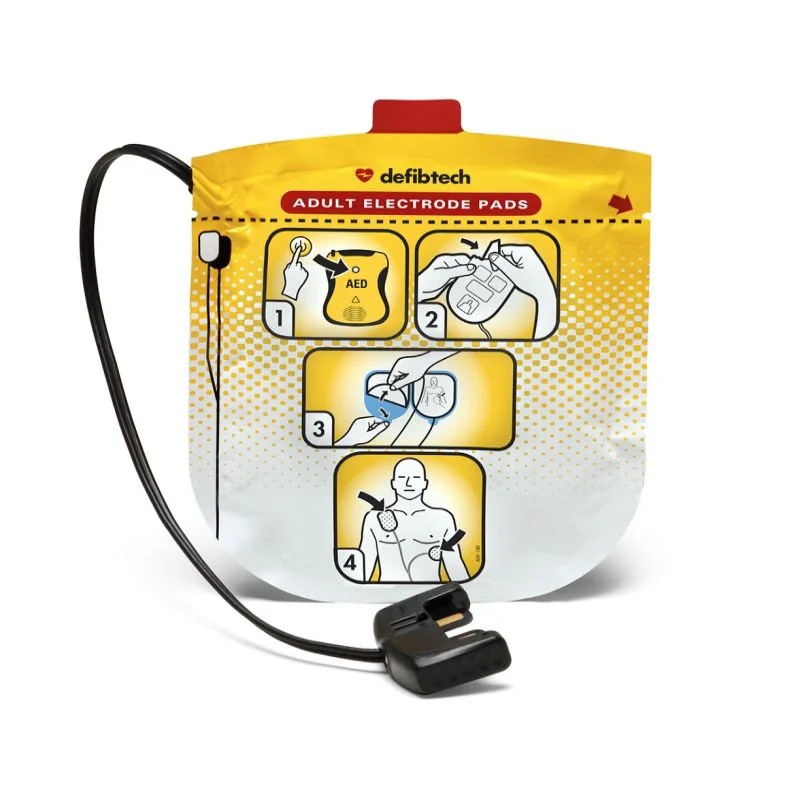 Defibtech Adult AED Pads
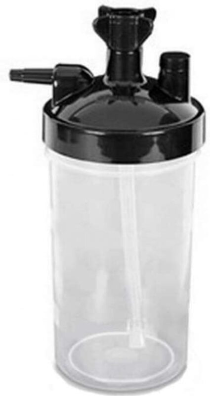 Humidifier for Oxygen Concentrator
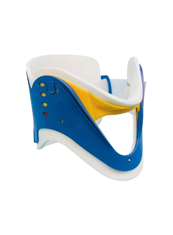 Cervical Collar for Adult
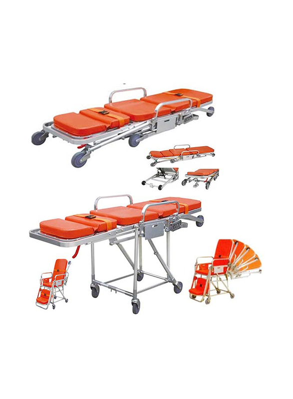 Automatic Loading Chair Stretcher
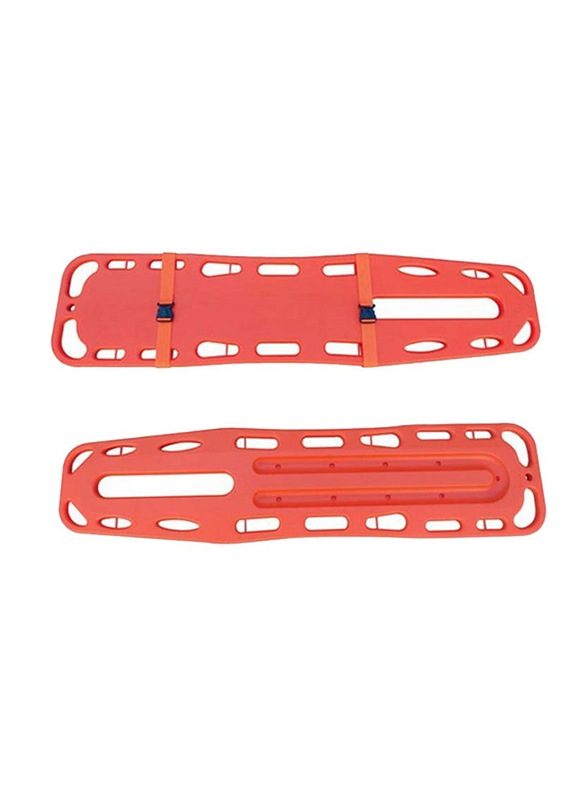 Spinal Board
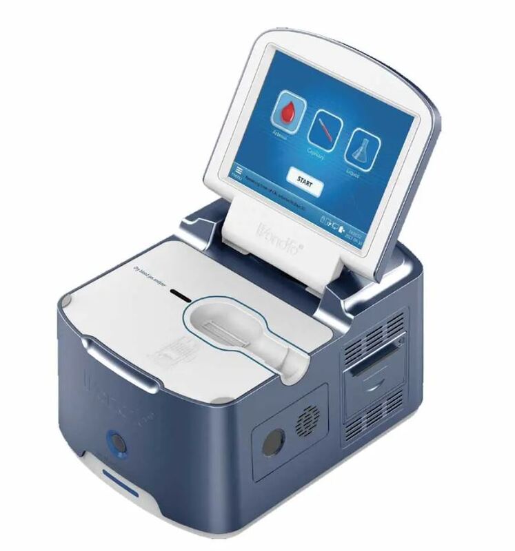 Blood Gas Analyzer
.jpg) Surgical Minor Kit
Surgical Minor Kit
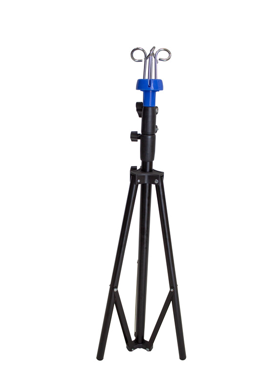 Foldable IV Stand
.jpg) Oxygen Regulator Full Set with Flow Meter Humidifier
Oxygen Regulator Full Set with Flow Meter Humidifier
.jpg) Foldable Stretcher With Wheels
Foldable Stretcher With Wheels
.png) Portable Breast Pump
Portable Breast Pump
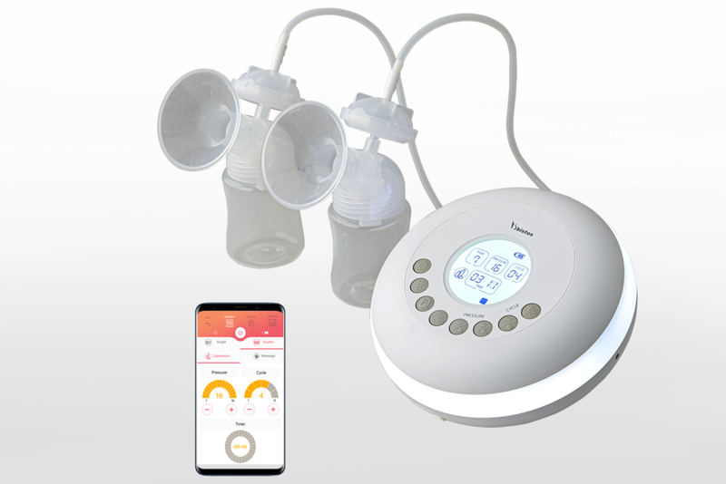 Desktop Breast Pump
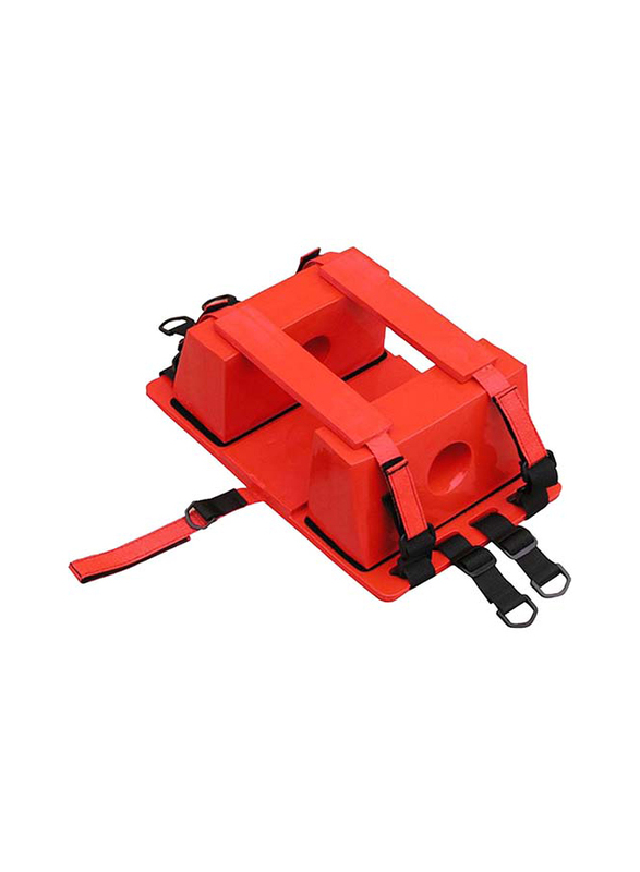 Head Immobilizer
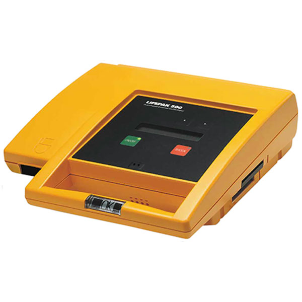 LIFEPAK 500 AED MACHINE Refurb
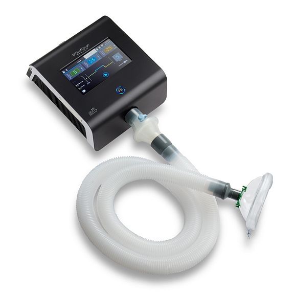 Cough Assist BiWaze
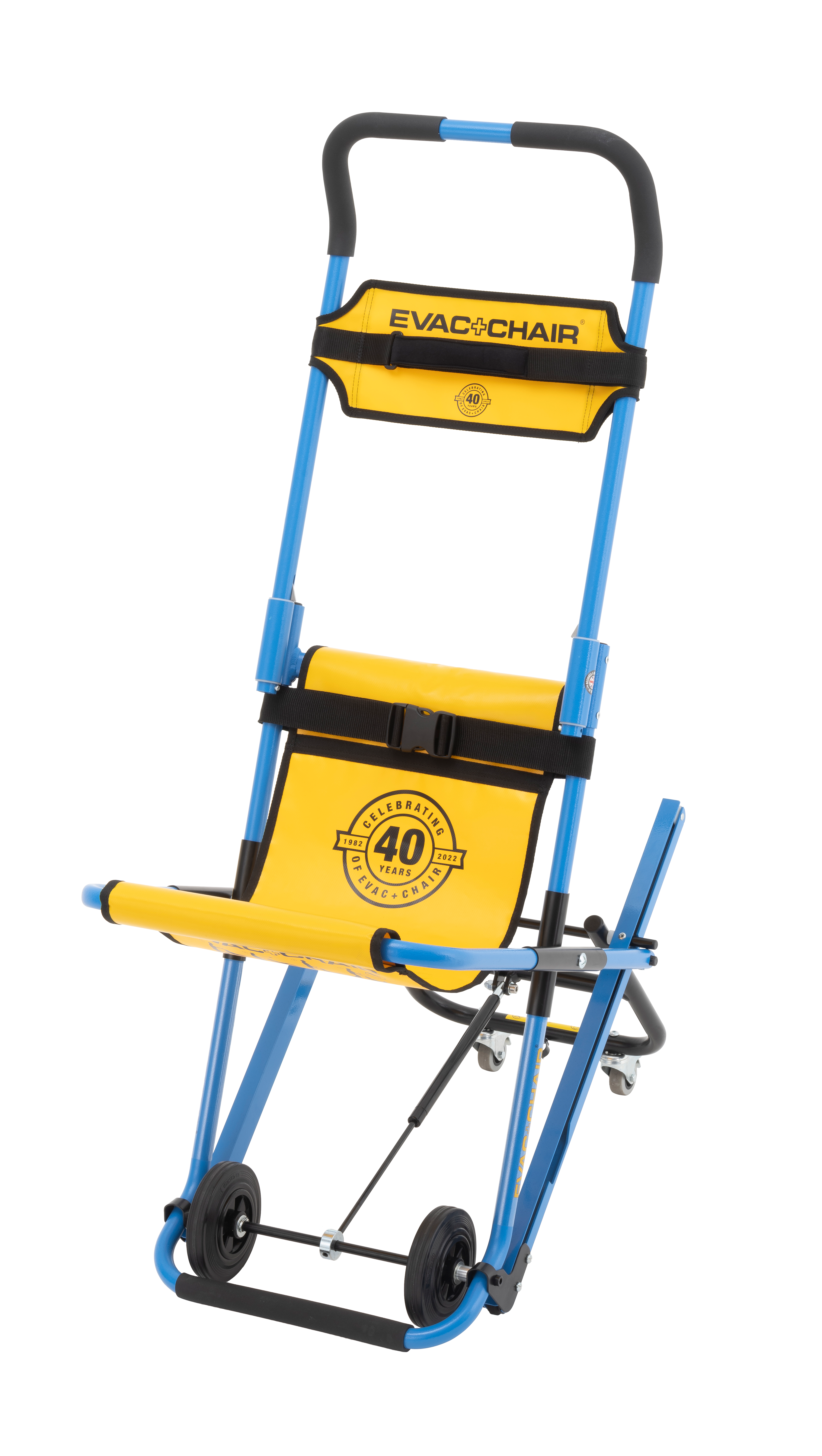 EVACUATION CHAIR
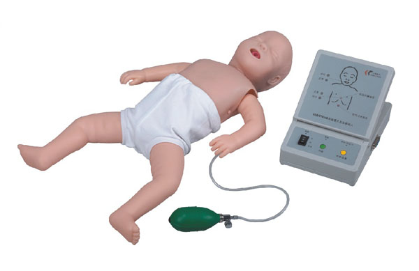 Infant Cpr Manikin
 GUTTA PERCHE POINTS
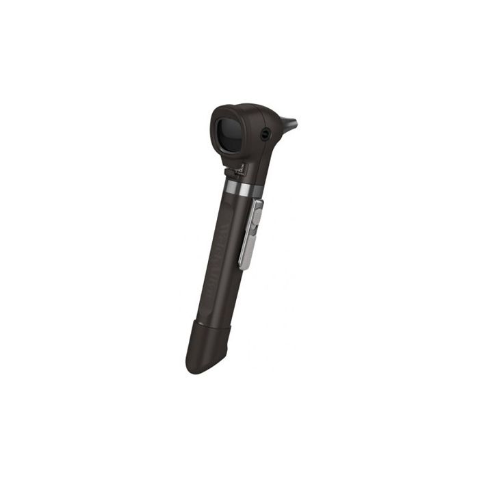 WelchAllyn Otoscope
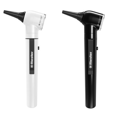 Riester Otoscope
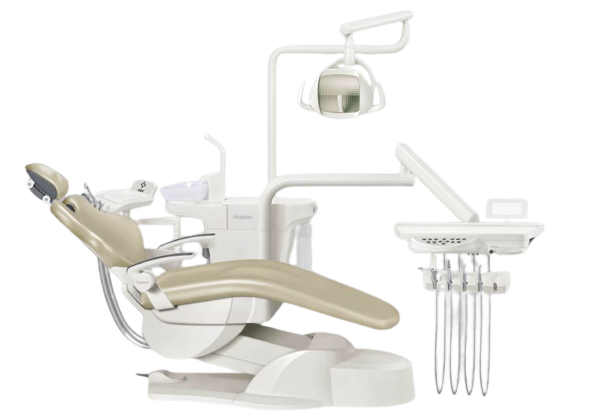 SUNTEM DENTAL CHAIR
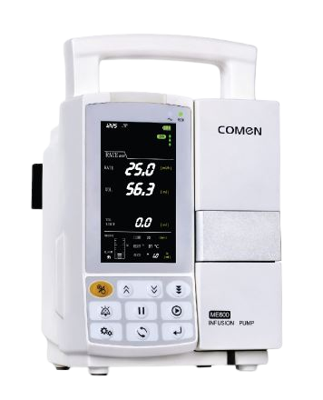 INFUSION PUMP COMEN
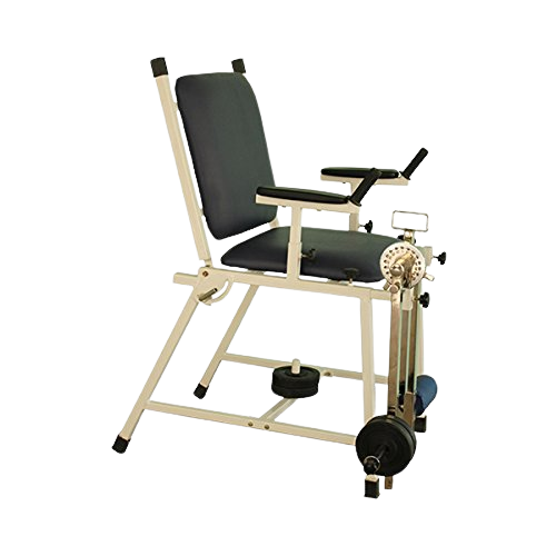 Quadriceps Chair
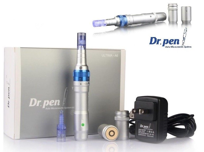 Dr pen Ultima A6- wireless dermapen
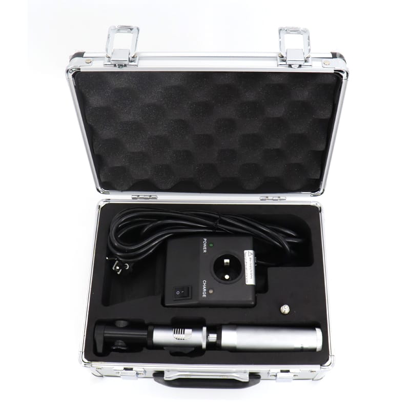 RETINOSCOPE Rechargable
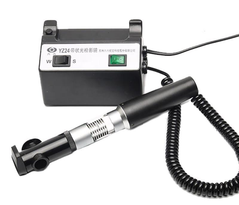 RETINOSCOPE
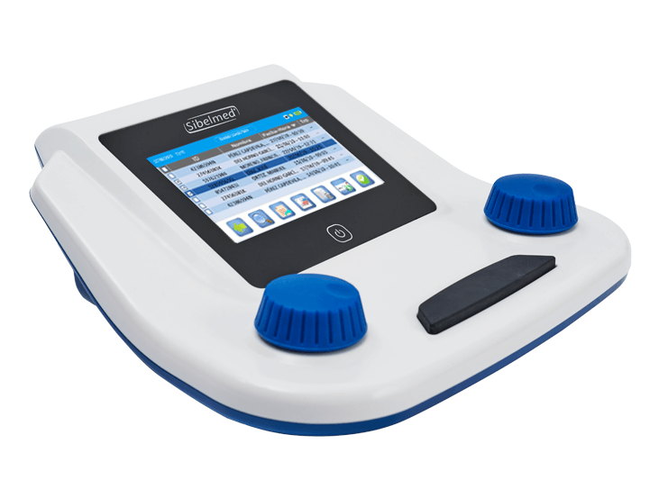 SIBELSOUND DUO Smart Audiometer
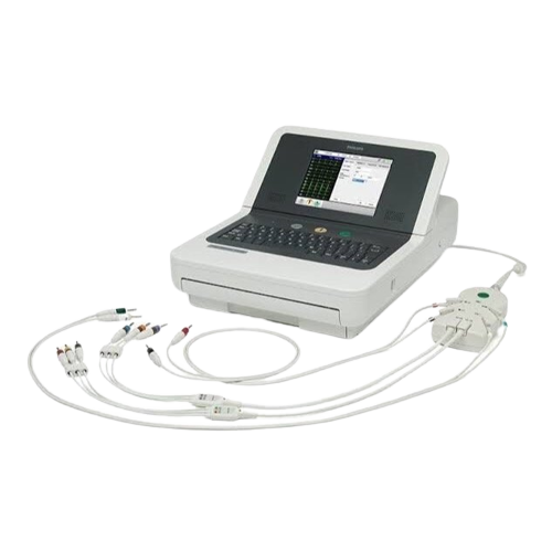 Philips ECG Machine
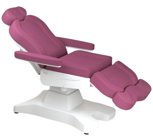 DERMA CHAIR
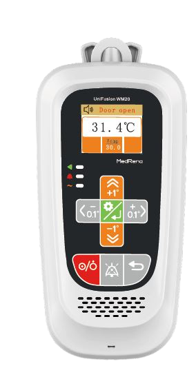 INFUSION WARMER
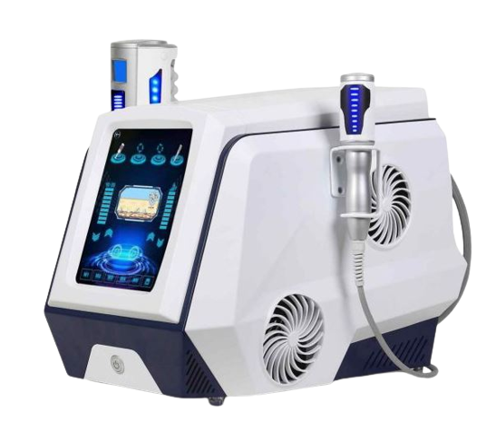 Inner ball roller machine
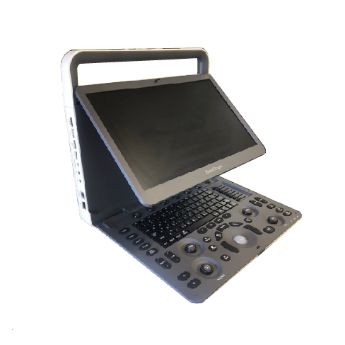 SONOSCAPE PORTABLE ULTRASOUND E1 EXP
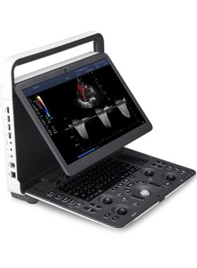 SONOSCAPE PORTABLE ULTRASOUND E2 EXP
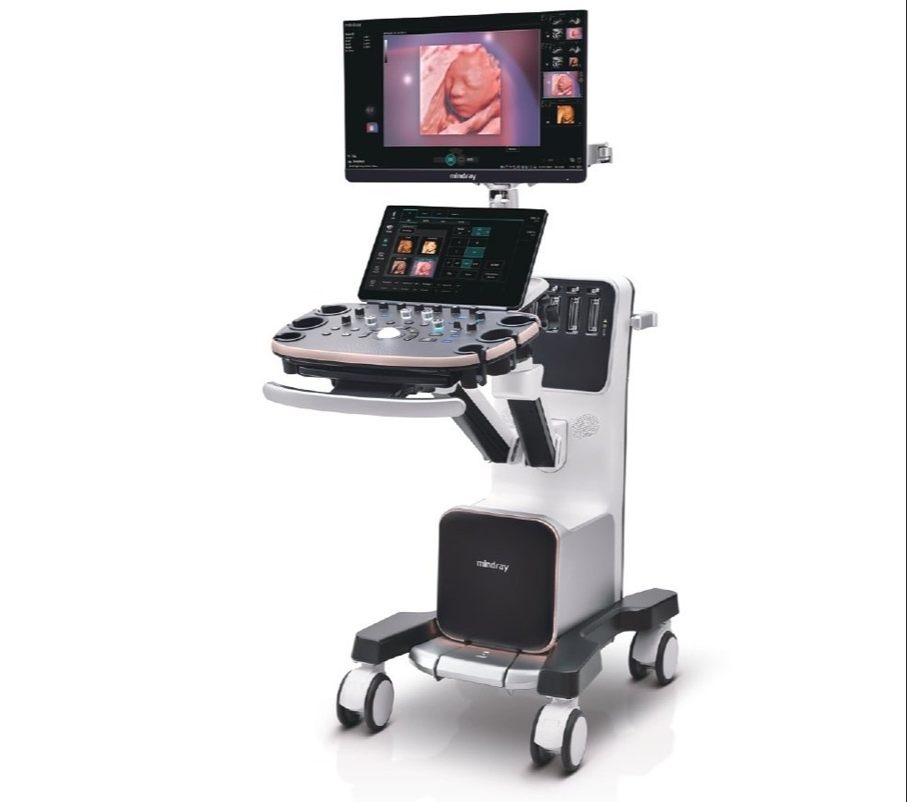 Mindray Nuewa I9 Diagnostic Ultrasound Machine 3D/4D
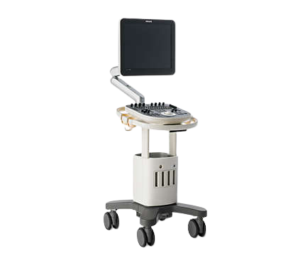 Philips Ultrasound ClearVue 650
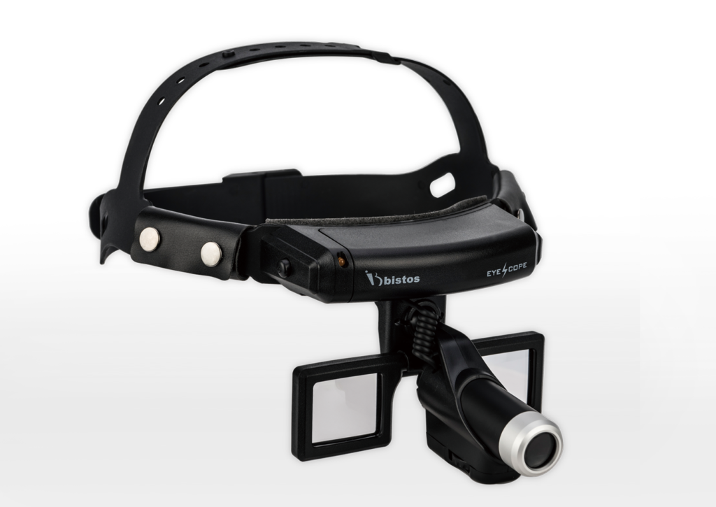 HEADLAMP BISTOS KOREA
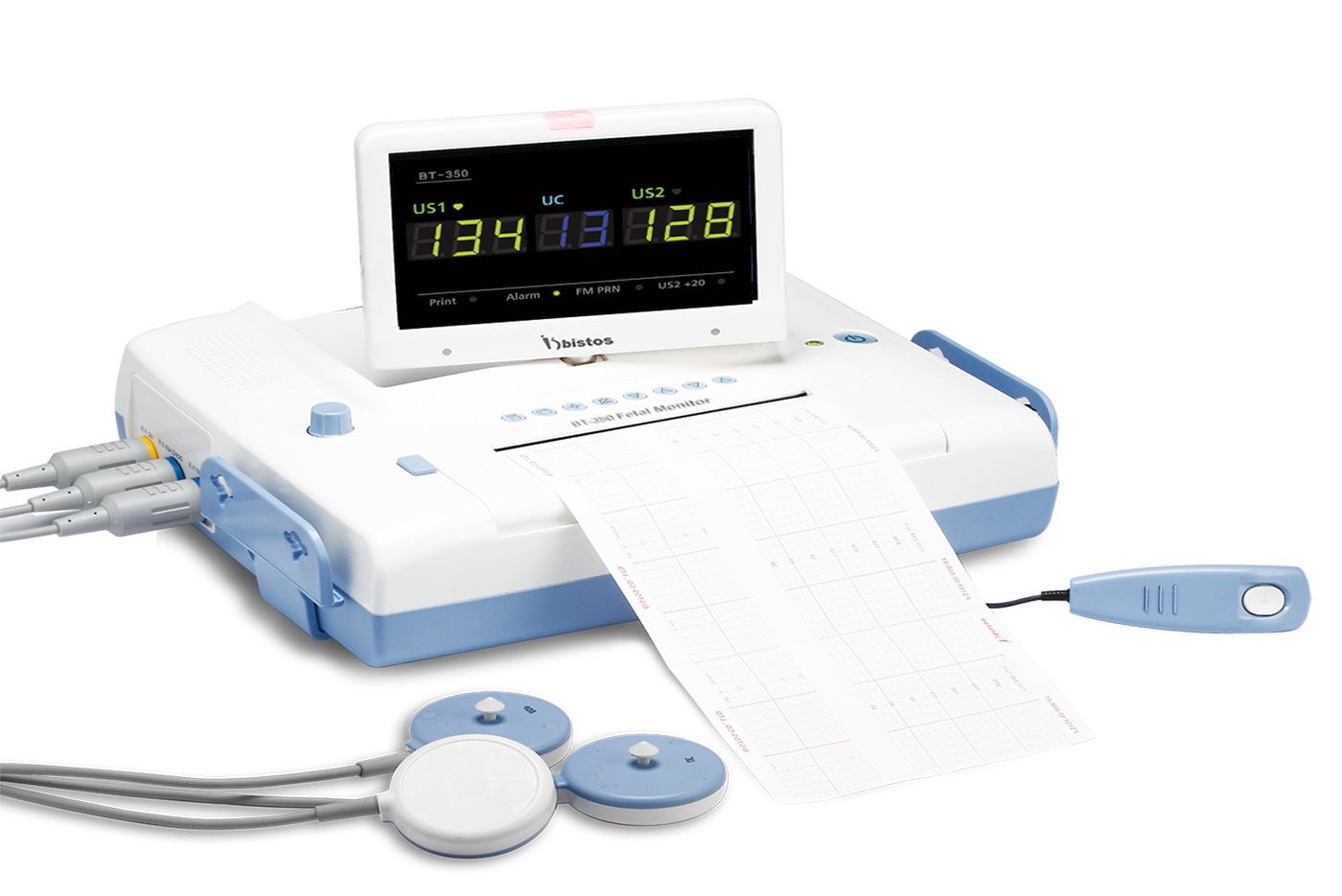 FETAL MONITOR BISTOS
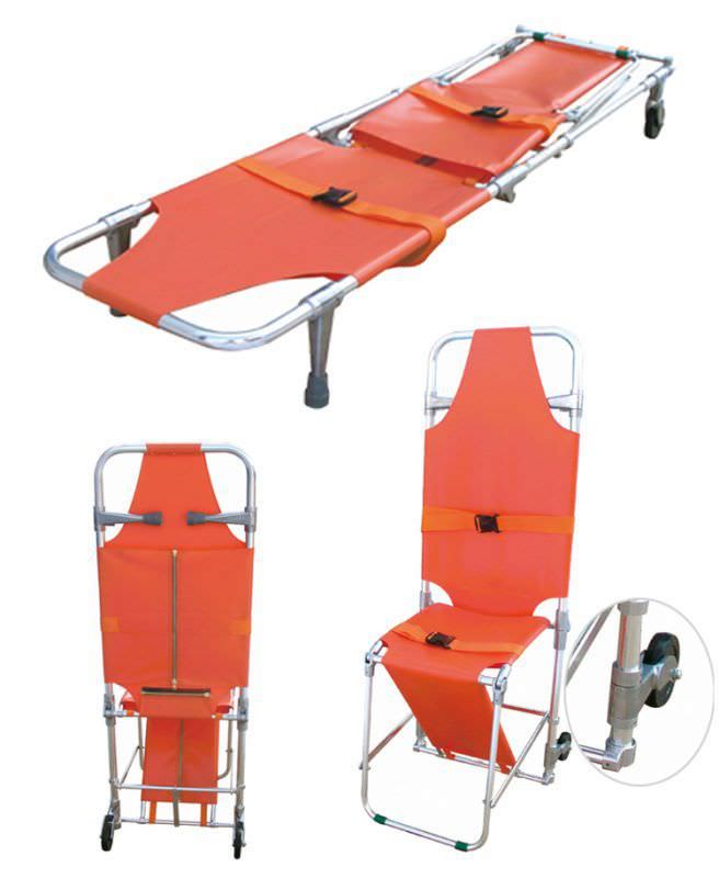 CHAIR STRETCHER
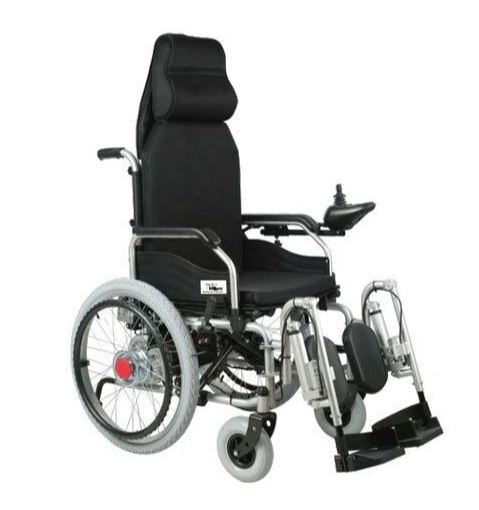 ELECTRIC WHEELCHAIR
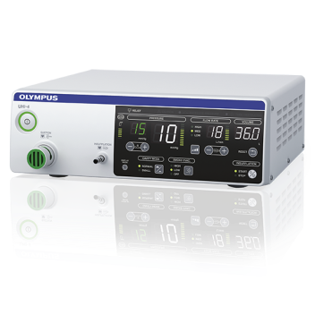 CO2 Insufflator Olympus
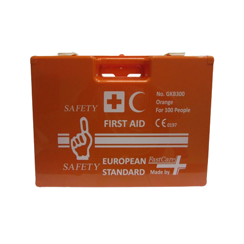 FIRST AID BOX FOR 100 PEOPLE
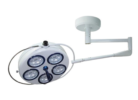 OT LIGHT Cold Light LED
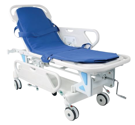 RESCUE STRETCHER
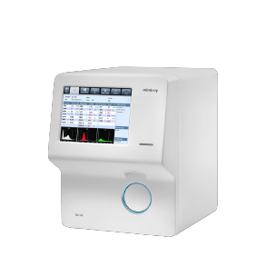 Auto Hematology Analyzer
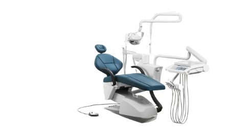 DENTAL CHAIR RUNNEYS care
.png) Bistos BT-740 Patient Monitor
Bistos BT-740 Patient Monitor
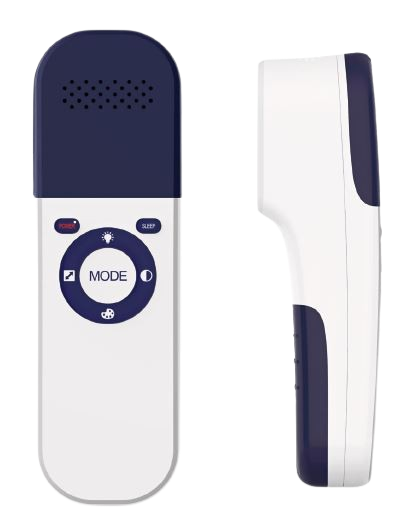 VEIN FINDER PULSE MED
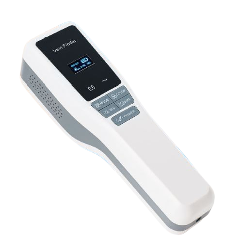 VEIN FINDER GY
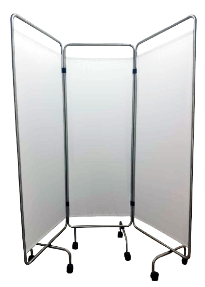 Ward screen Three Fold Premium
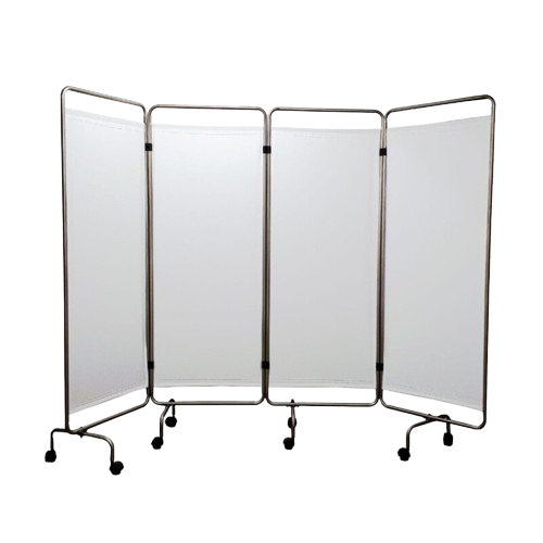 Ward screen Four Fold Premium
.png) Emergency Crash Cart Five Drawer
Emergency Crash Cart Five Drawer
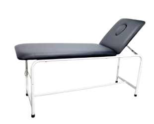 Examination Bed With Nose Cut
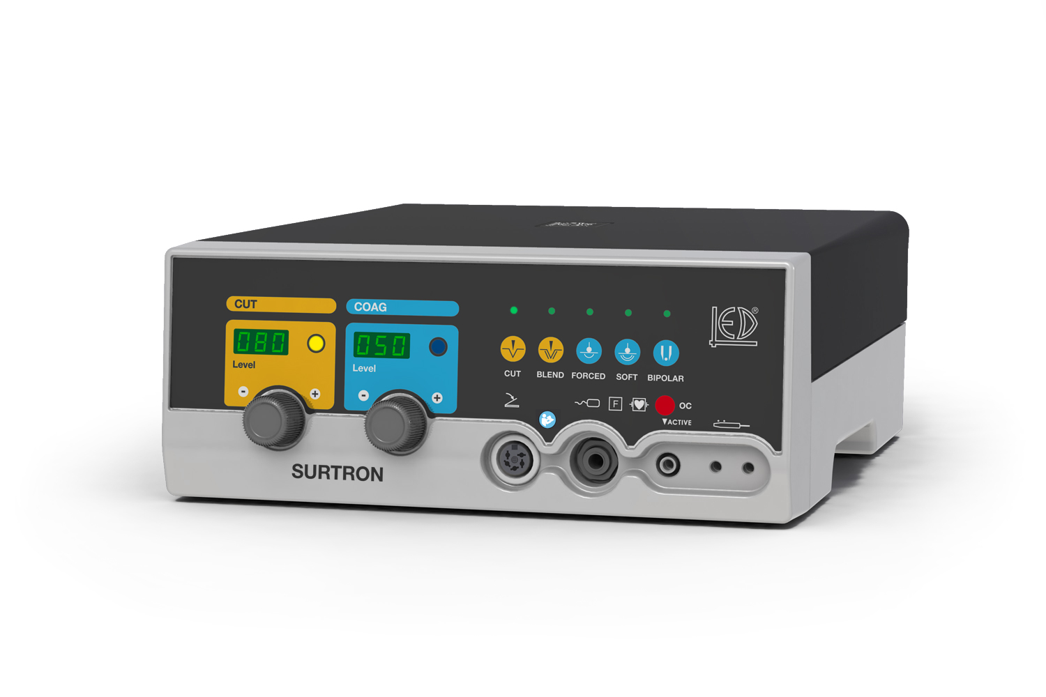 Electrosurgery Unit Surtron 80
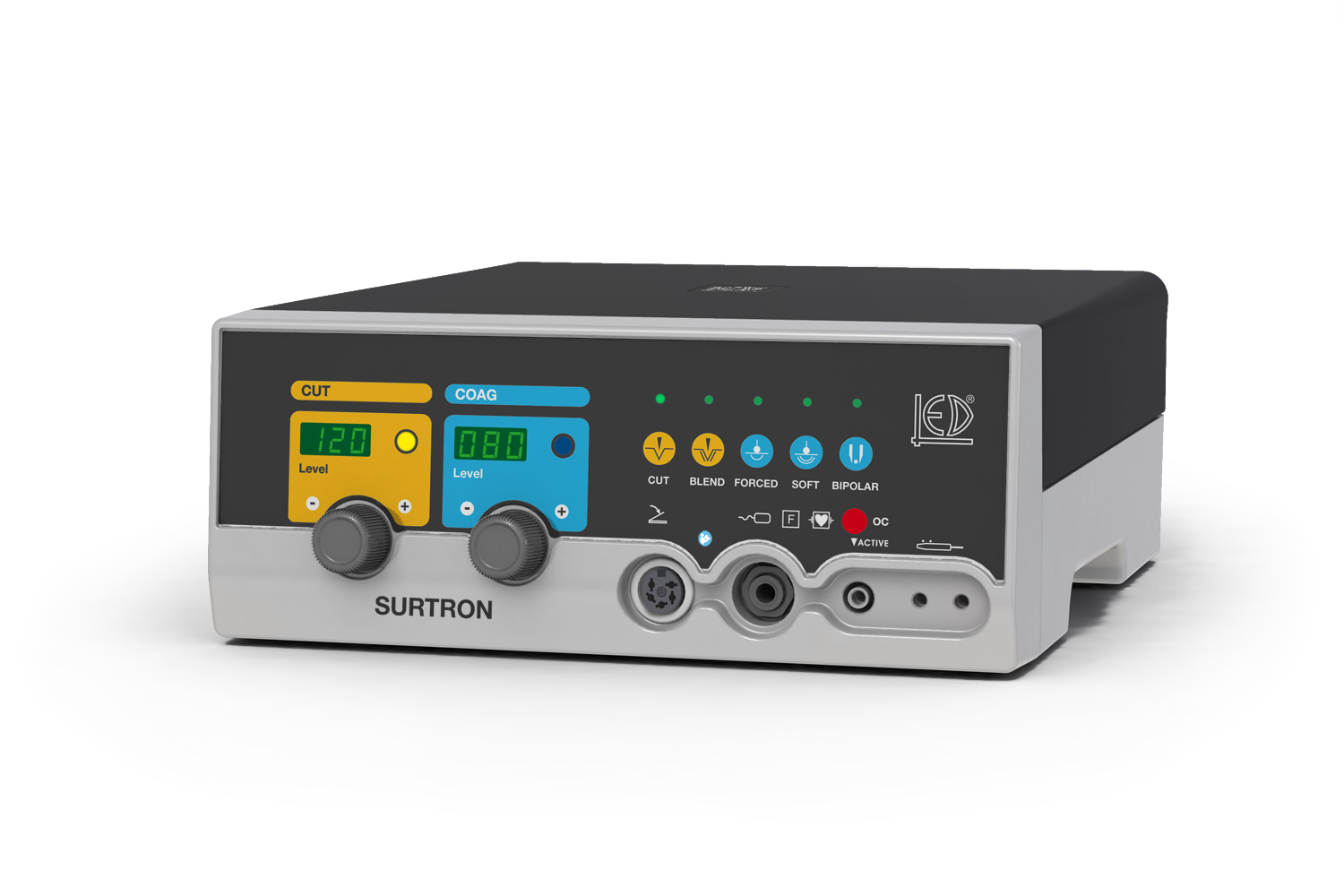 Electrosurgery Unit Surtron 120 LED SpA
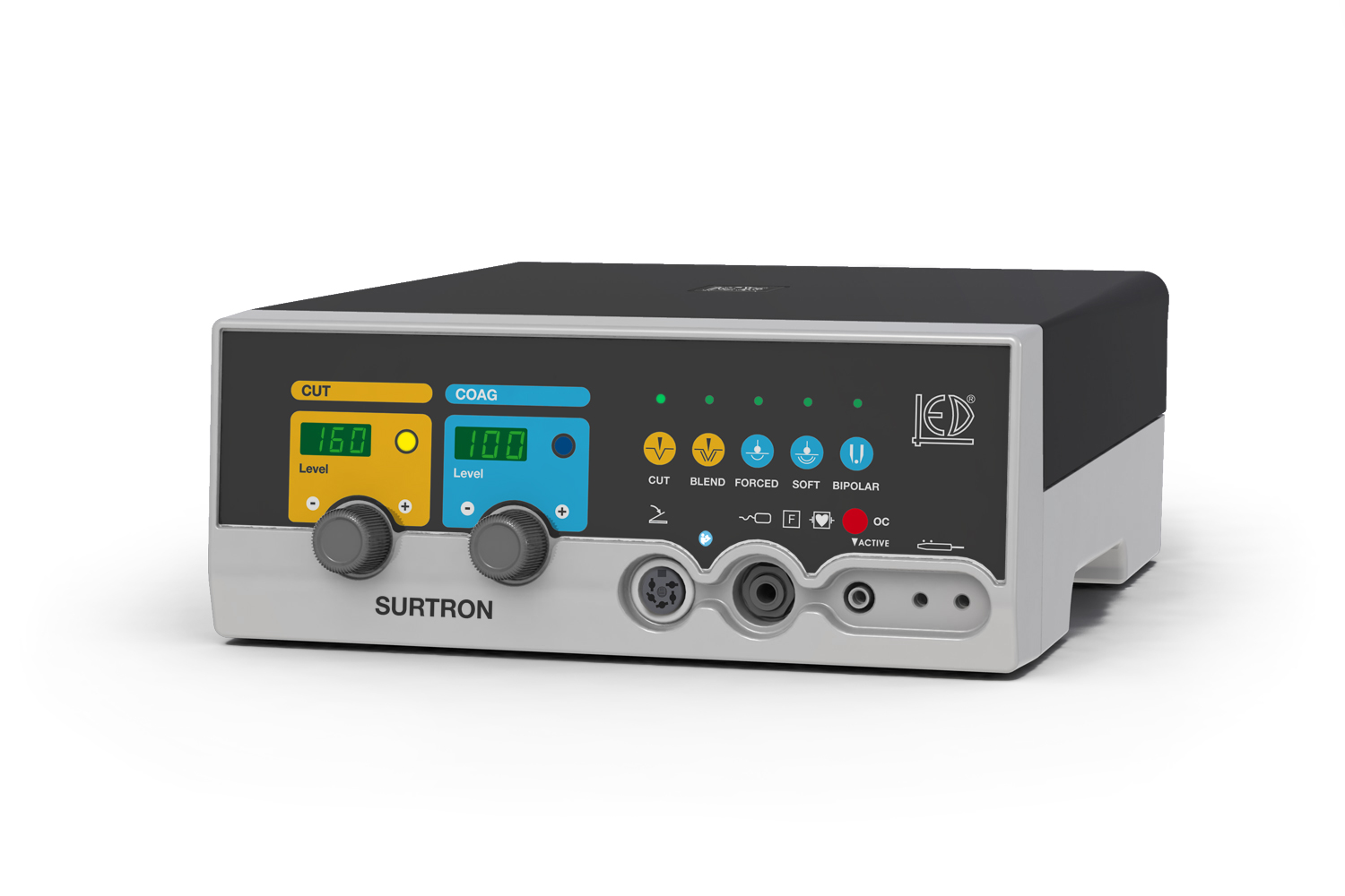 Electrosurgery Unit Surtron 160 LED SpA
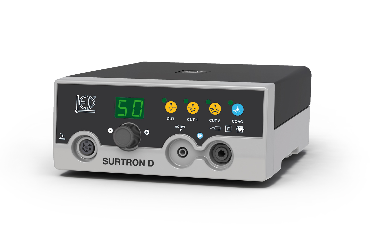 Electrosurgery Unit Surtron 50D LED SpA
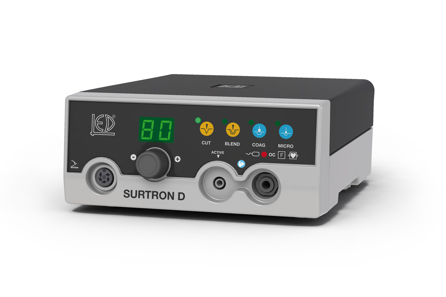 Electrosurgery Unit Surtron 80D LED SpA
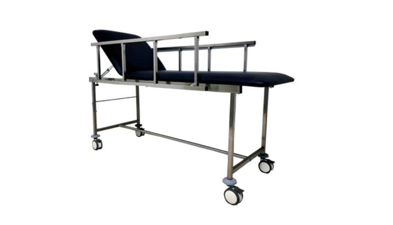 Examination couch With Wheels
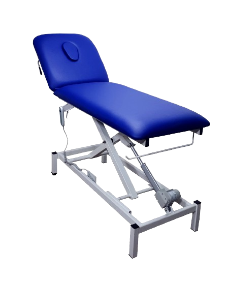 Electric Examination Couch 2 Section
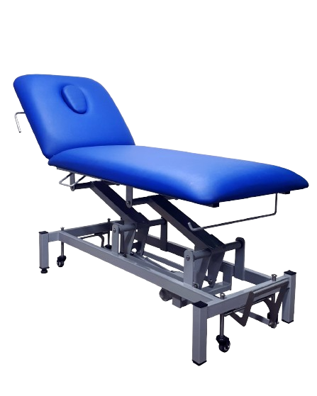 Electric Examination Couch 2 Section Ppremium Quailty
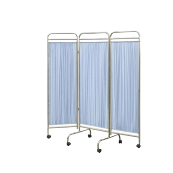 Ward Screen Three Folds
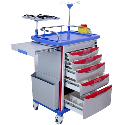 Medical Crash Cart
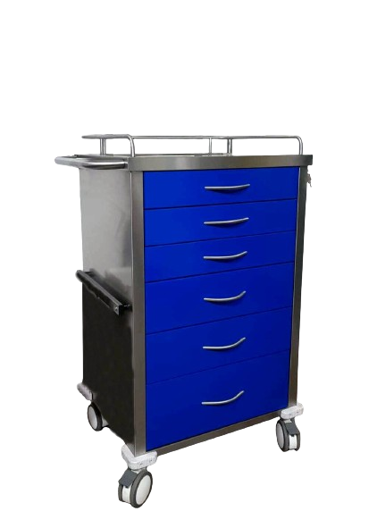 Medical Isolation Carts
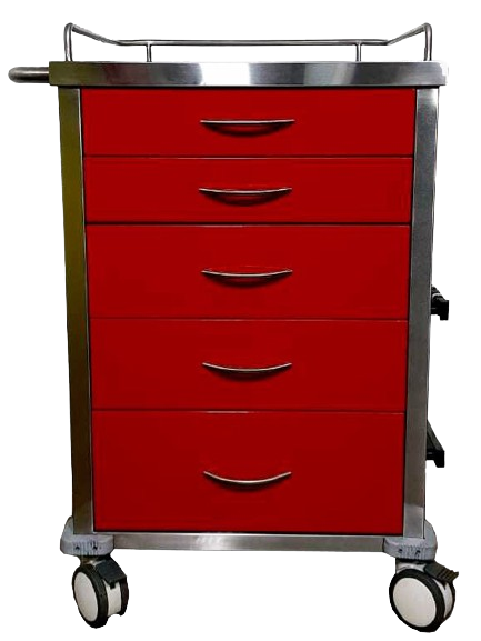 Pediatric crash cart
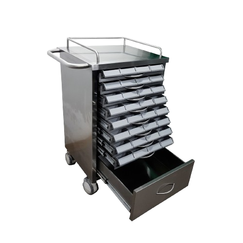 MEDICATION CART
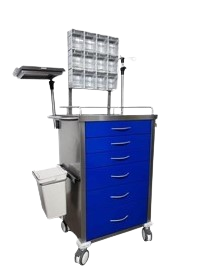 Anesthesia Trolley
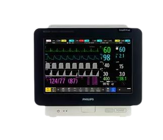 Philips IntelliVue MX450 Patient monitor
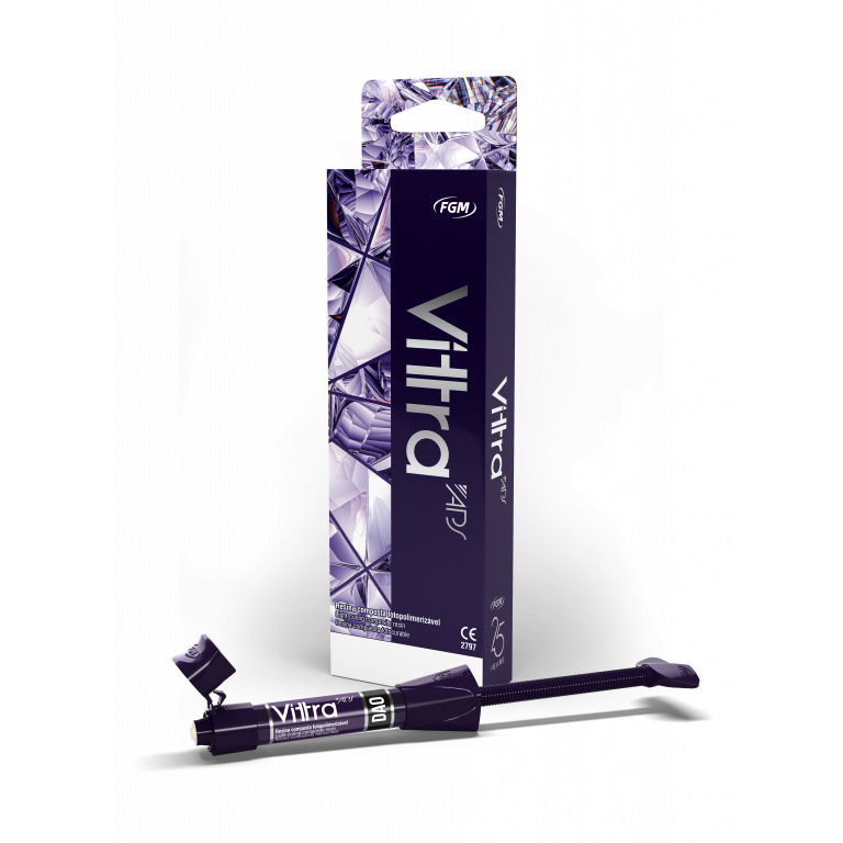 Vittra APS
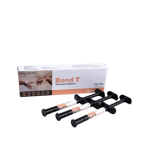 Bond T
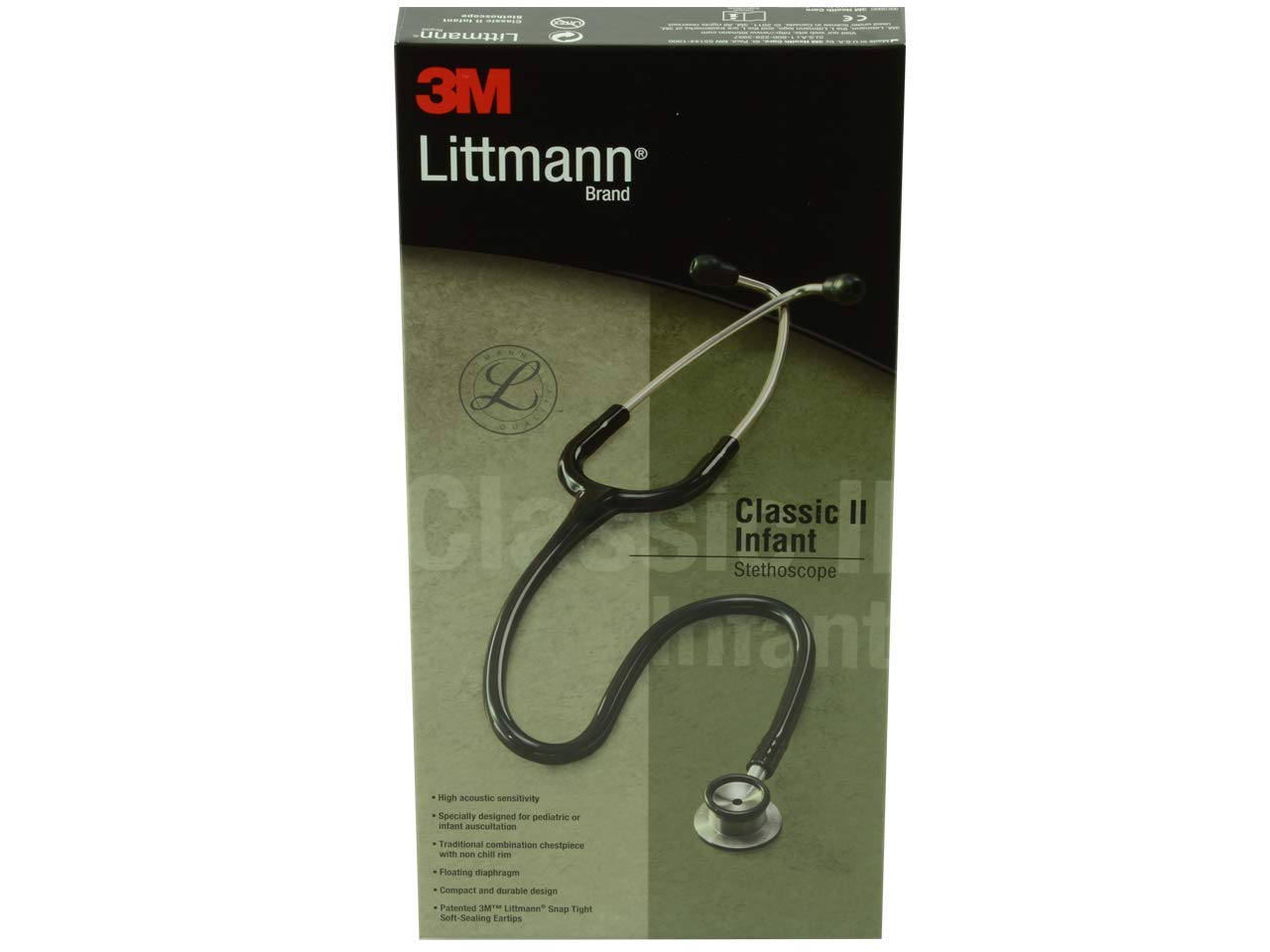 Littmann Classic II Infant Stethoscope
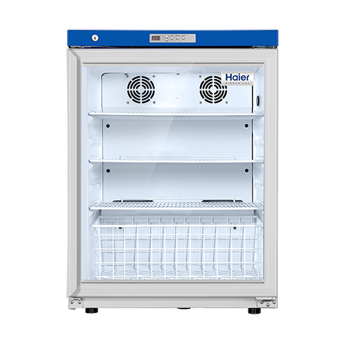 Pharmacy Refrigerator 118L Haier Biomedical
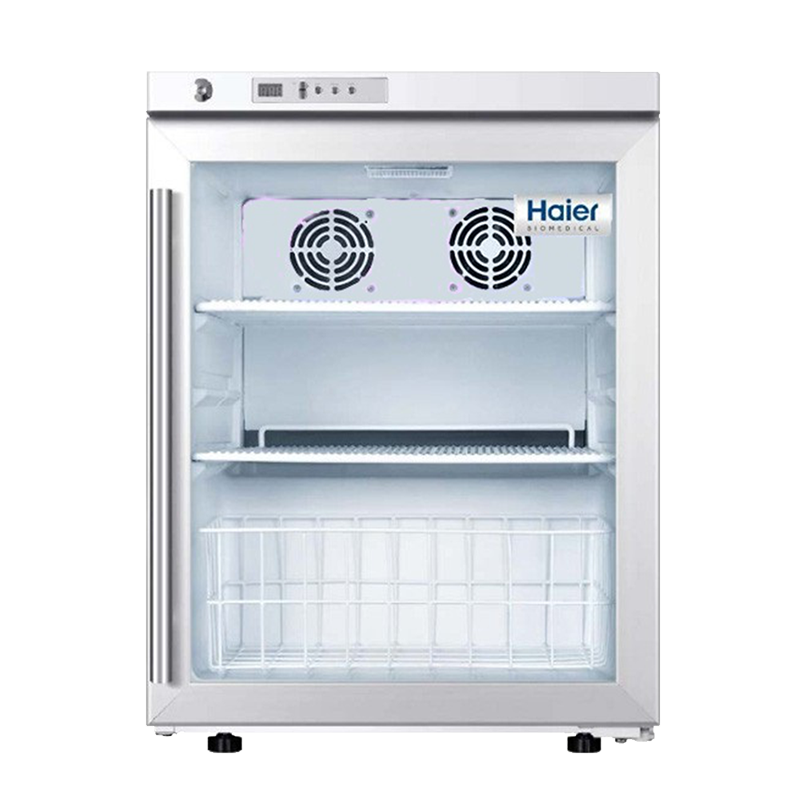 Pharmacy Refrigerator 68L Haier Biomedical 68A
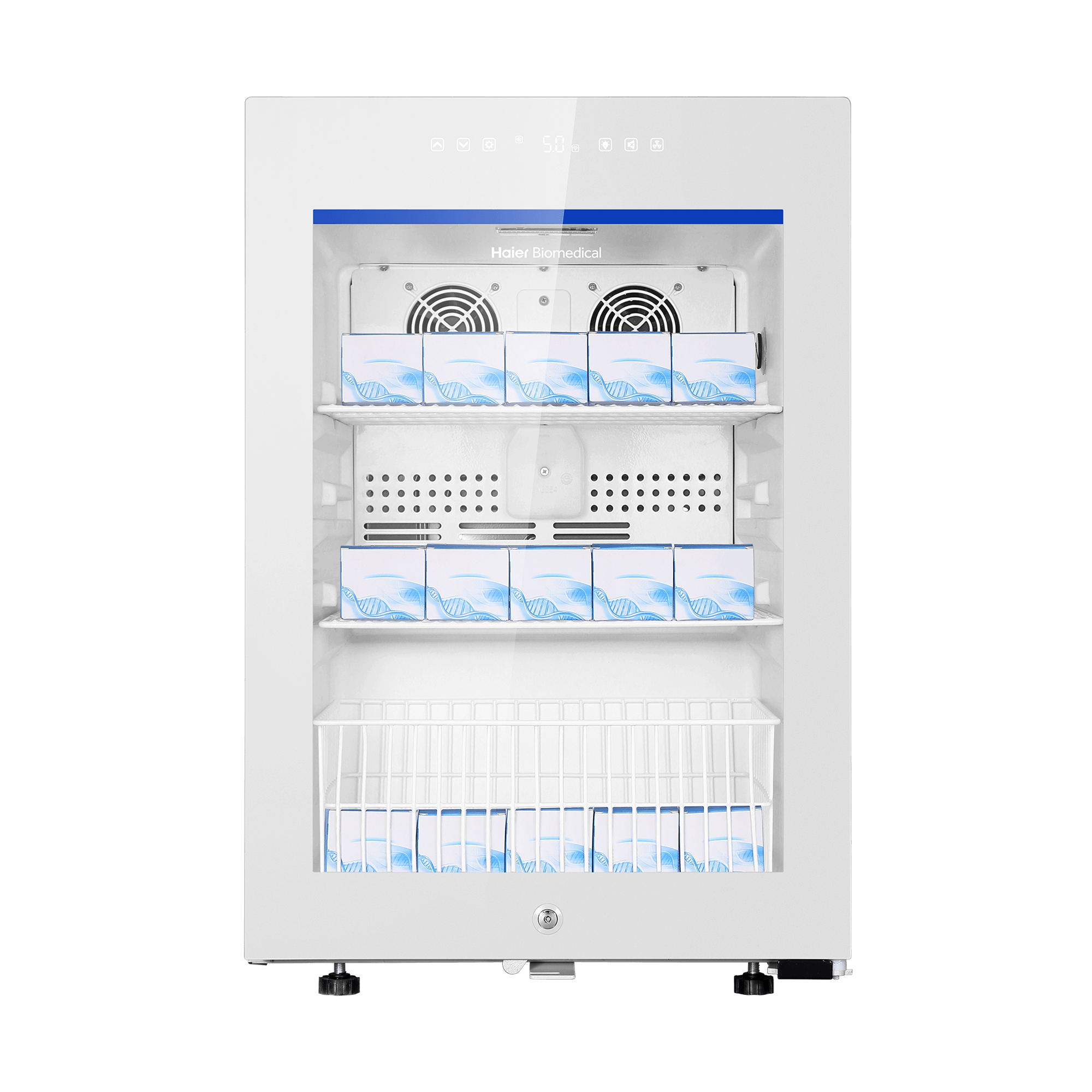 Pharmacy Refrigerator 85L Haier Biomedical 85GD
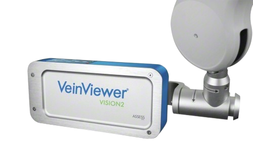 Vein Viewer Vision 2 B Braun
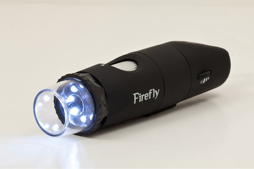 Dermatoscope Firefly DE350
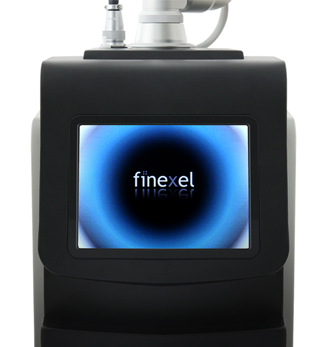 Co2 Laser machine
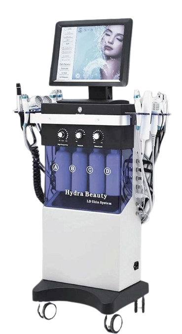 Hydrafacial machine
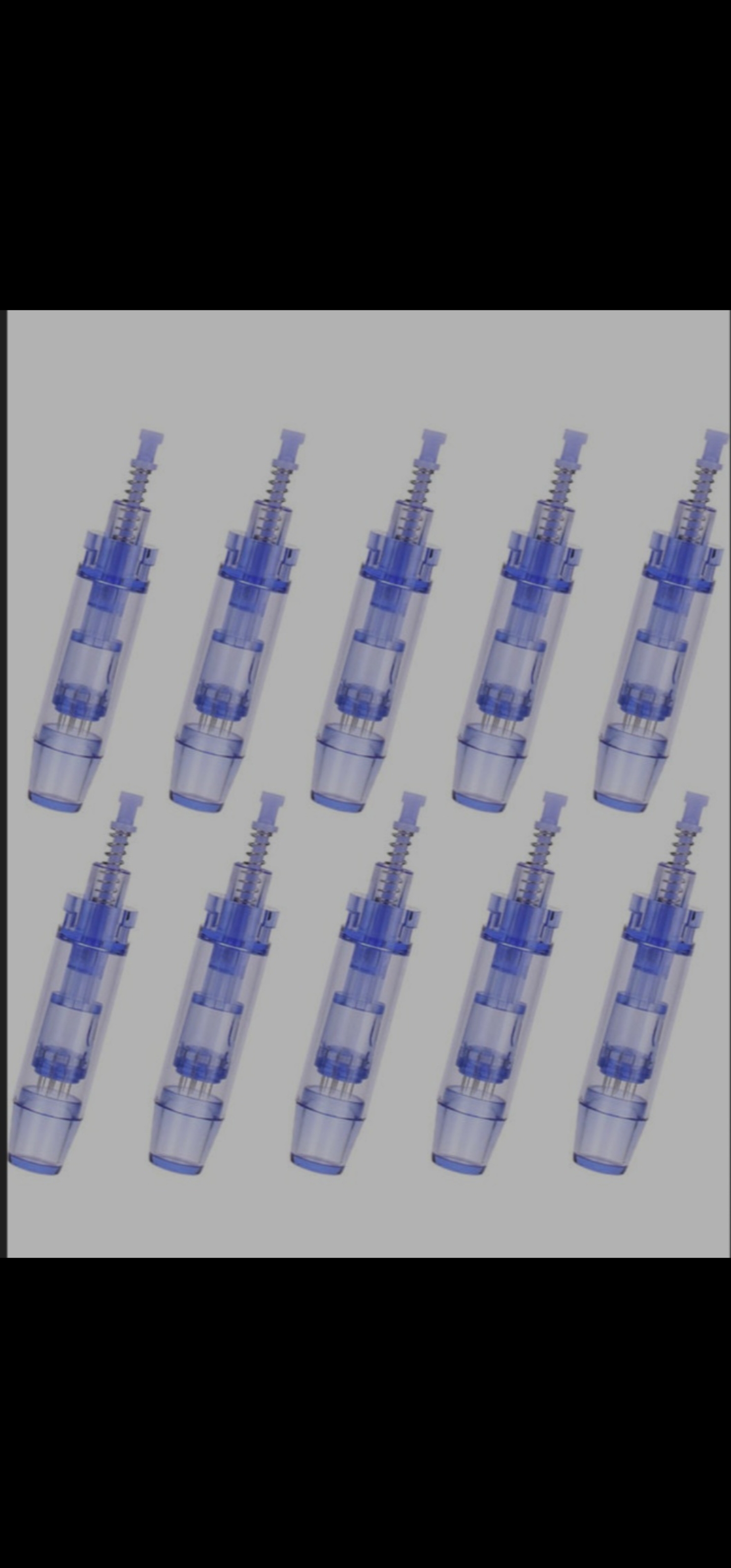 Microneedling
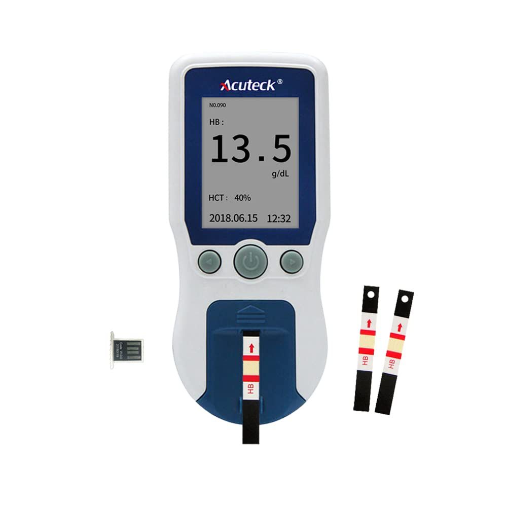 HEMOGLOBINOMETER
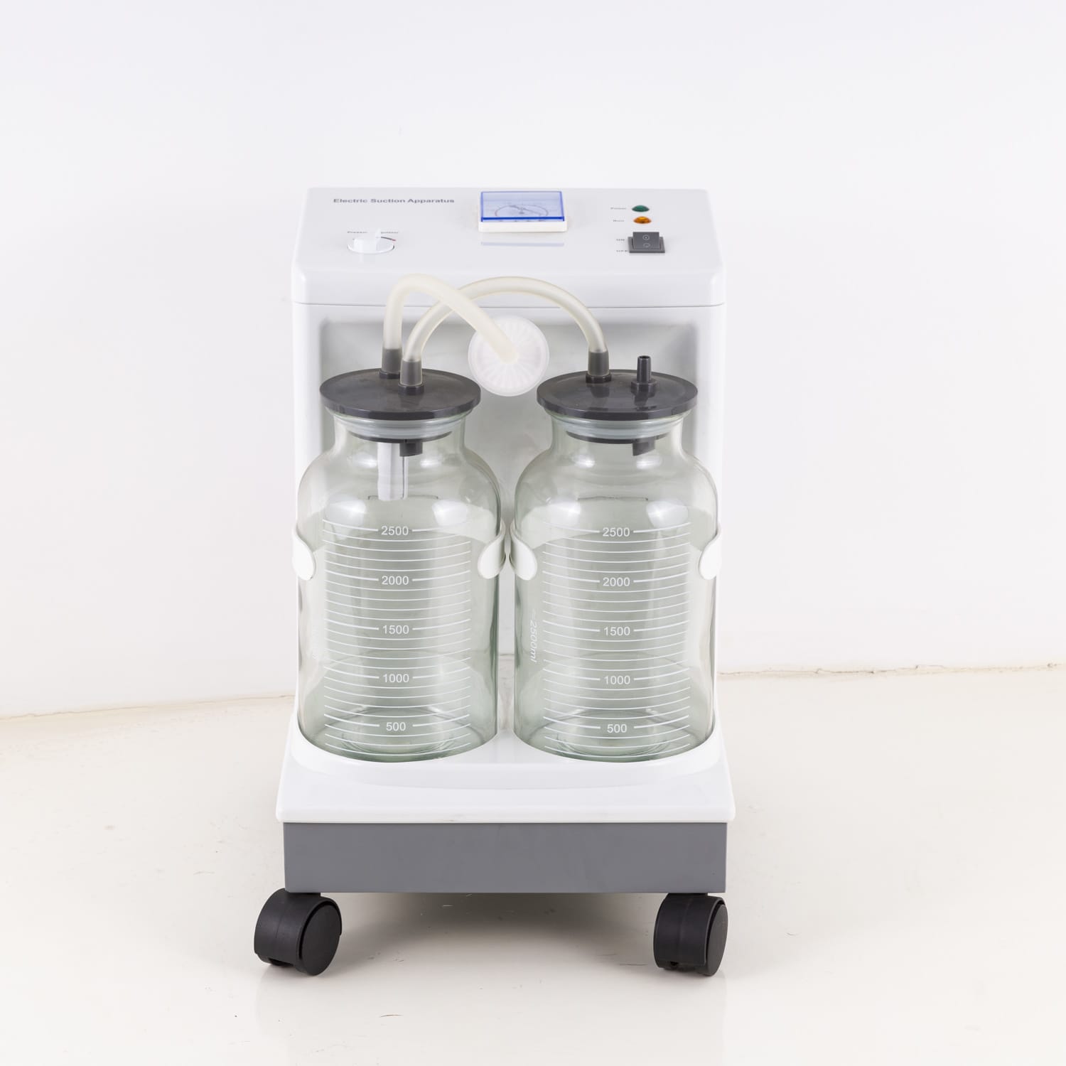 ELECTRIC SUCTION UNIT SS-8A
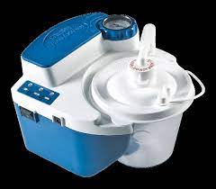 VacuAide QSU 7314
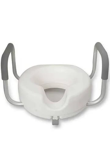 RAISED TOILET SEAT WITH HAND
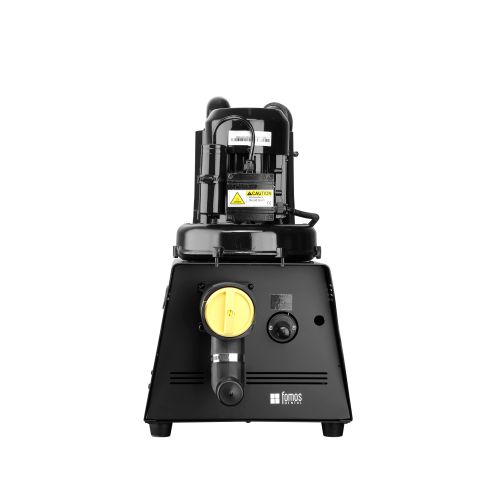 DENTAL SUCTION UNIT
 AIR PROPHY UNIT
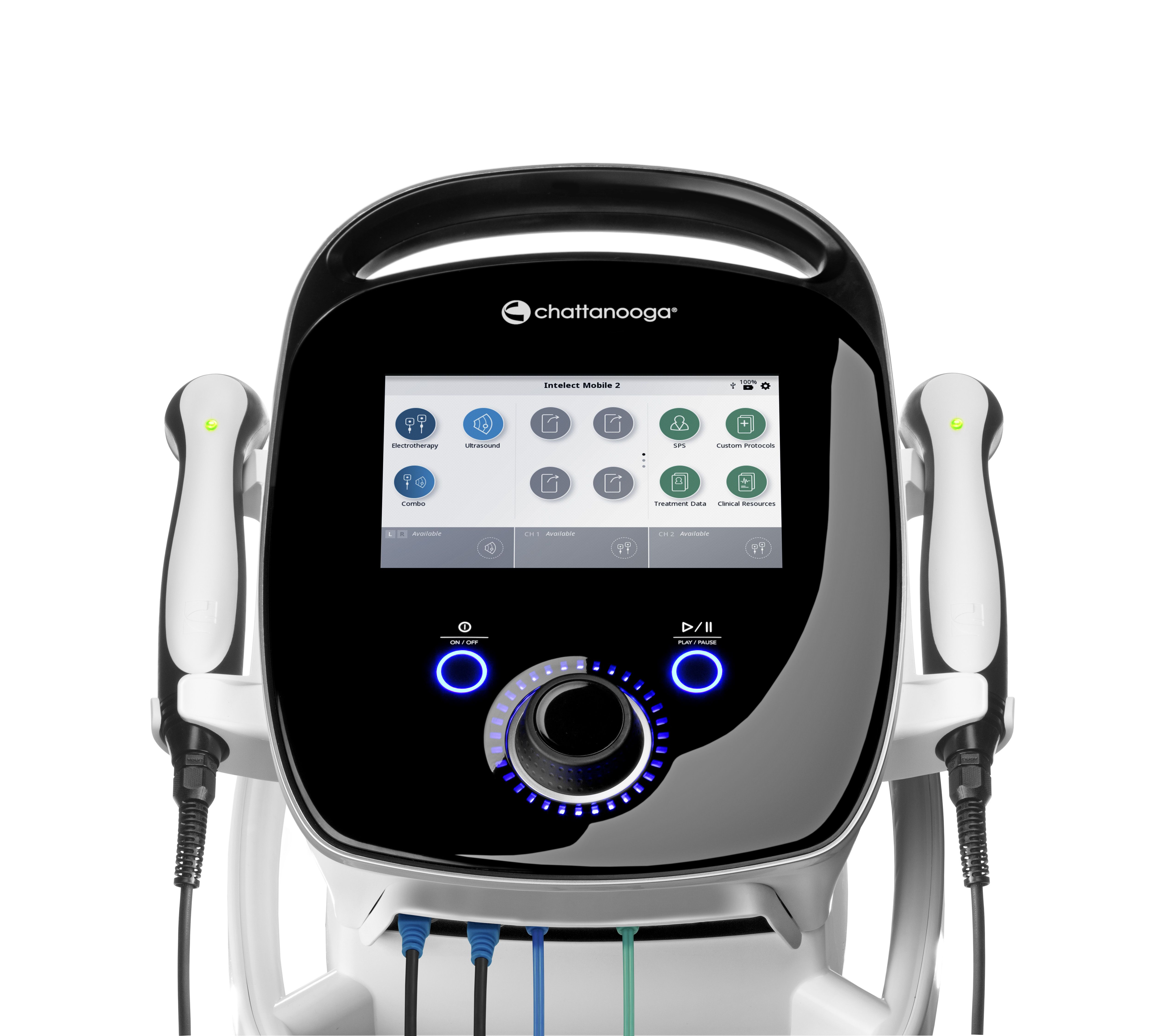 INTELECT MOBILE 2 COMBO
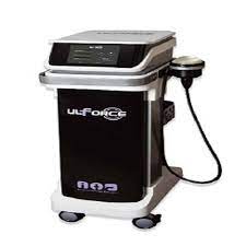 UL Force Radial Shockwave
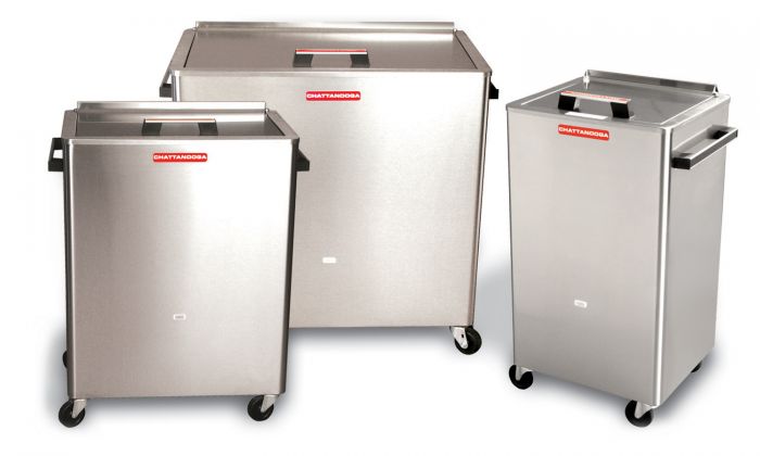 Chattanooga Hydrocollator 8M SS-2
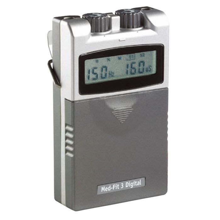 Digital Tens Dual Channel
 Cando Go Band
 HYDROCOLLATOR 8M SS2
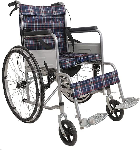 COMMOD WHEEL CHAIR
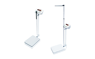 HEIGHT AND WEIGHT SCALE
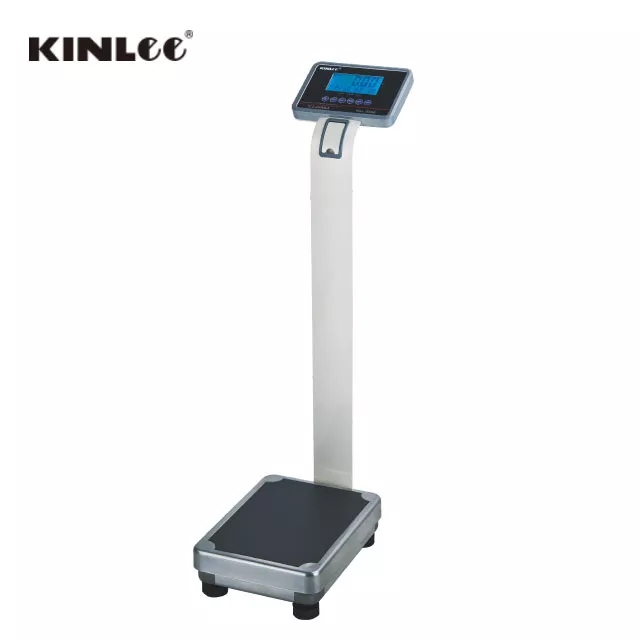 Electronic Digital Platform Scale with LCD Display
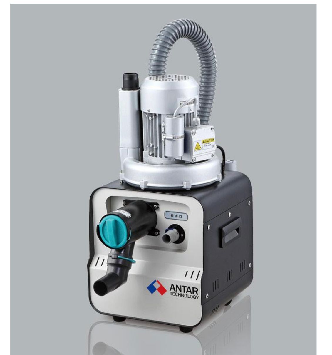 Dental suction 750 L
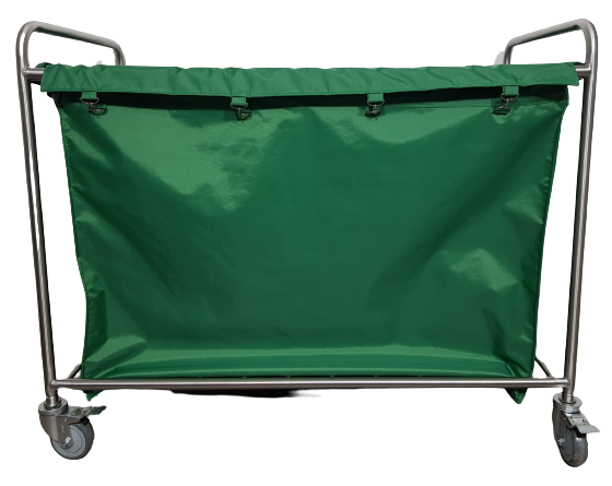 LINEN transport
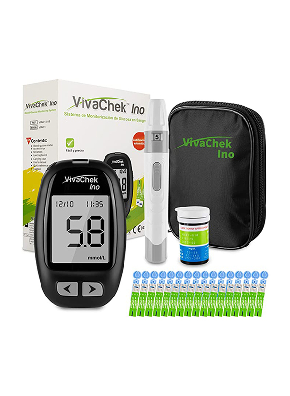 VivaCheck Ino Glucometer
.jpeg) Wave balance BP Moniter
Wave balance BP Moniter
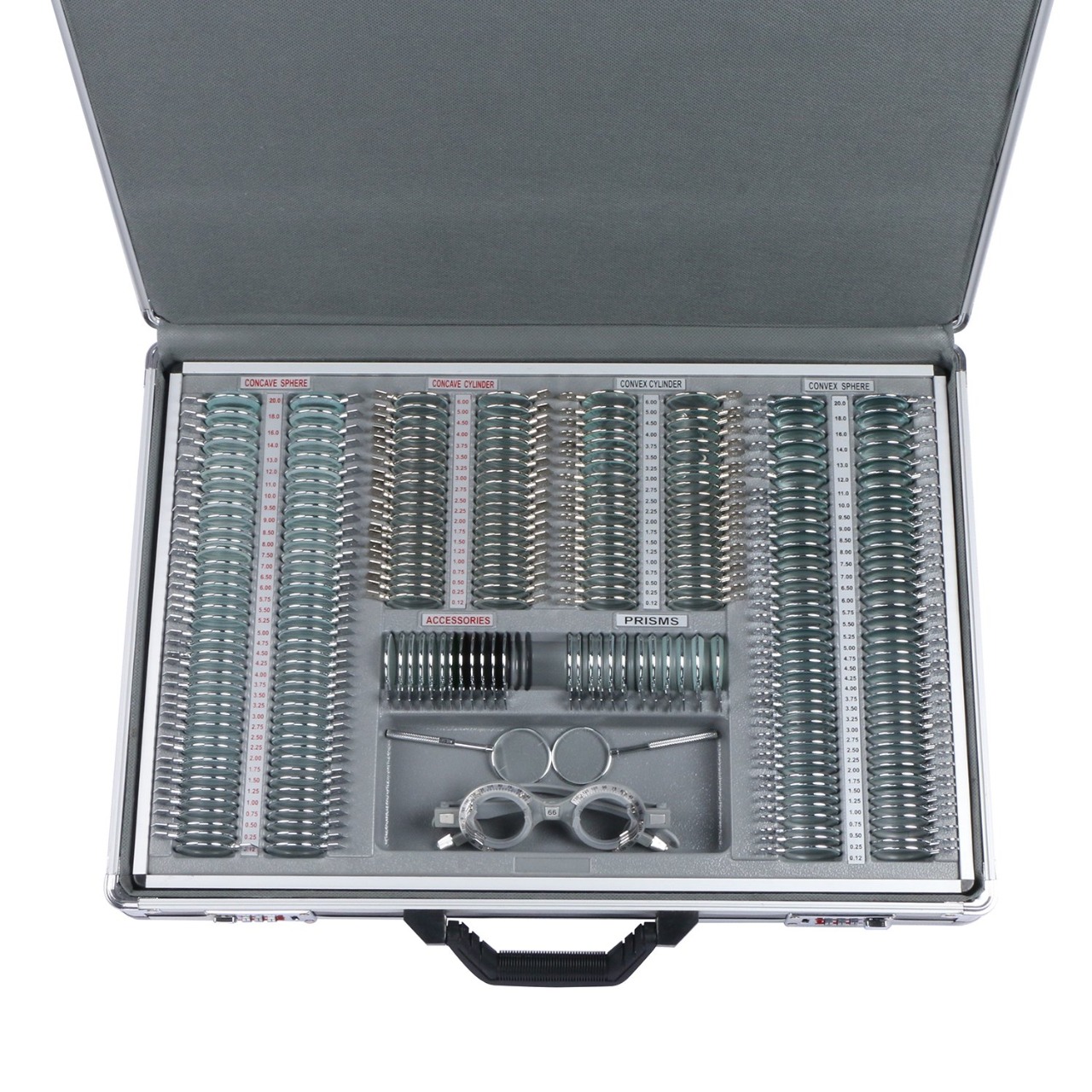 METAL TRIAL CASE
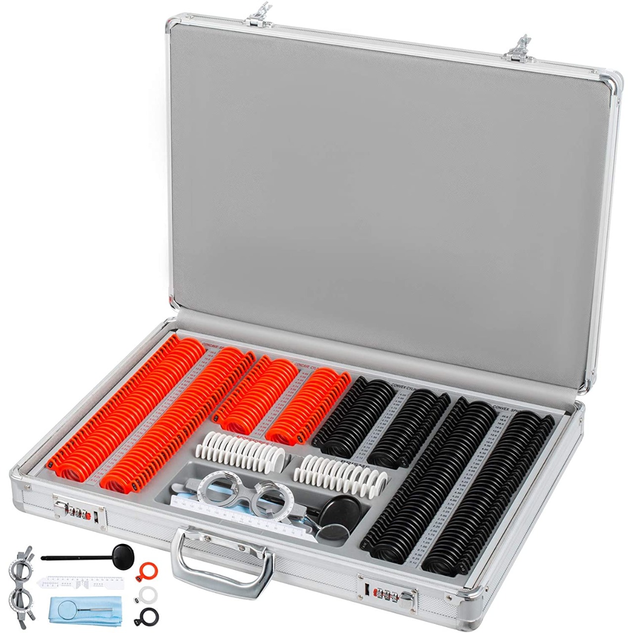 PLASTIC TRIAL CASE
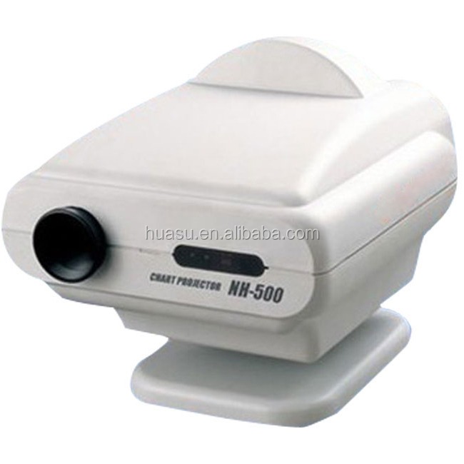 VISION CHART PROJECTOR
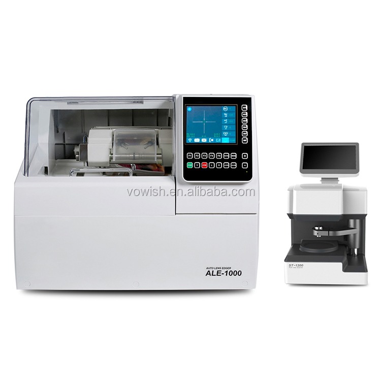 AUTOMATIC LENS EDGER
 RM 9000
 RMK150
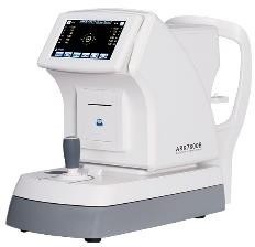 7800B AUTOREFRACTOMETER
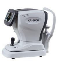 KR9600 AUTOREFRACTOMETER
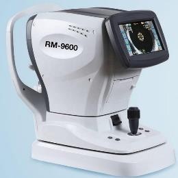 RM9600 AUTOREFRACTOMETER
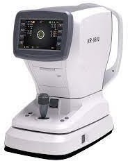 KR9800 AUTOREFRACTOMETER
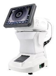 ARK 1 AUTOREFRACTOMETER
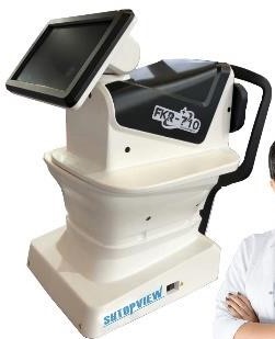 FRK 710 AUTOREFRACTOMETER
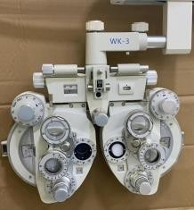 WK3 PHOROPTER
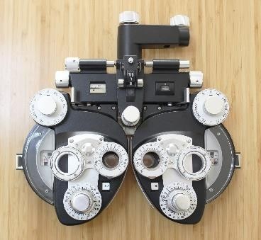 WK10 PHOROPTER
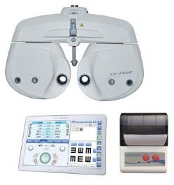
 7600CV AUTO PHOROPTER
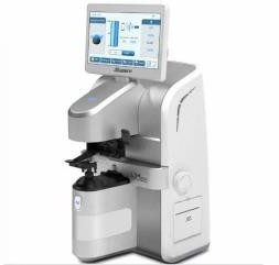
 7200CV AUTO PHOROPTER
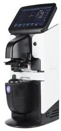
 LM 800 LENSMETER
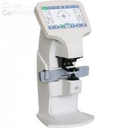
 2600A LENSMETER
 FL 8600 LENSMETER
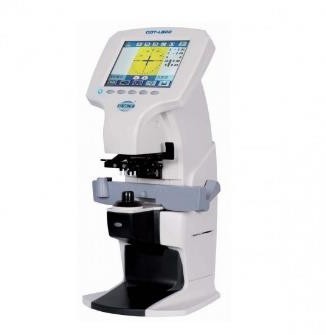 COT800 LENSMETER
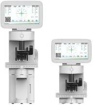 JR LM002 WITHOUT BLUETOOTH PRINTER
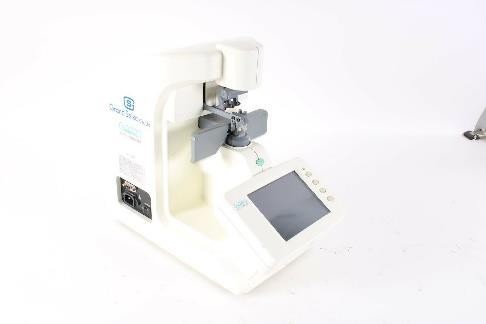 GL 7000 GRAND SEIKO
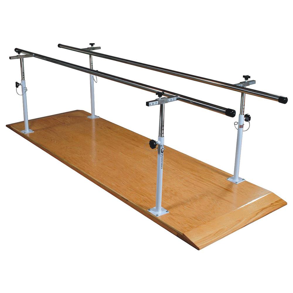 SL PB 100 PARALLEL BAR WITH WOODEN PLATFORM
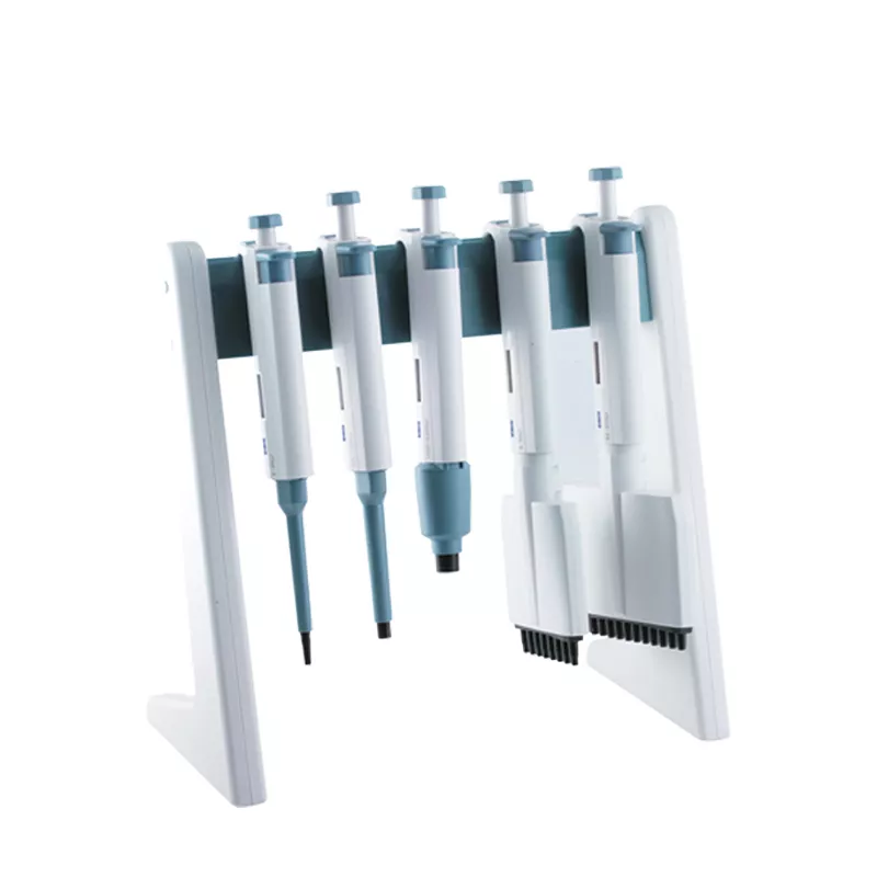 PIPPETTE STAND
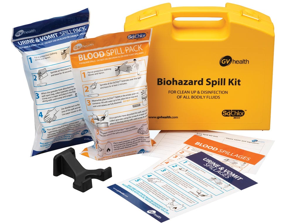 SPILL KIT
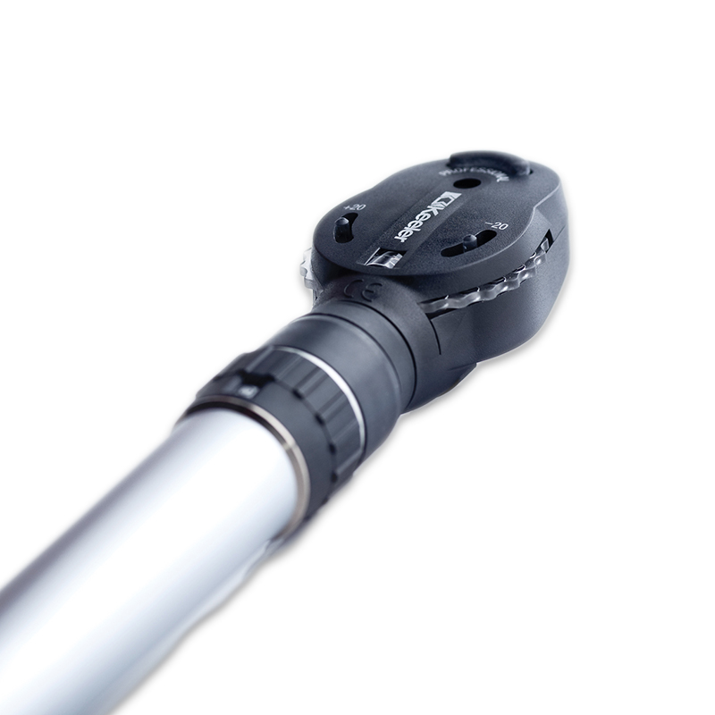 OPTHALMOSCOPE
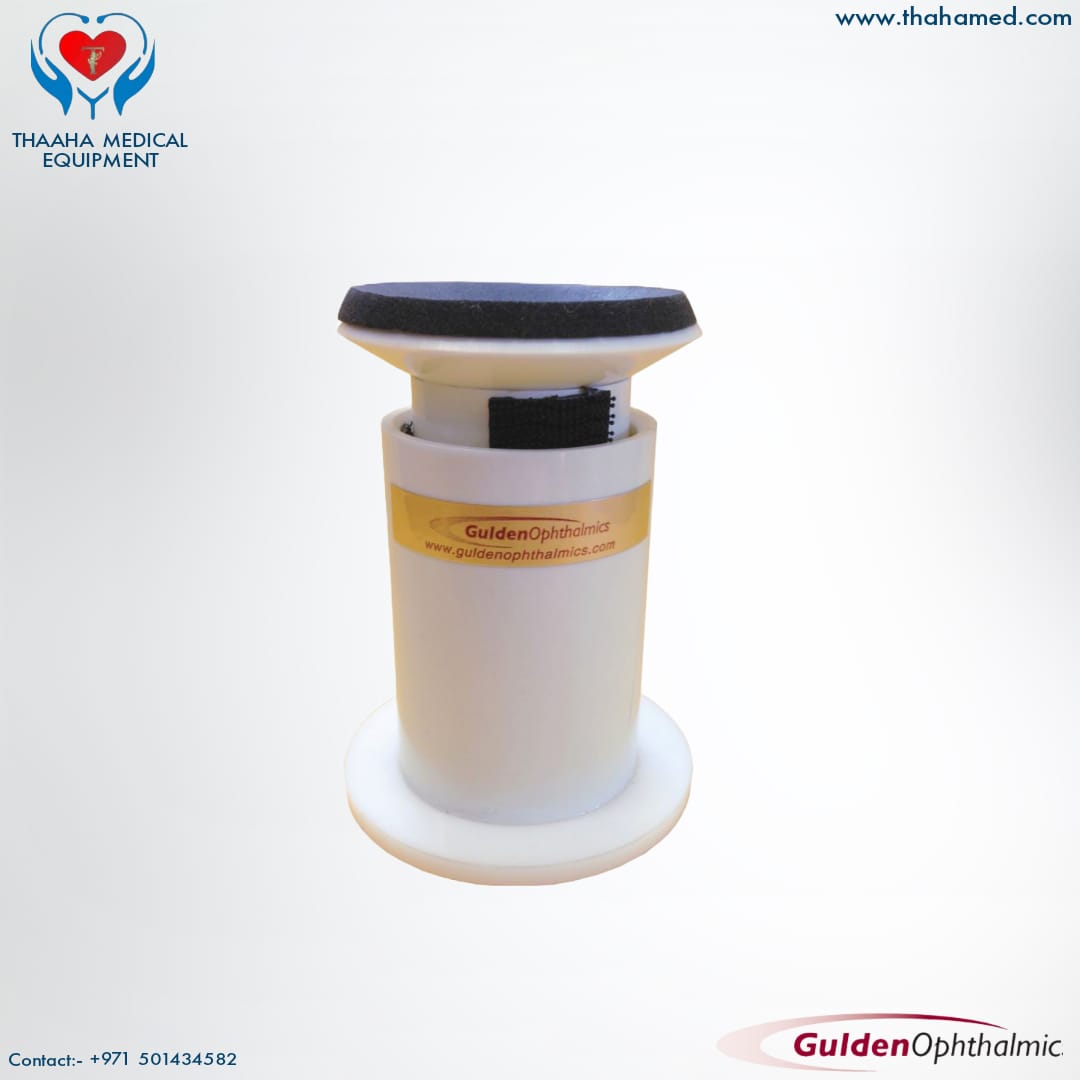 ELBOW SLIT LAMP REST
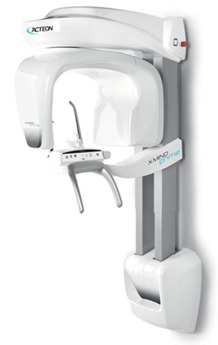 OPG UNIT WALL MOUNT 2D X RAY MACHINE
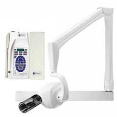 WALL MOUNT X-RAY
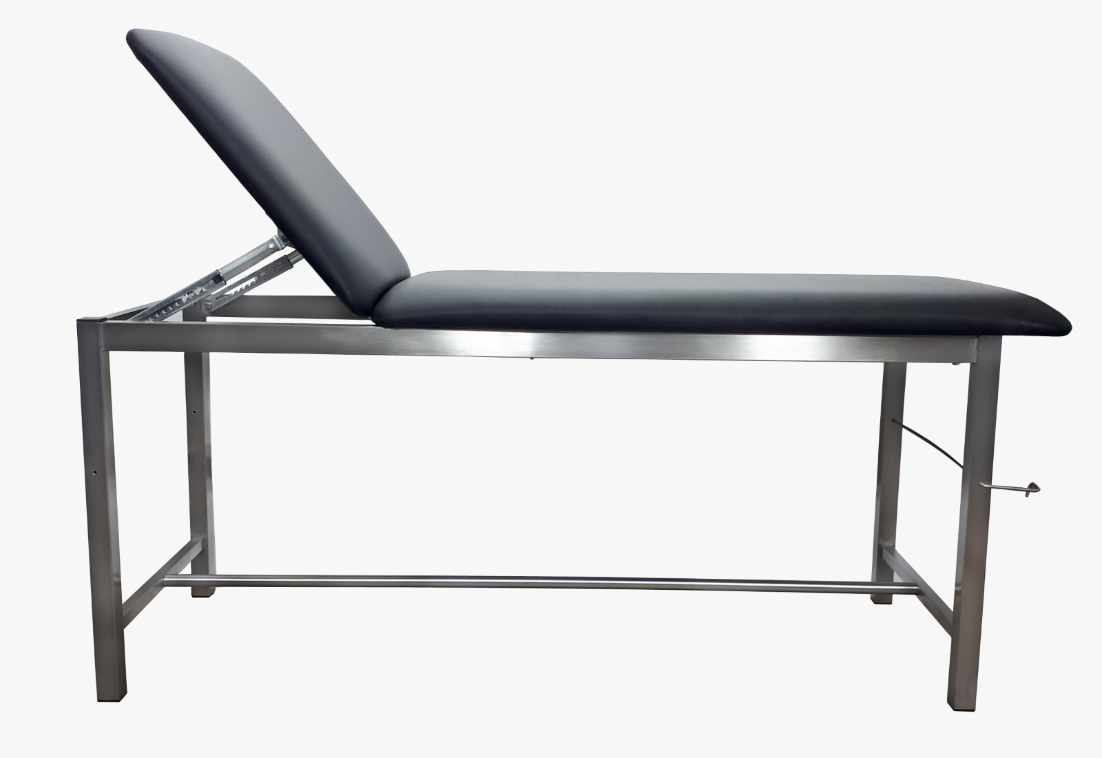 EXAMINATION COUCH
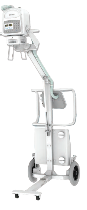 PORTABLE X RAY SYSTEM
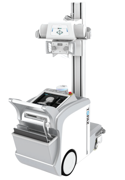 MOBILE DR SYSTEM
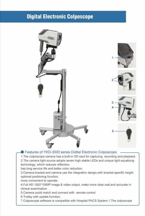 DIGITAL COLPOSCOPE
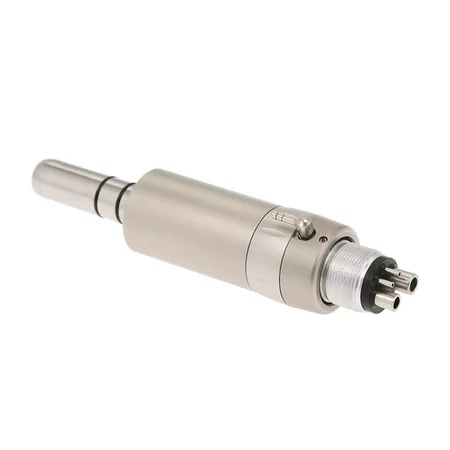 Low Speed Air Motor Handpiece
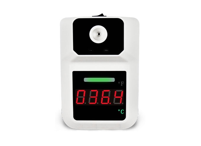 Infrared Thermometer wall mound
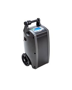 OXLIFE INDEPENDENCE PORTABLE OXYGEN CONCENTRATOR
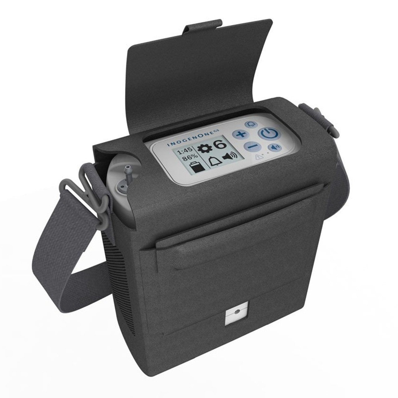 Inogen One G5 Portable Oxygen Concentrator System
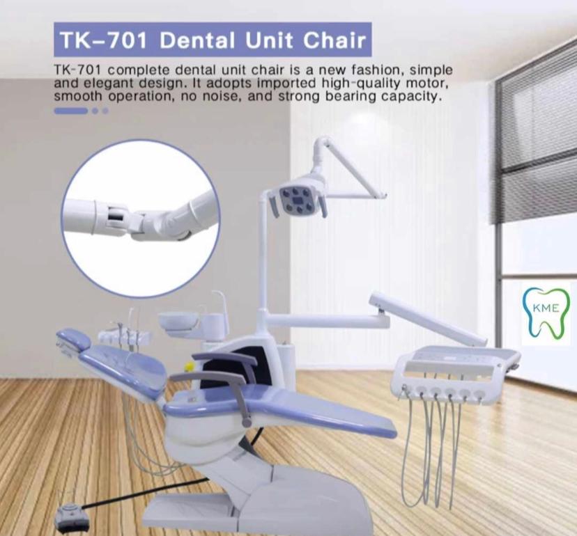 DENTAL CHAIR TK 701
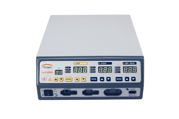 Orion HA400 Electrosurgical Unit
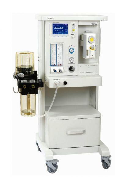 Anesthesia workstation AM832 - (1 Vaporizer)
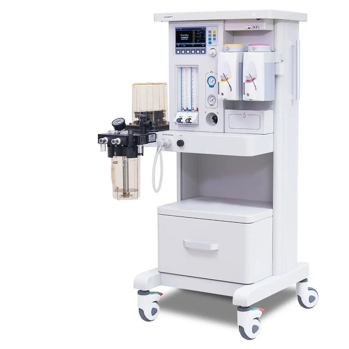 Anesthesia workstation AM832
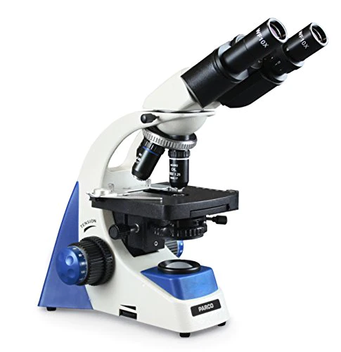 Binocular Microscope
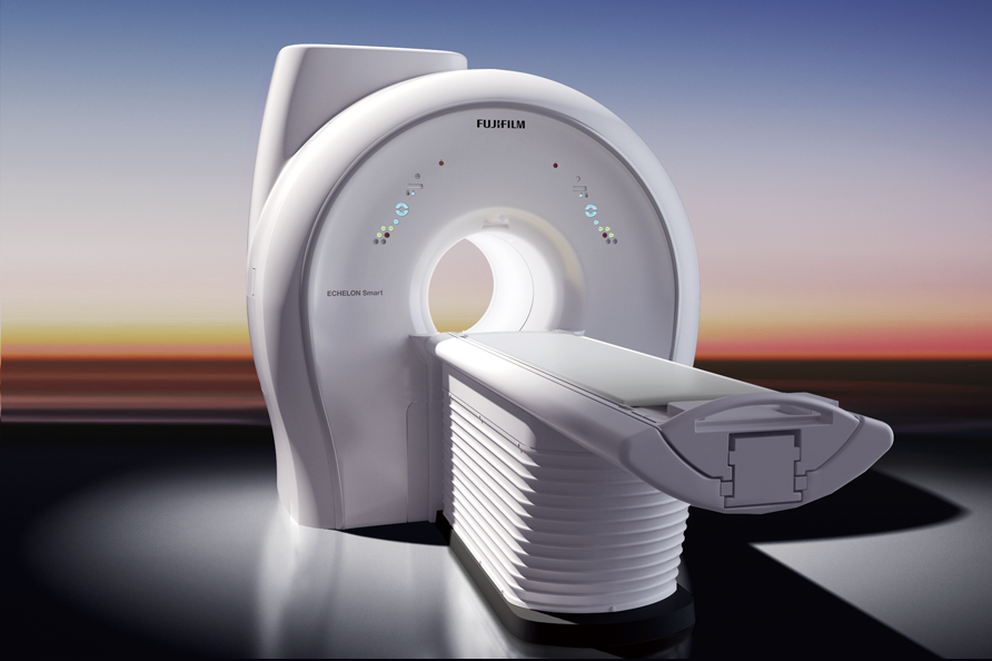 FUJIFILM ECHELON Smart 1.5T
 Light Cure
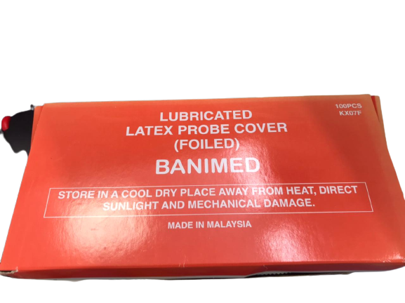 Lubricated Latex Probe Cover
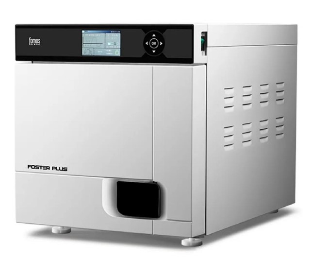 Autoclave
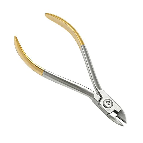 Hard Wire Cutter
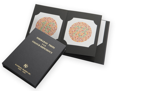 Ishihara Book
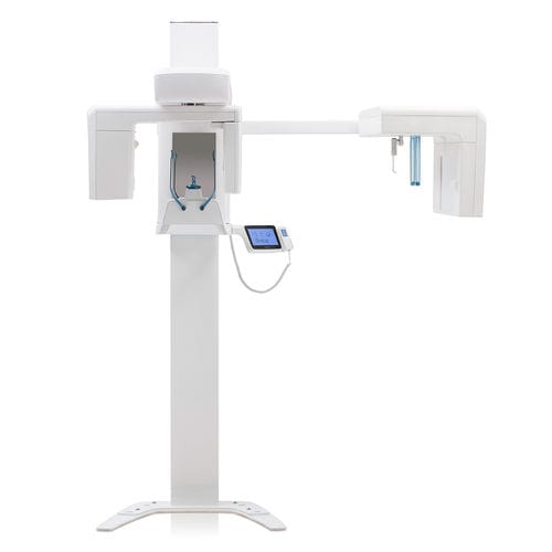 Panoramic X-ray system
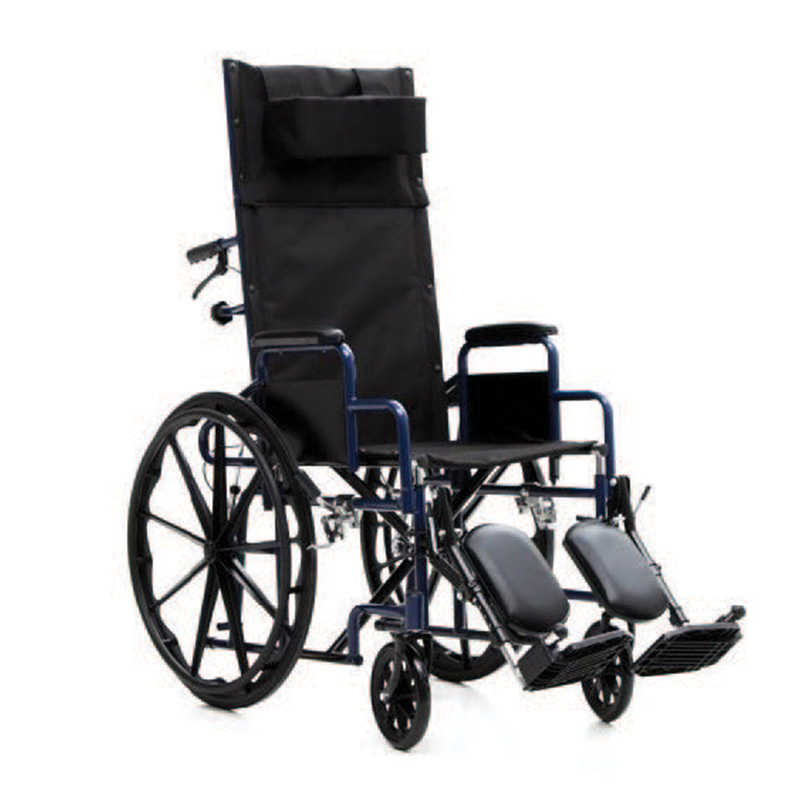 Recline wheel chair
 THERMOPHORE CLASSIC DEEP-HEAT THERAPHY
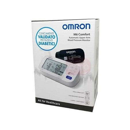 M6 COMFORT AUTOMATIC UPPER ARM BLOOD PRESSURE MONITORE
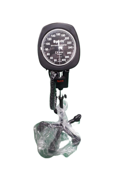 Spirit Blood Pressure apparatus with trolley
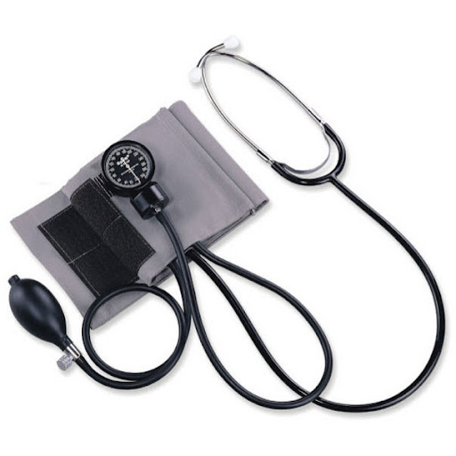 Spirit Aneroid Bp Monitor With Stethoscope
 Accu Chek Active, 50 Test Strips
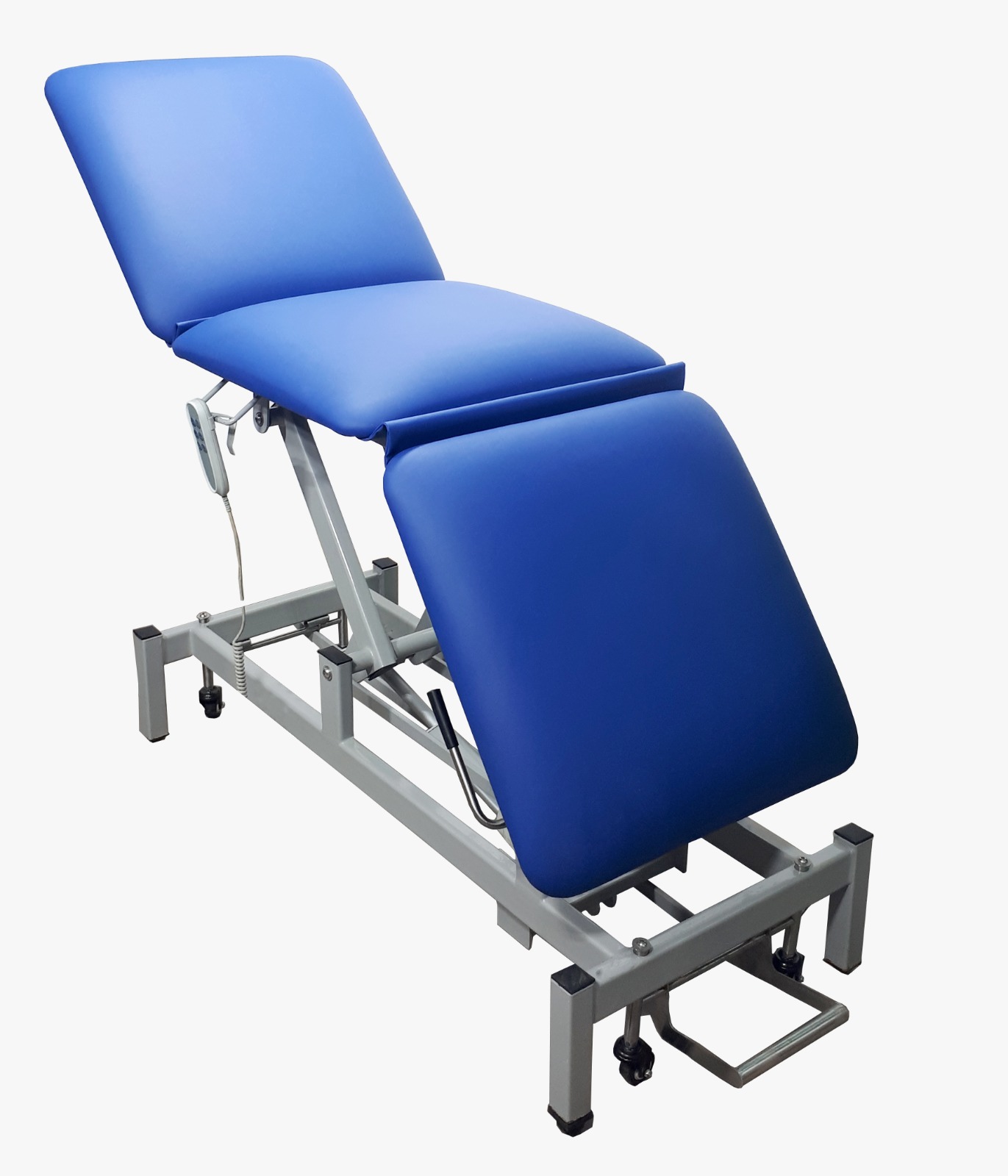 Electric examination couch 3 section
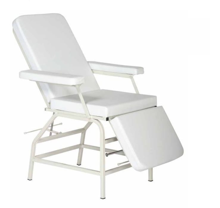 Blood donation chair manual (TURKEY)
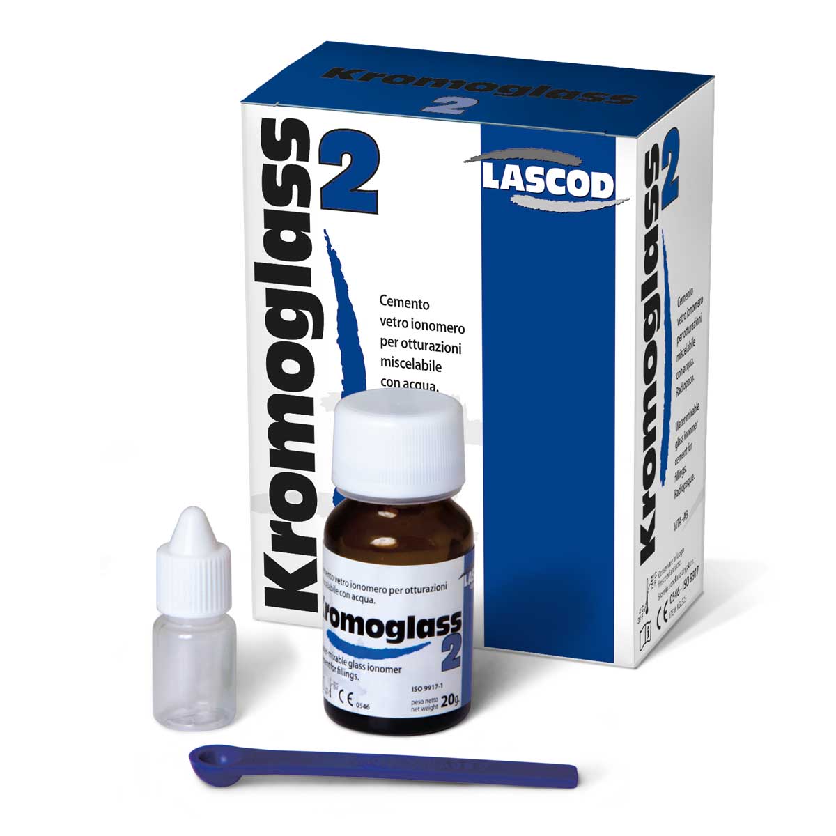 Kromoglass2 Permanent glass ionomer cement
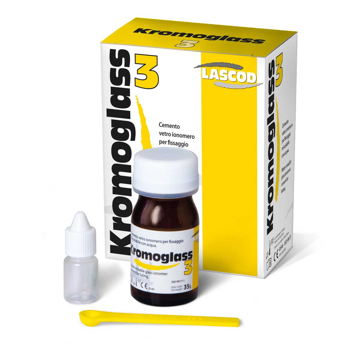 KROMOGLASS 3 (Glass Ionomer Cement Lutting)
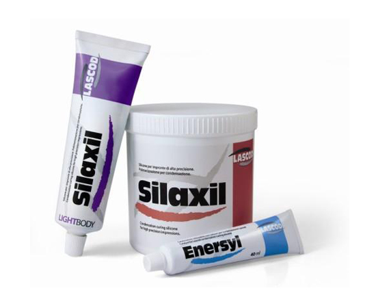 SILAXIL KIT (Putty/Activator/Light body)
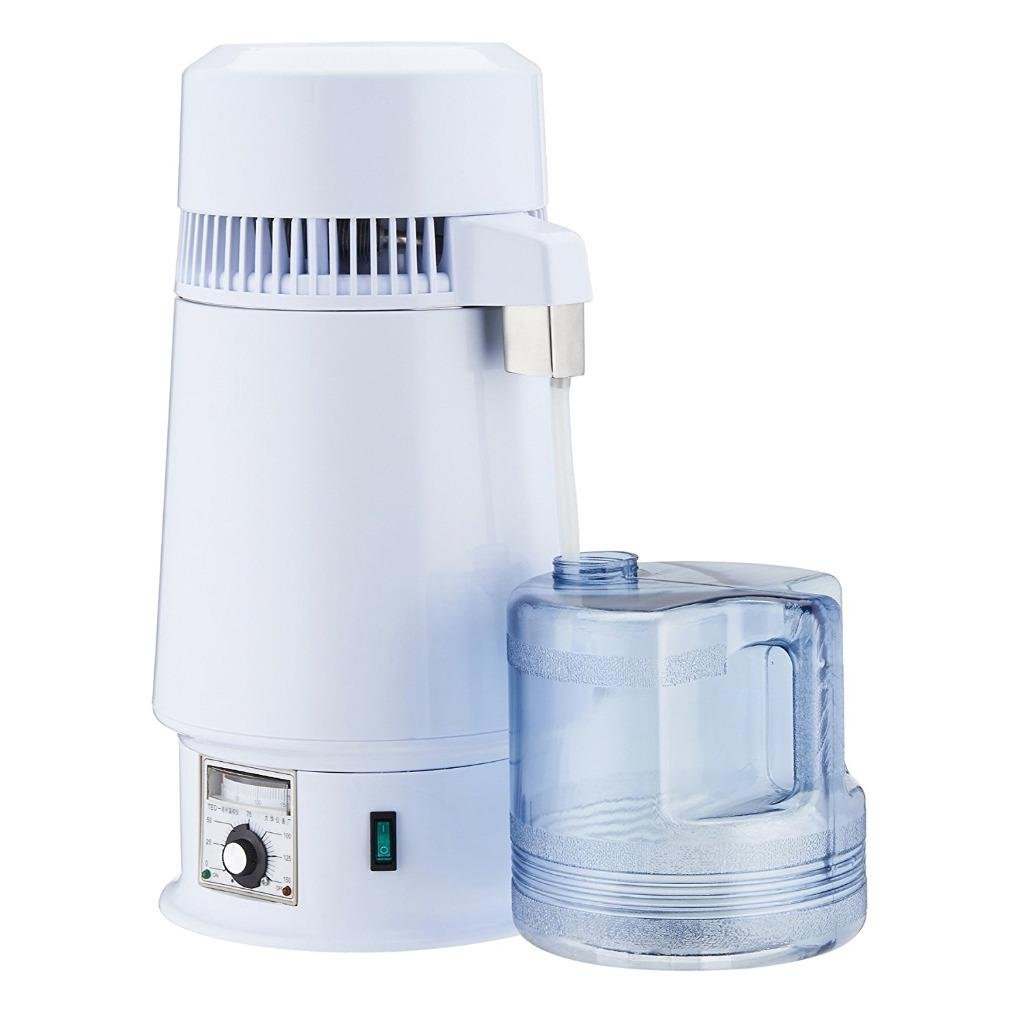 Water Distiller Machine
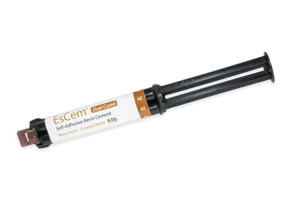 SPIDENT EsCem Resine Cement
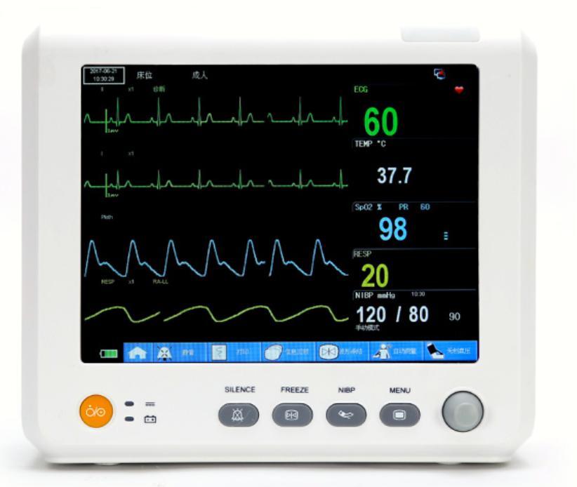 Patient Monitor M8
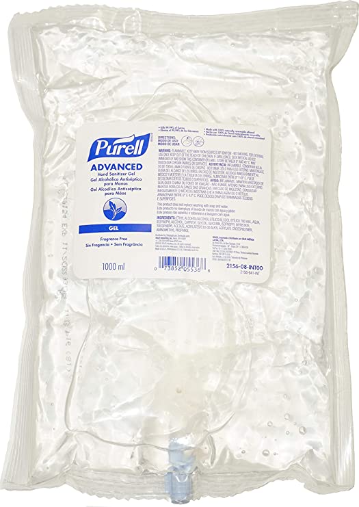 PURELL NXT Advanced hand sanitizer refill 1000ml
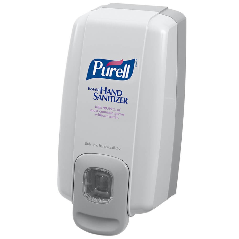 PURELL NXT space saver push type dispenser – wall mounted
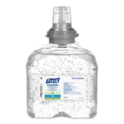 Purell 5476-04 Advanced Hand Sanitizer Refill 1200 mL
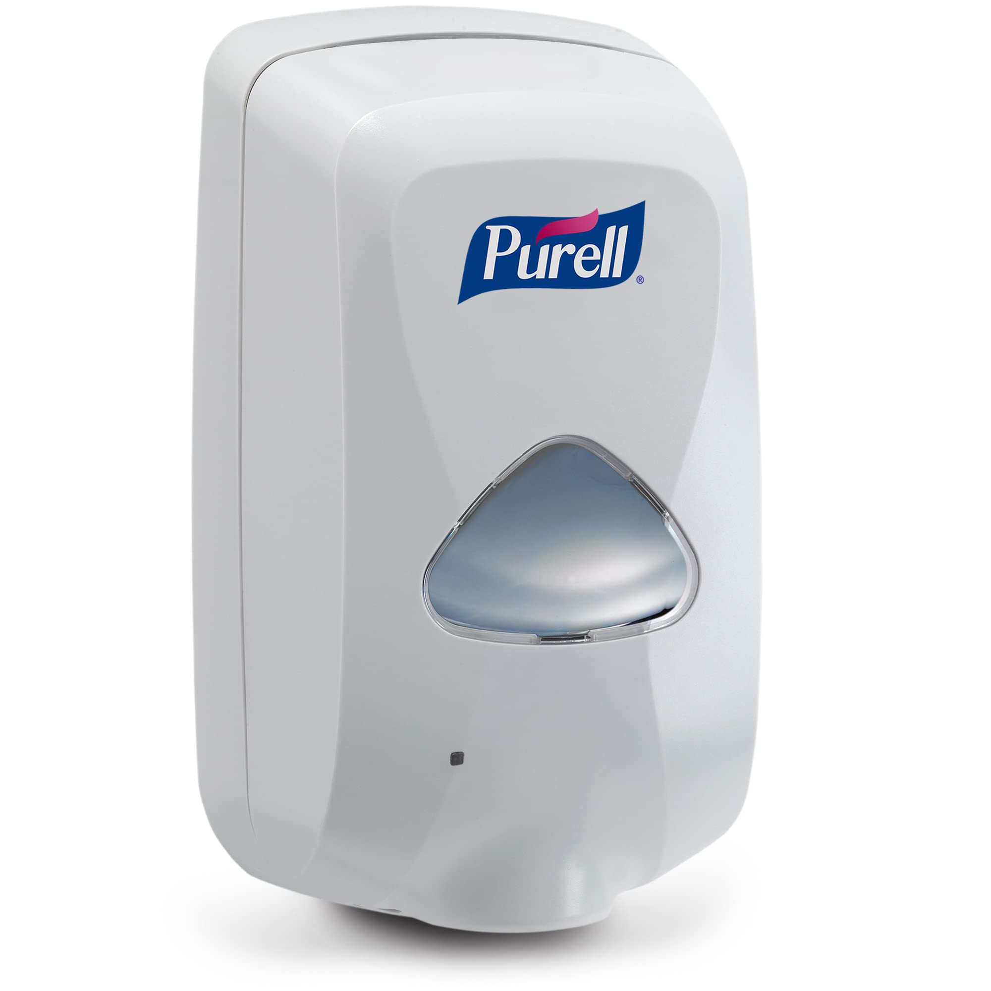 Purell® 2720-12 TFX™ Touch-Free Hand Sanitizer Dispenser
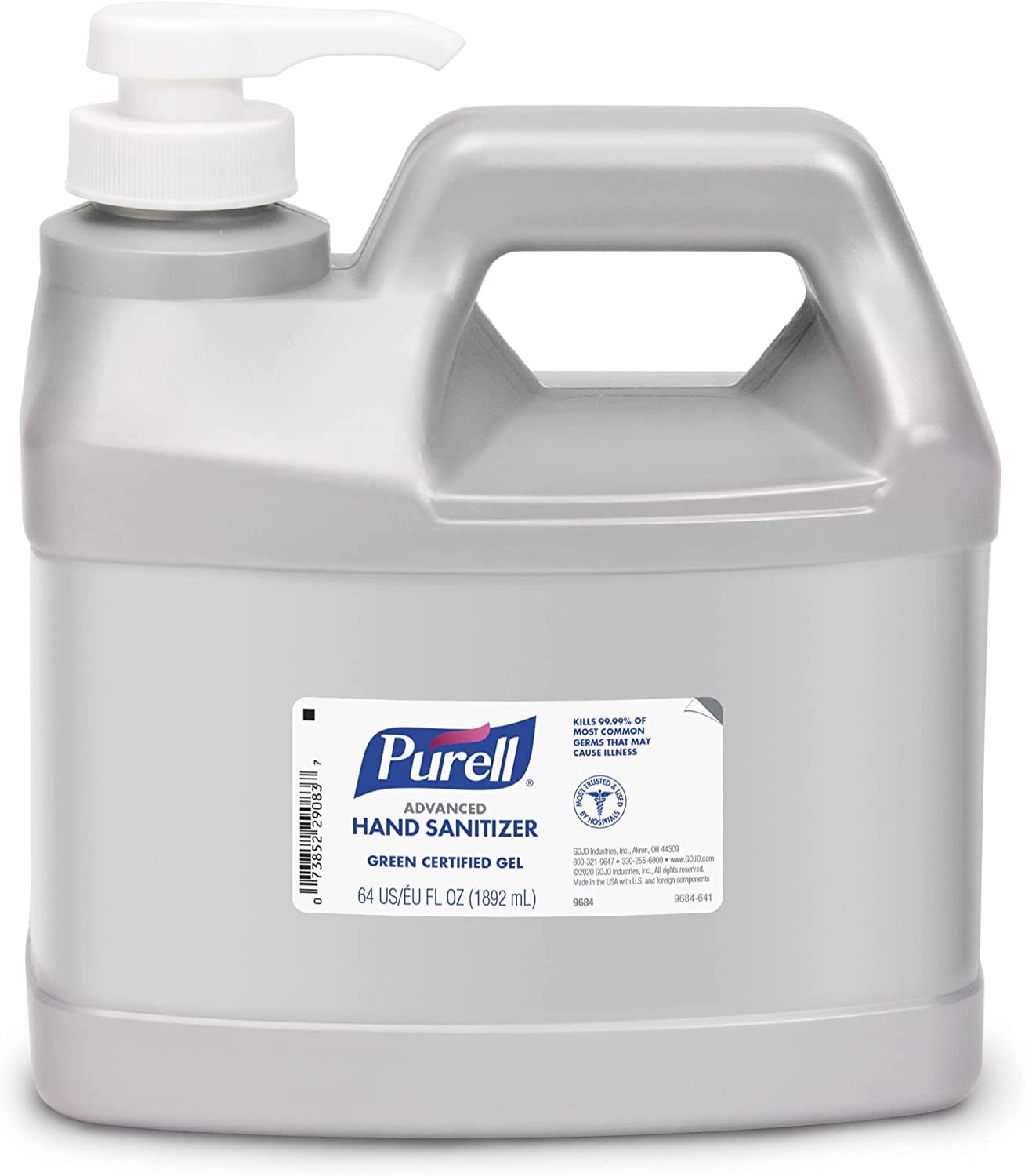 PURELL advanced hand sanitizer 1.89L – pump type
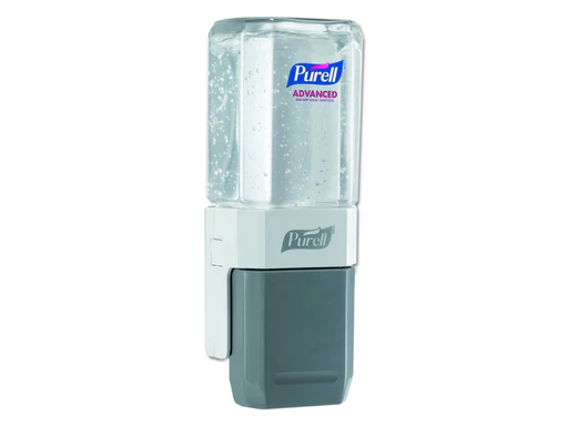 Purell Everywhere System Starter Kit (Base and Refill)
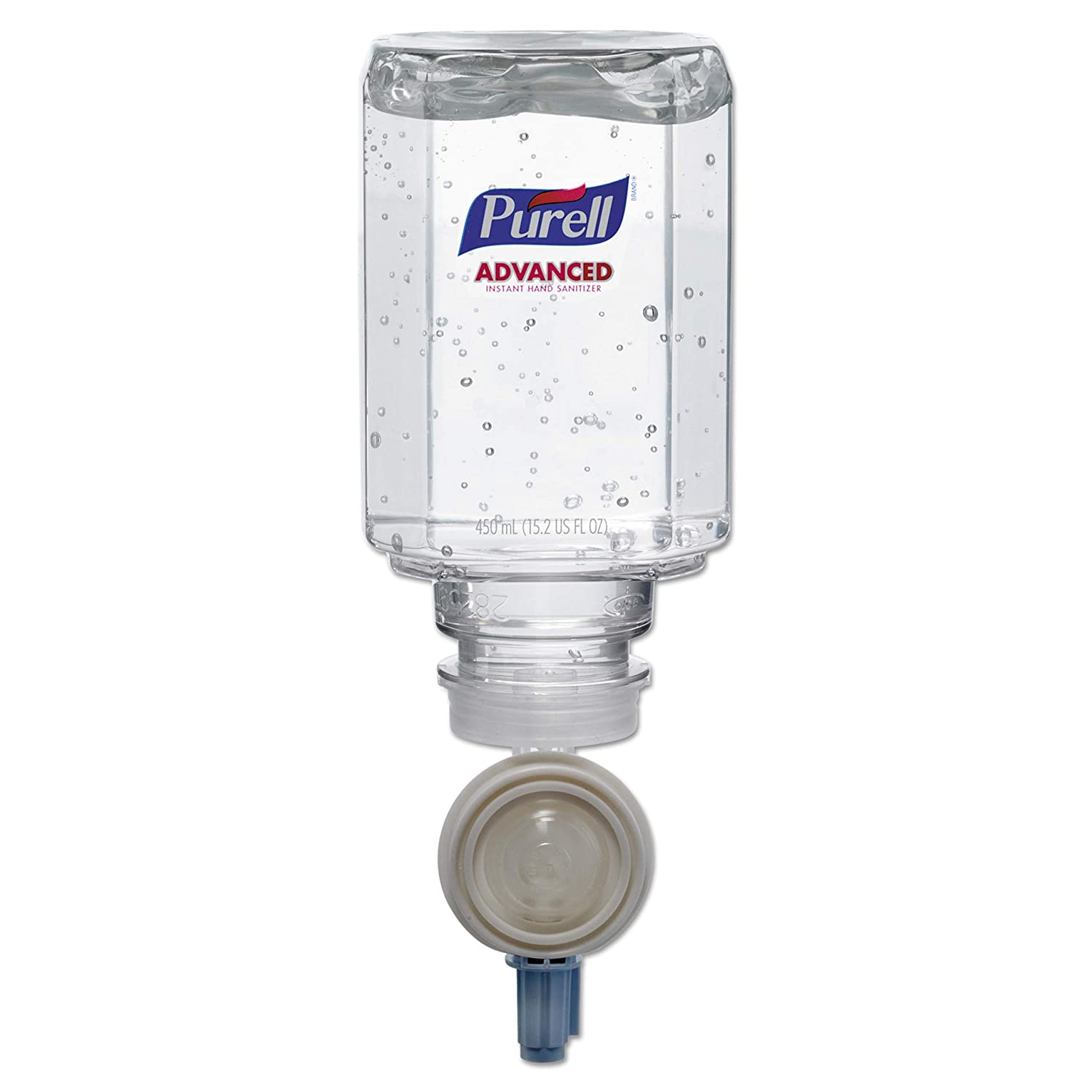 Purell® 1450-06 Hand Sanitizer Gel 450ml Refill for Purell ES® Everywhere System
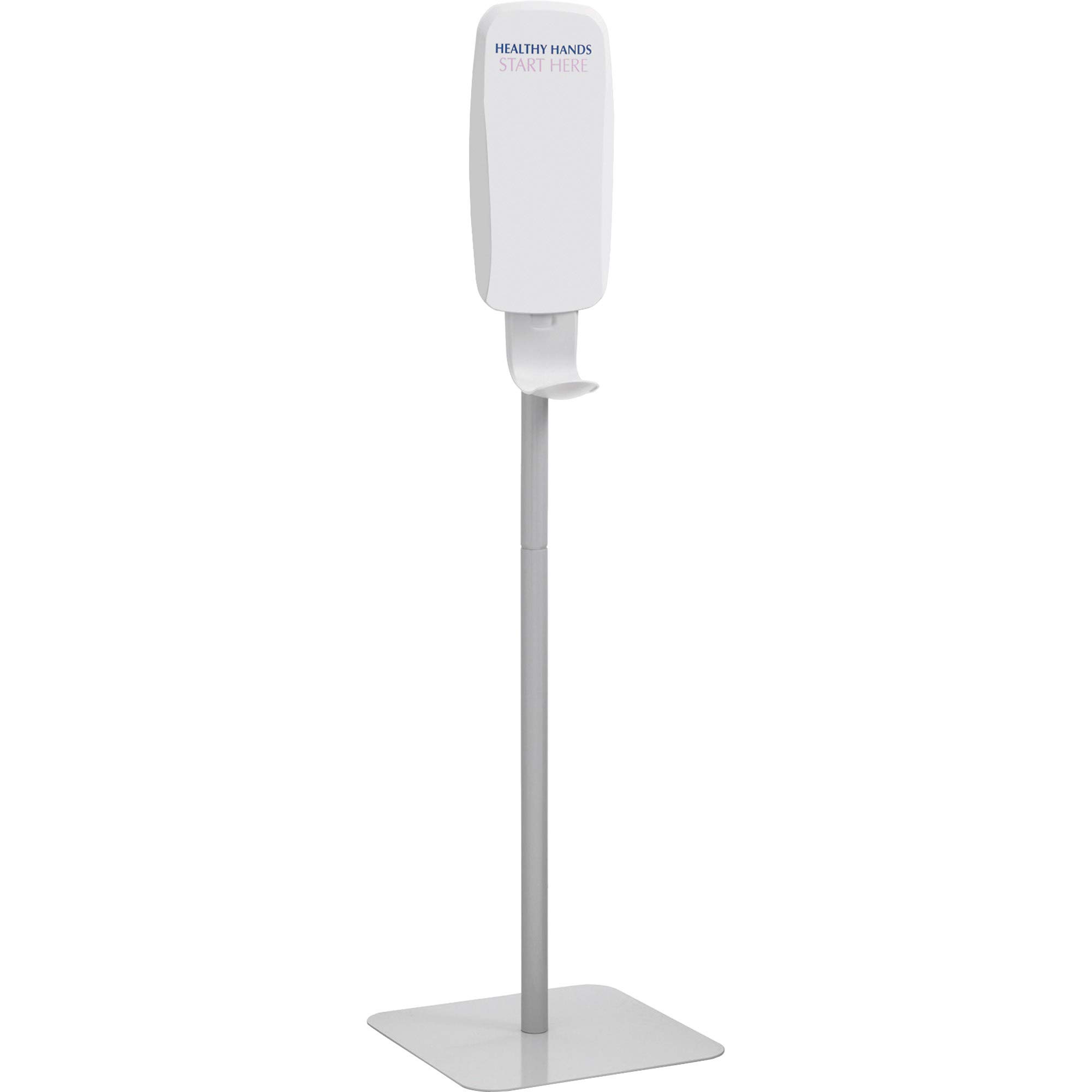 PURELL Floor Stand for TFX Touch Free Dispenser, Light Gray
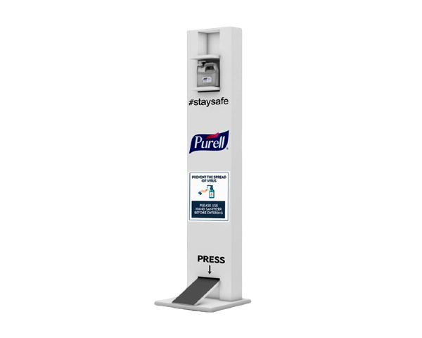 PURELL Hand sanitizer foot operated stand
 PURELL advanced hand sanitizer 1 L bottle with pump
 PURELL advanced hand sanitizer 354ml – pump bottle
 PURELL advanced hand sanitizer 236ml – pump bottle
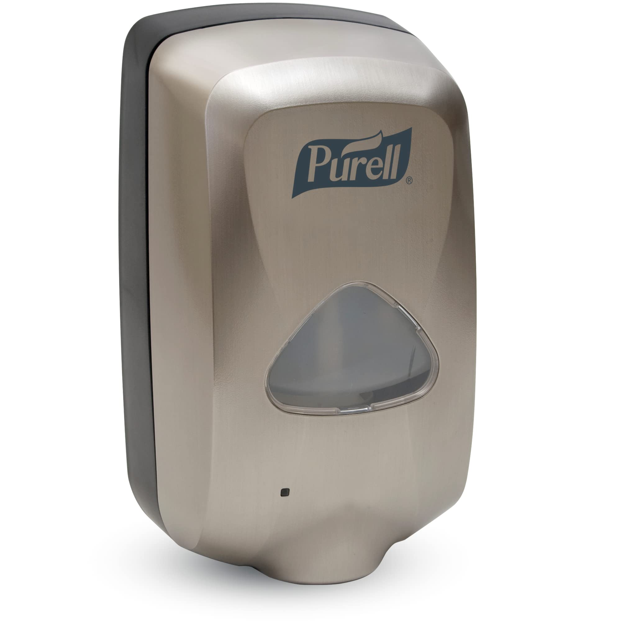 PURELL TFX touch free dispenser Nickel color – wall mounted
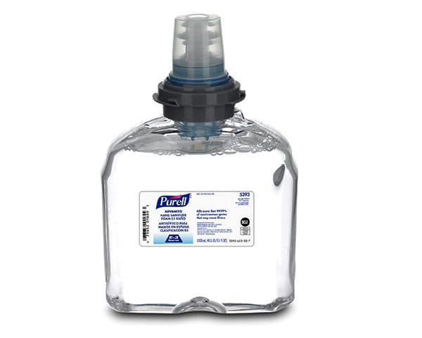 PURELL E3 hand sanitizer TFX refill 1200ml
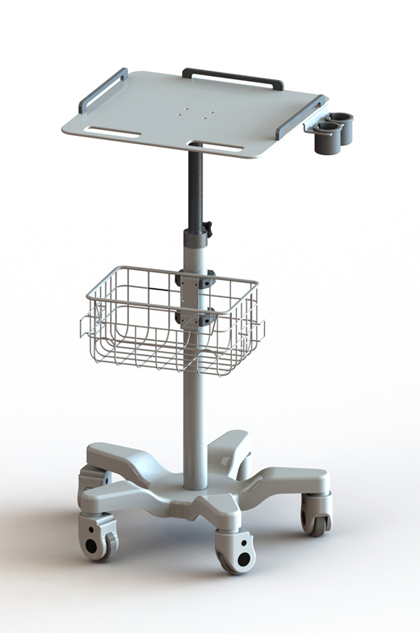 ECG Trolly
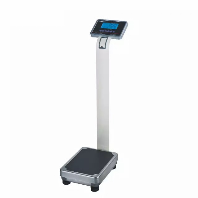 Weighing Scale with Height scale (Adult )
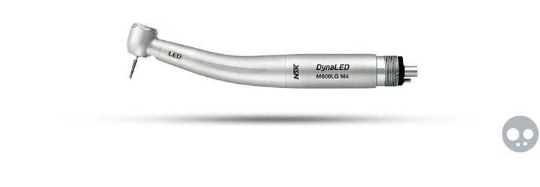 Dental Handpiece With LED Light
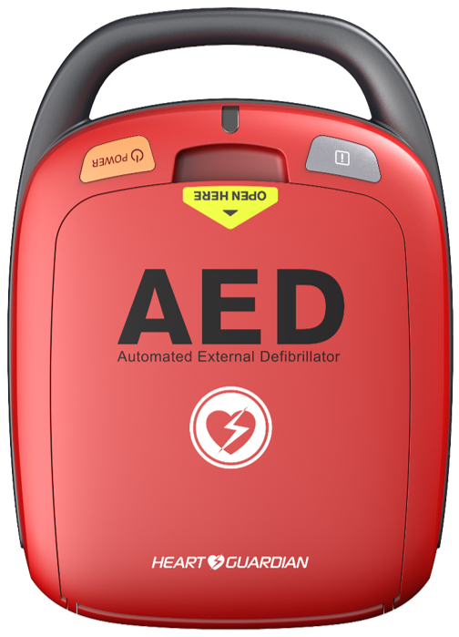 AED Defibrillator - HR-501 Heart Guardian - RED
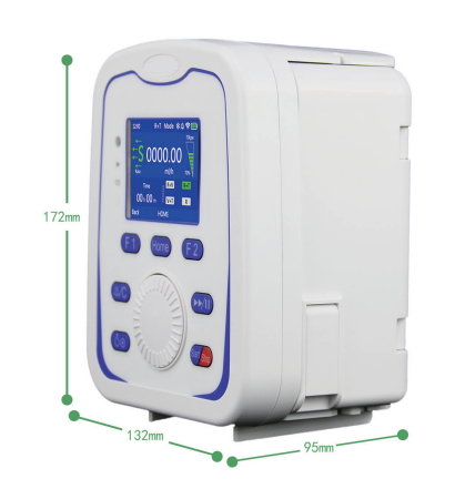 Infusion pump
.jpeg) Syringe Pump
Syringe Pump
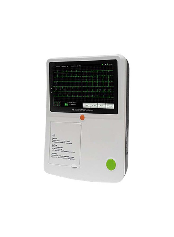 ECG Machine - 3 Channels
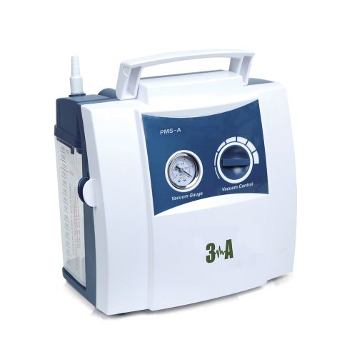 Portable Suction Machine PMS-A
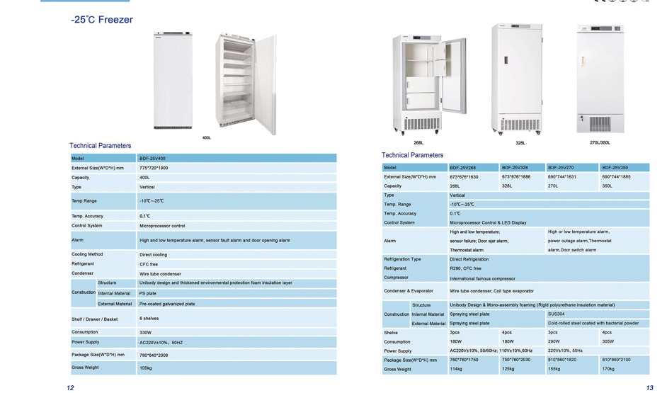 -25℃ Freezer BDF-25V 270
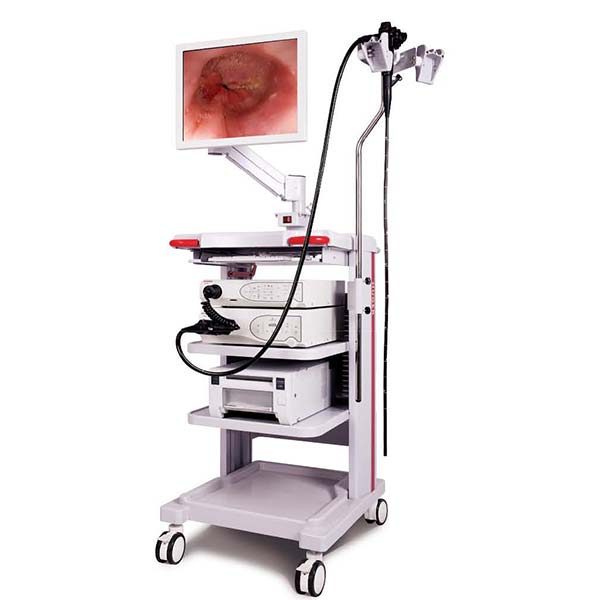 Huger Endoscopy system Refurbished
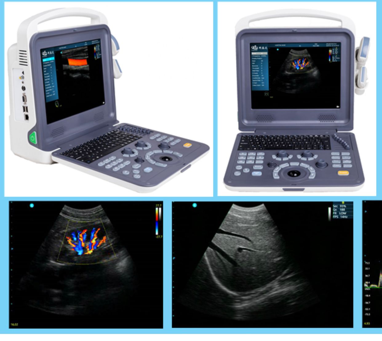 Portable Color doppler ultrasound with two probes
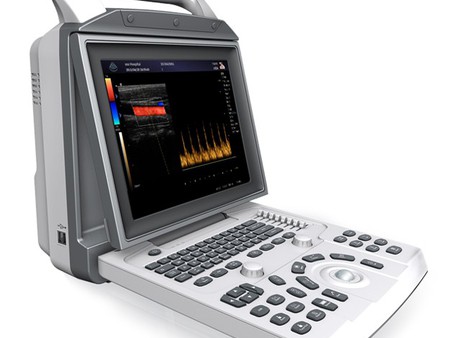 Portable Color doppler ultrasound with two probes
.jpg) Laboratory refrigerator -5V250 .
Laboratory refrigerator -5V250 .
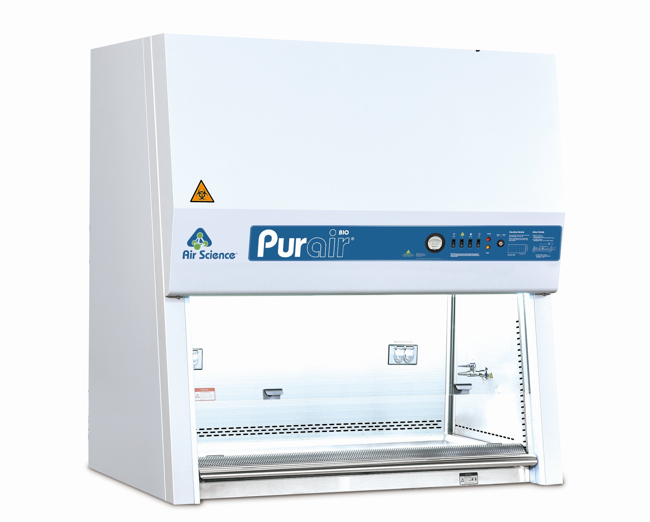 Biological Safety Cabinet Bcs-1100A2-X
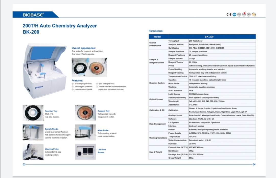 Chemistry Analyzer Fully Automatic BK-200
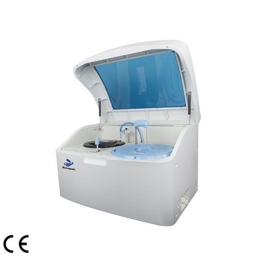 Fully-auto Biochemistry Analyzer, BA-A-120
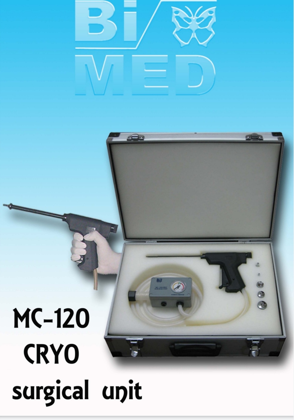 Cryosurgical unit Turkey
.jpg) Patient bed single crank
Patient bed single crank
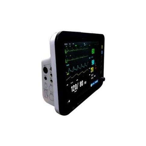 CARDIAC MONITOR
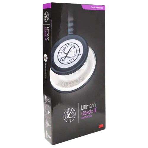 Littmann Classic - 3 stethescope
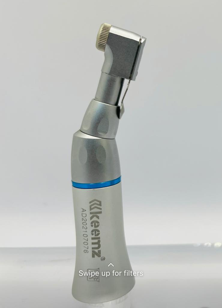 Dental contra angle
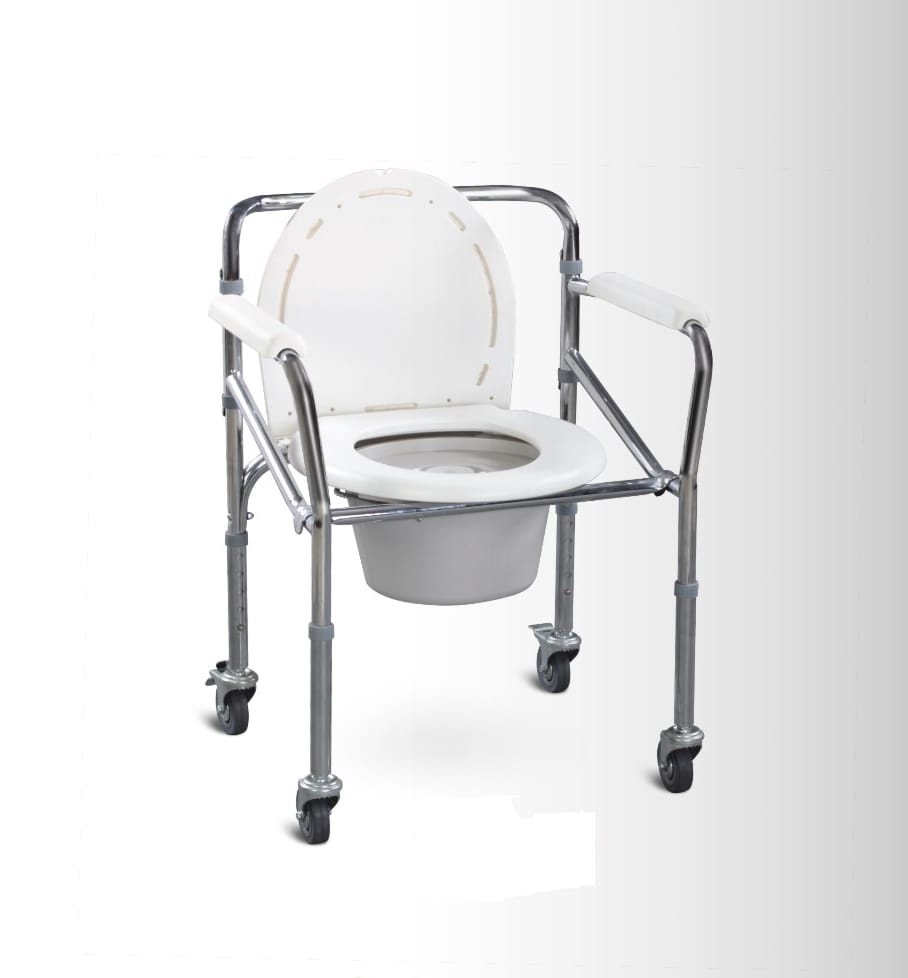 Commode chair
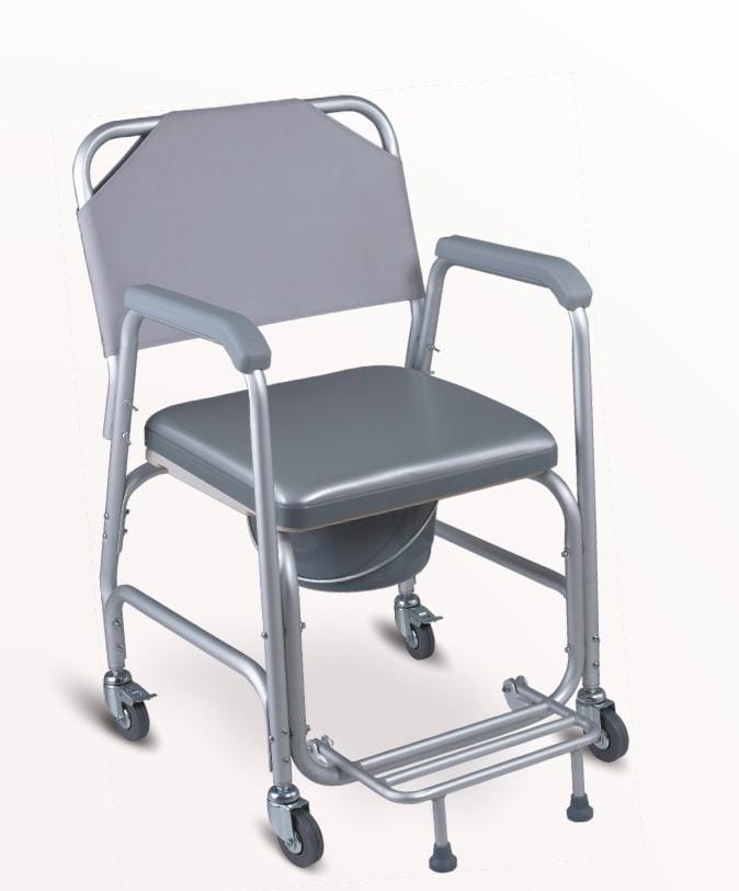 COMMODE CHAIR
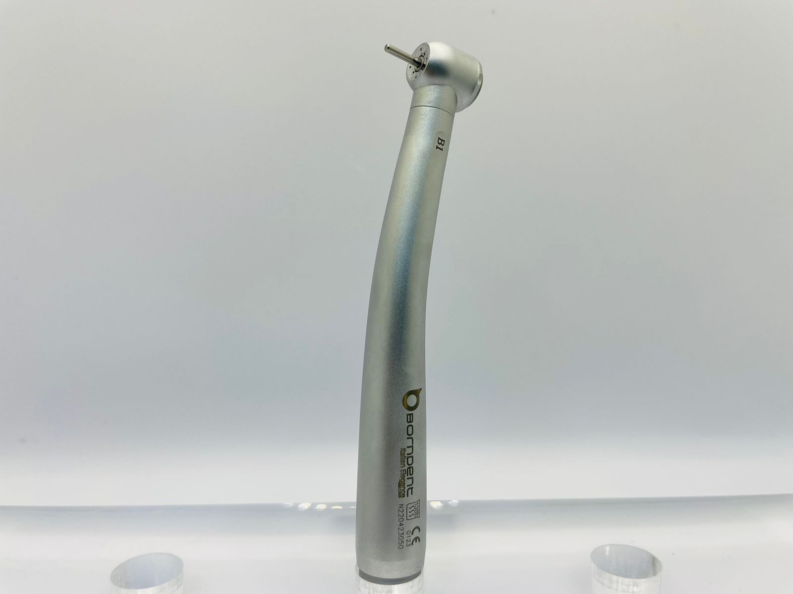 Dental hand piece
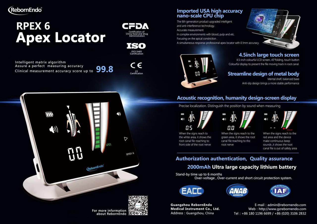 Apexlocater
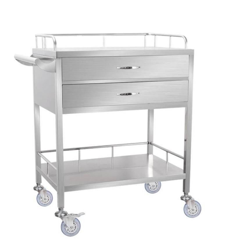 Dressing Trolley Two drawer
.jpg) Ear&forehead infrared thermometre
Ear&forehead infrared thermometre
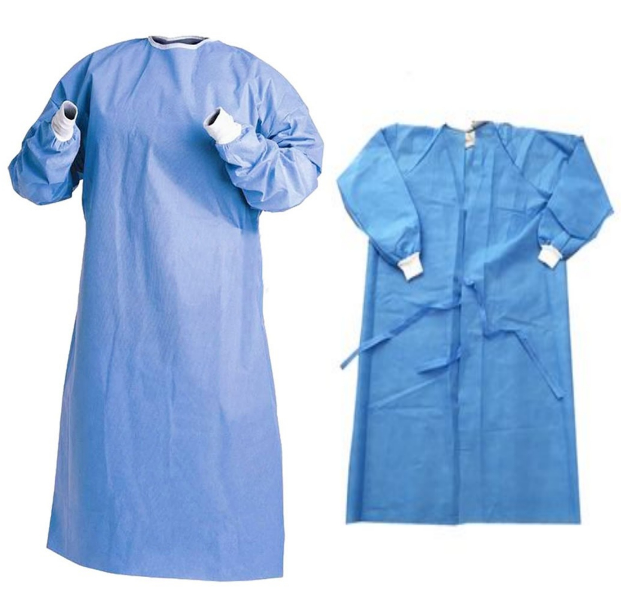 Surgical gown
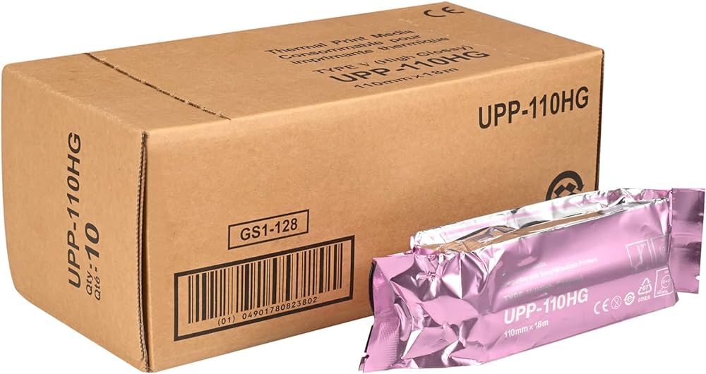 Sony Ultrasound Paper Film/Media
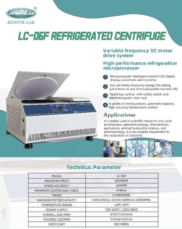 refrigerated centrifuge for blood bags
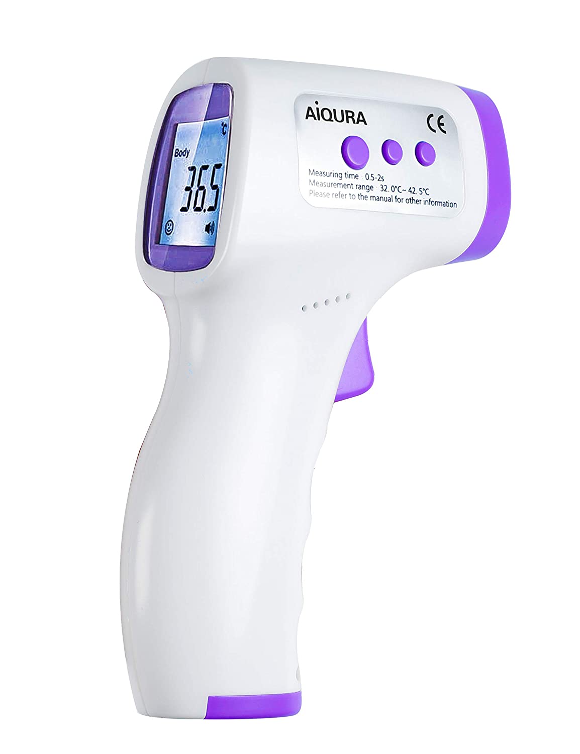 Infrared Forehead Thermometer
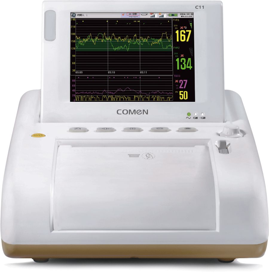 Comen C11 Specialized Fetal Monitor
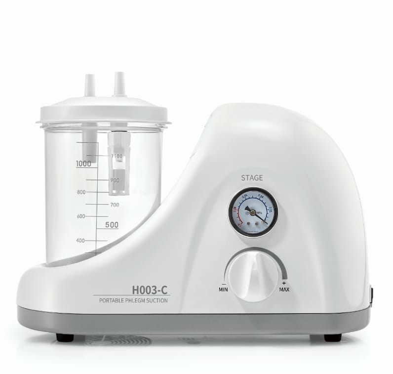 Suction machine
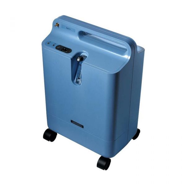 Everflow Oxygen Concentrator 5L
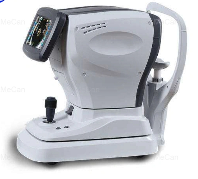 Keratometer
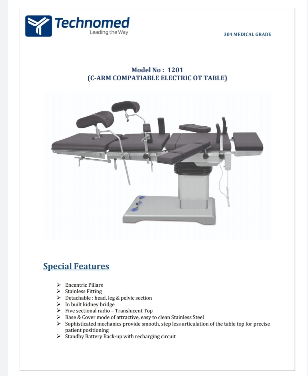 ELECTRIC OT TABLE
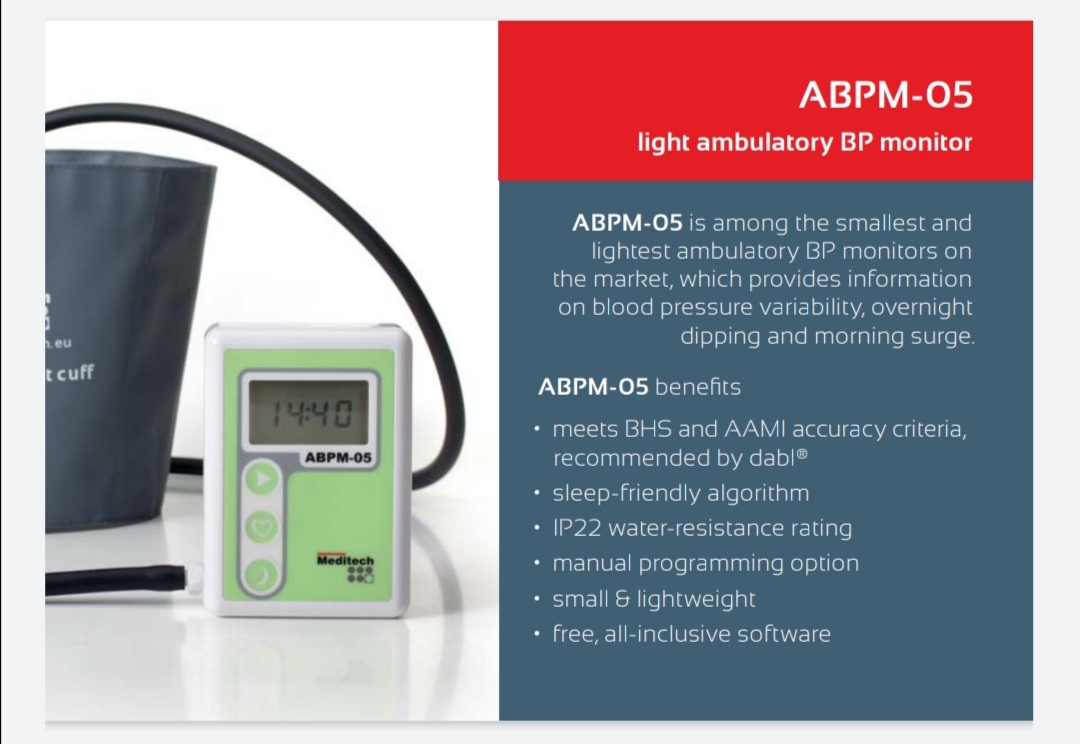 ABPM Meditech,Hungary
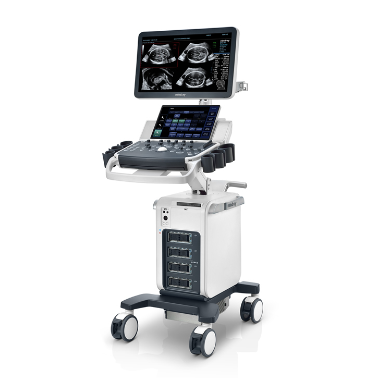 Mindray DC 70 X Insight 4D Ultrasound with three Probes
.png) Ultrasound Sonoscape P9 mobile color Doppler with two probes
Ultrasound Sonoscape P9 mobile color Doppler with two probes
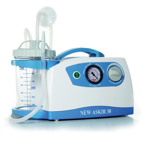 Suction machine Cami/Italy. Askir30
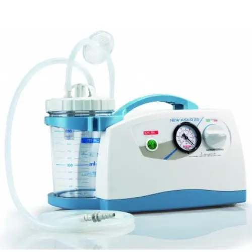 Suction Machine Askir20. Cami/Italy
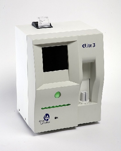 Haematology analyser Erba Elite3
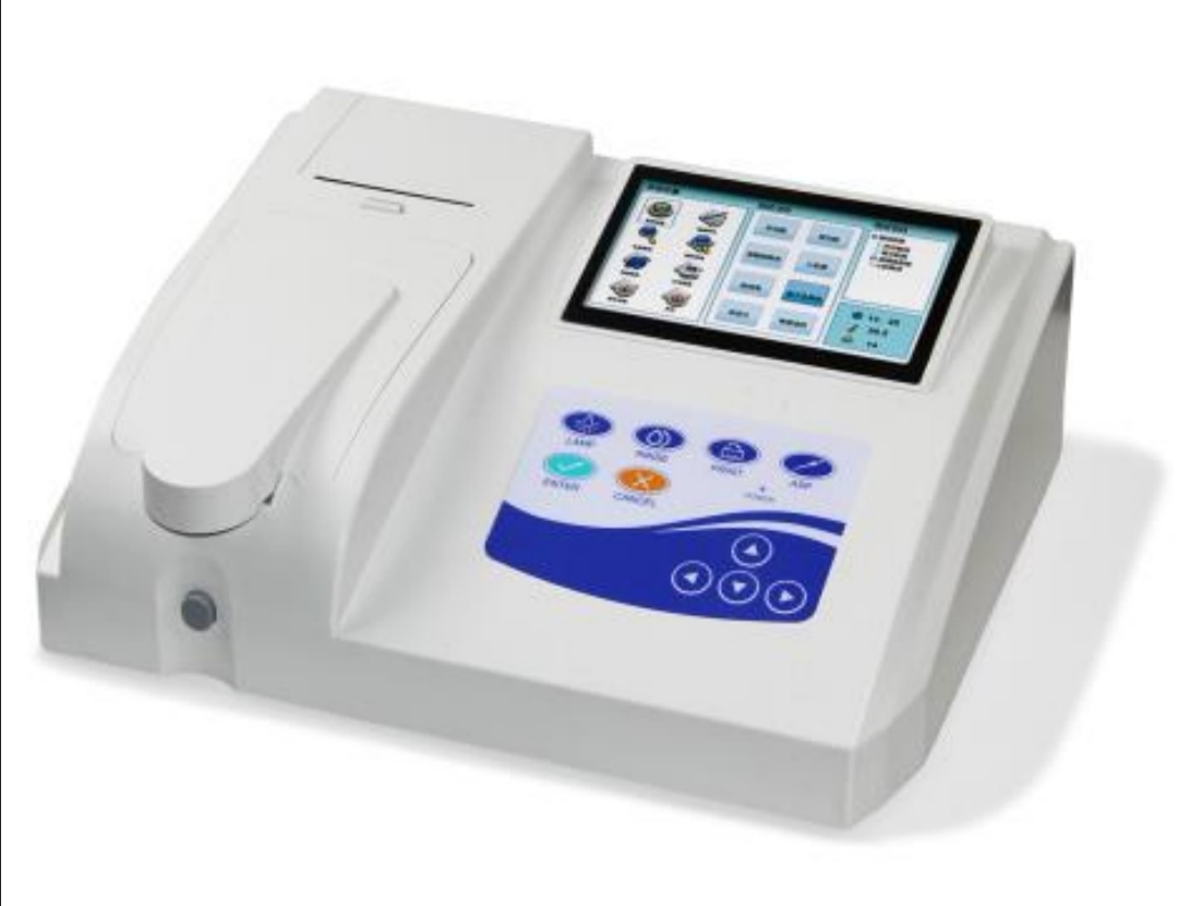 Chemistry analyser semi auto
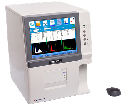 HEMATOLOGY ANALYZER
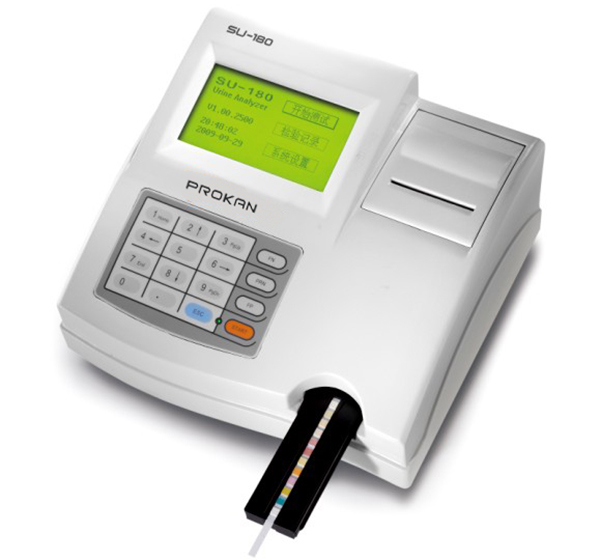 Urine Analyser
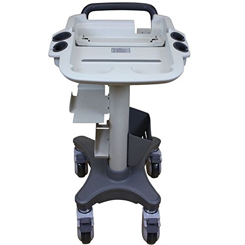 Ultrasound Trolley
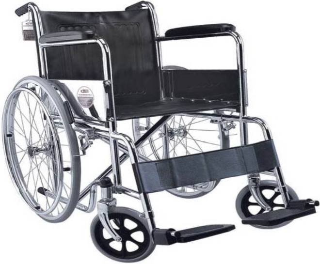 Wheel chair
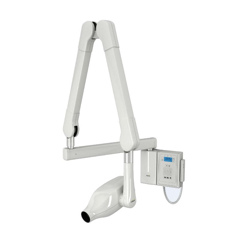 Intra oral xray
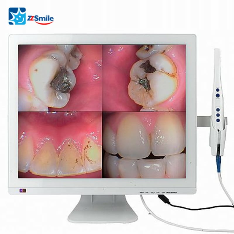 Intra Oral Camera With Screen
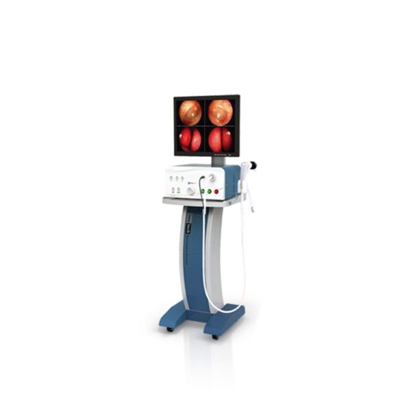 ENT diagnostic Endoscpe INV250
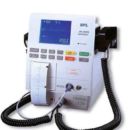 Biphasic defibrillator BPL India
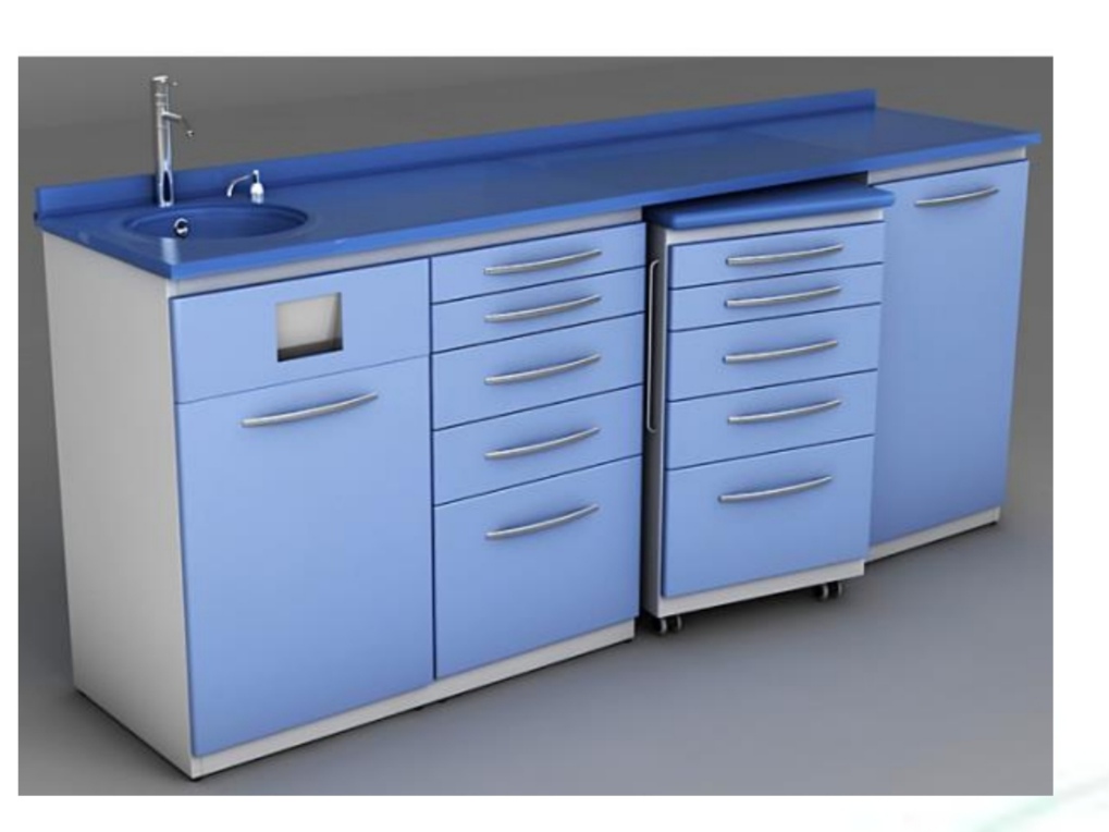 Dental floor metal cabinet
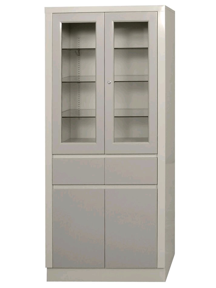 Narcotic Cabinet
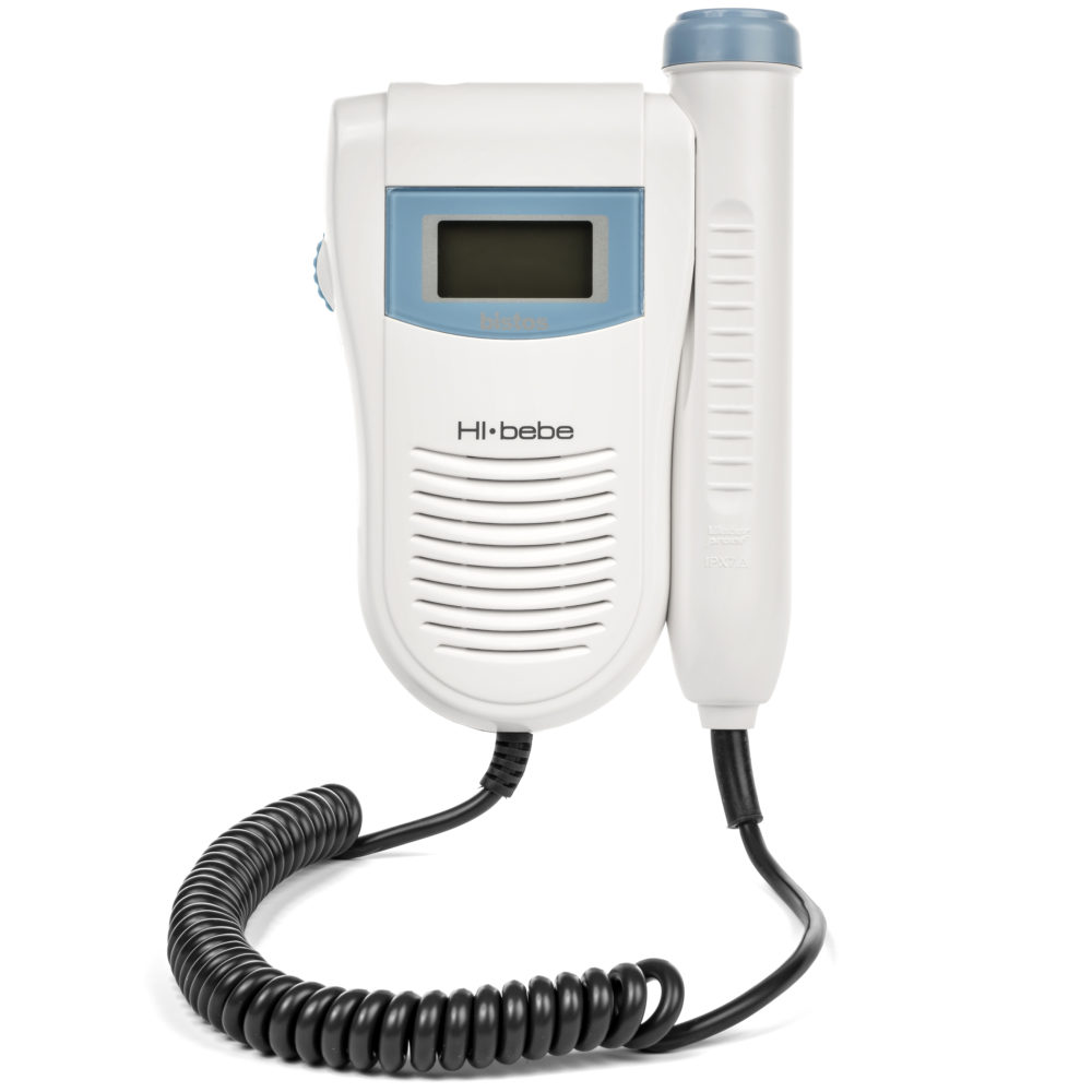 FETAL DOPPLER
.png) Ultrasound Sonoscape P20
Ultrasound Sonoscape P20
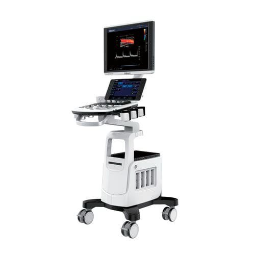 KIRAN SonoRad V10 Color Doppler System
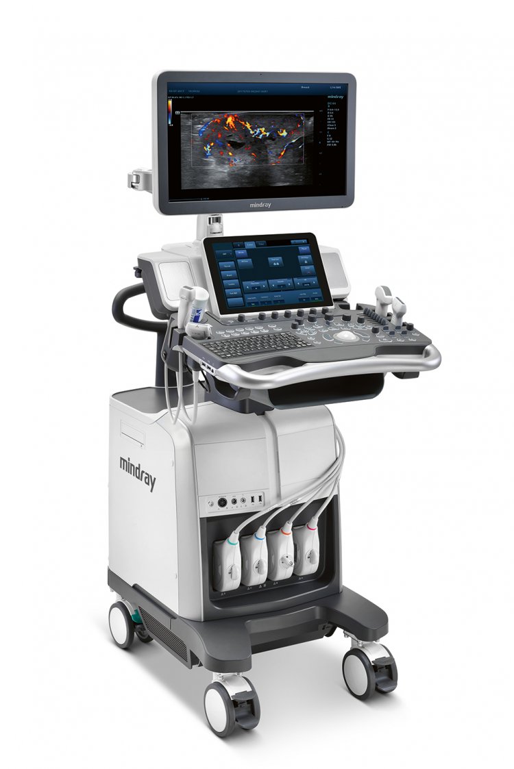 MINDRAY DC 80 X INSIGHT ULTRASOUND
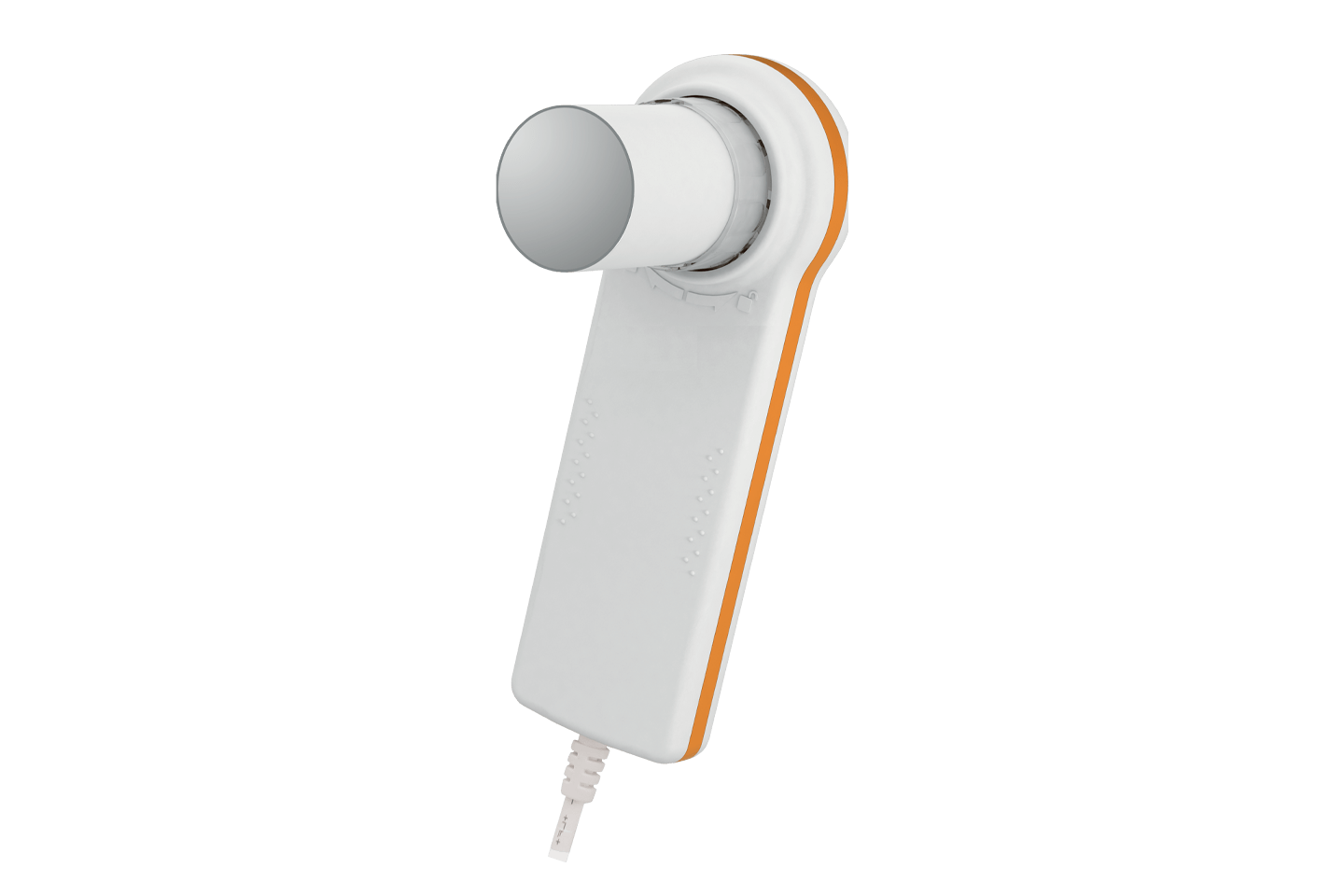 Spirometer handheld Mini spir Light MIR/Italy
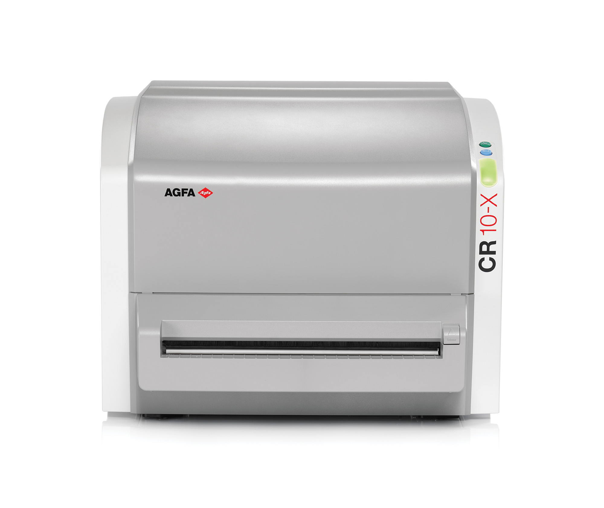 AGFA CR10-X-Digitizer
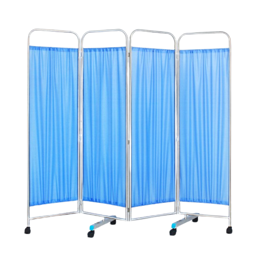 Ward Screen Four Fold
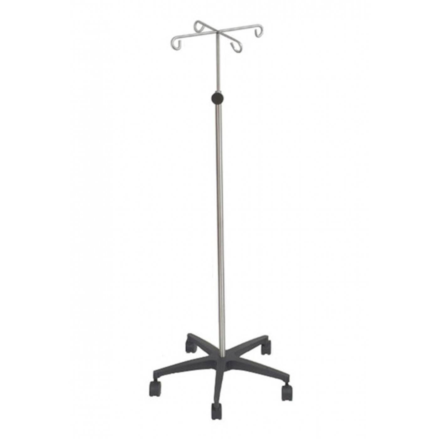 IV Stand with 4 hooks
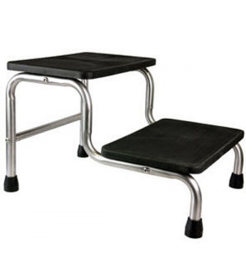 Double foot step
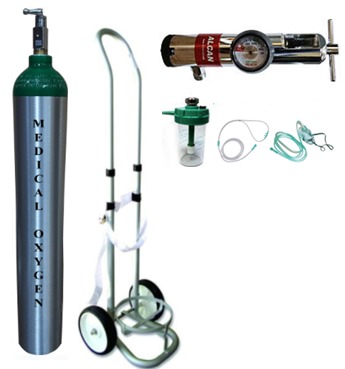 Portable oxygen cylinder 10L with trolley and regulator
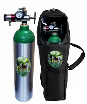 2.5L Portable aluminium light weight oxygen cylinder full set
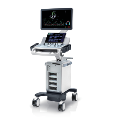 Mindray DC 60 Echo Ultrasound machine
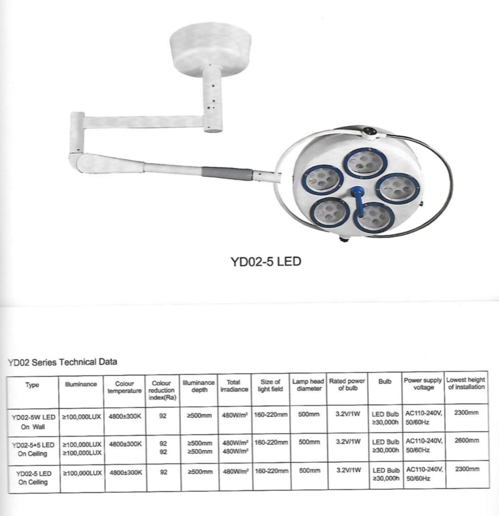 OT light Single
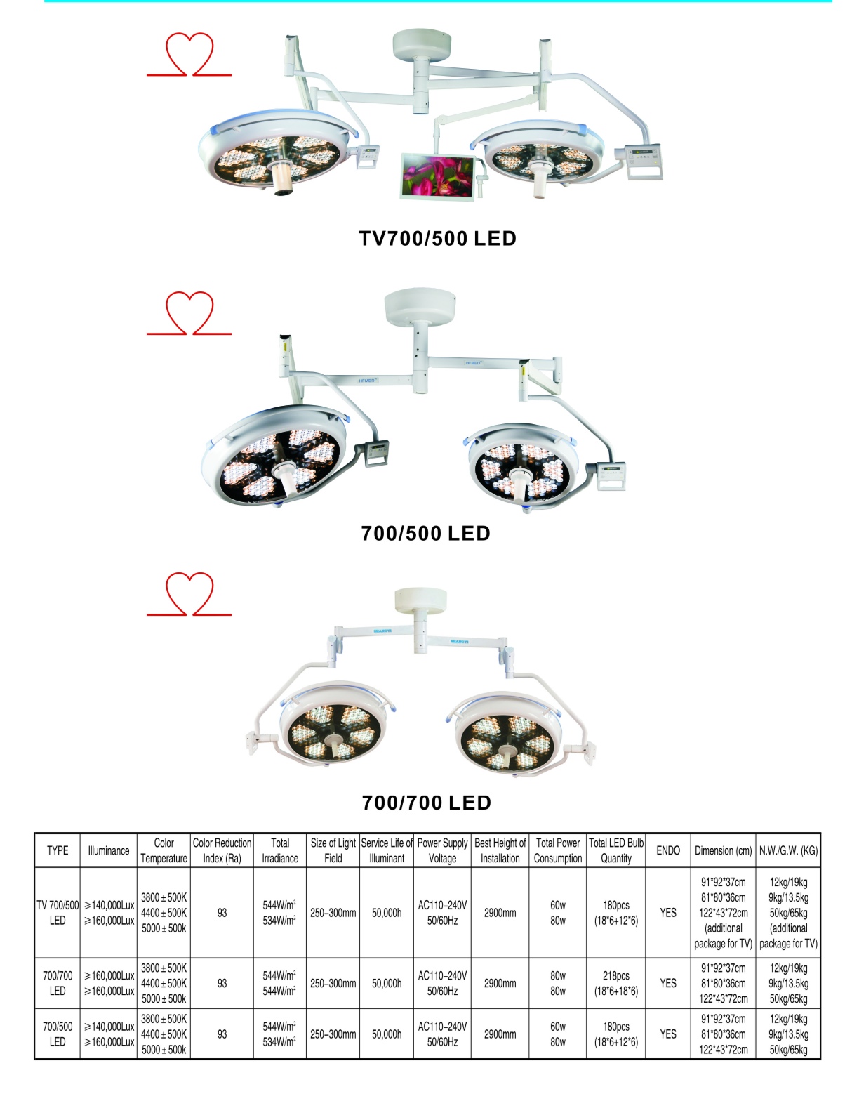 OT light Double
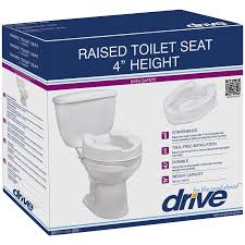 Raised Toilet Seat 4" inch height without armrest
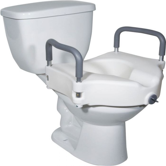 Raised Toilet Seat 5" inch height
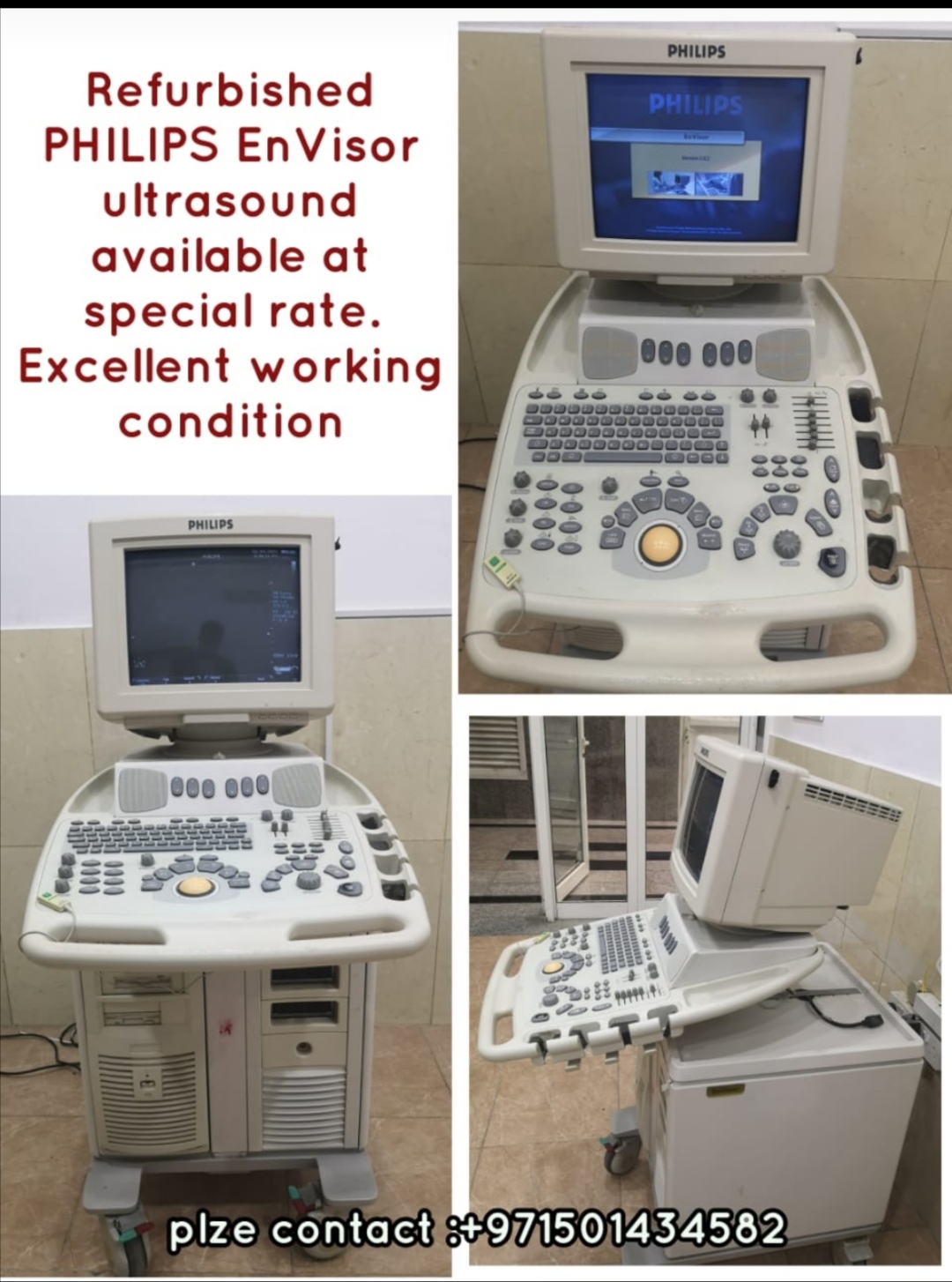 Refurbished PHILIPS EnVisor ultrasound
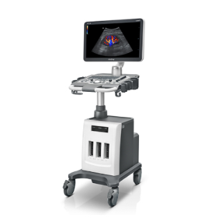 Mindray DC 30 Full HD Ultrasound System With Convex &Transvaginal probes
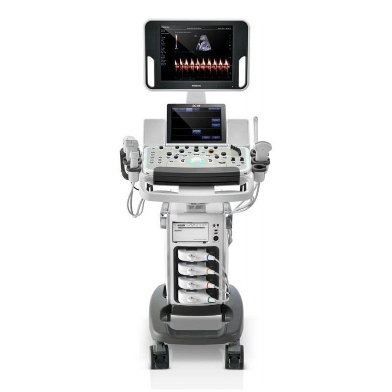 Mindray DC 40 Crystal Ultrasound System
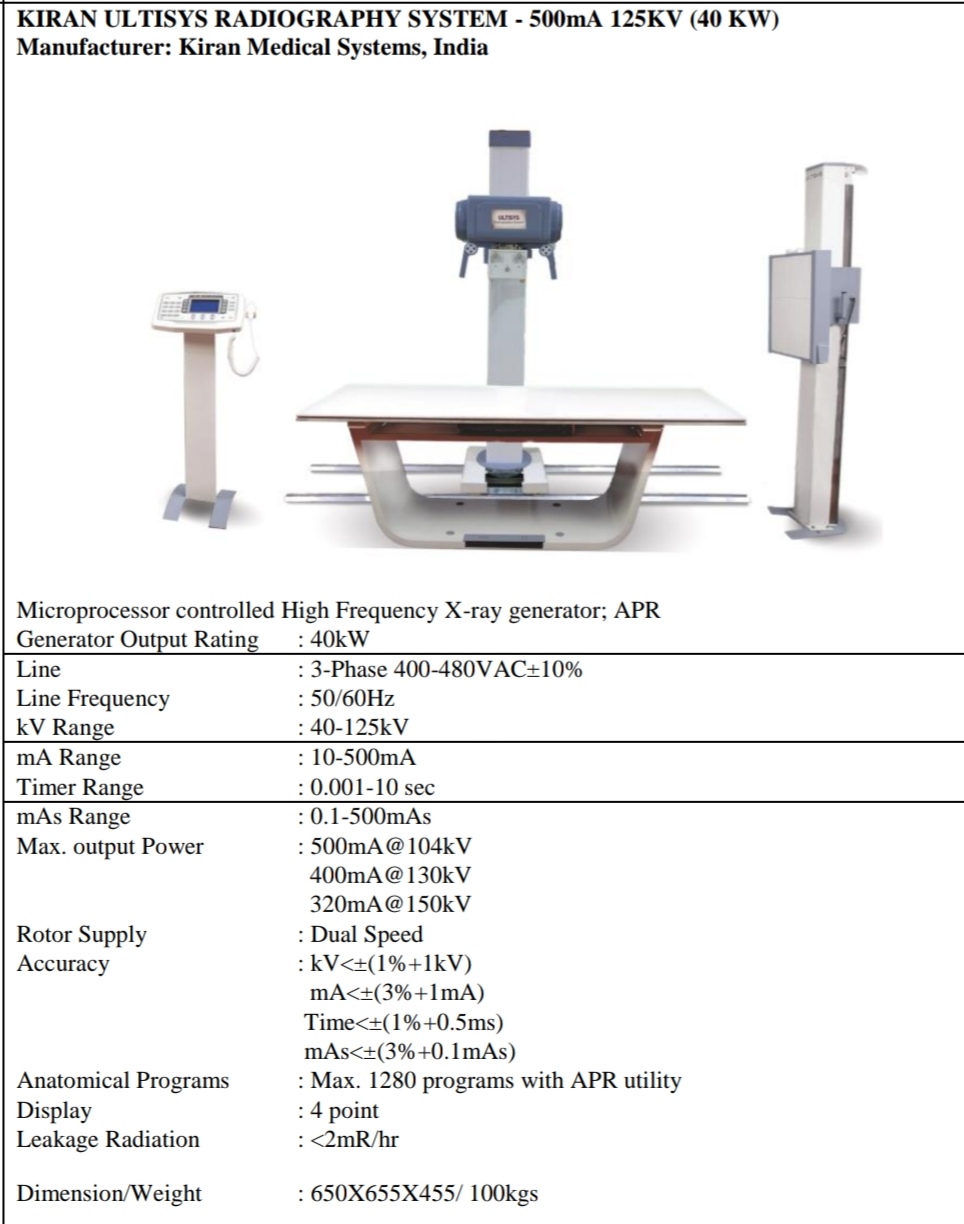 KIRAN ULTISYS RADIOGRAPHY SYSTEM - 500mA 125KV (40 KW)
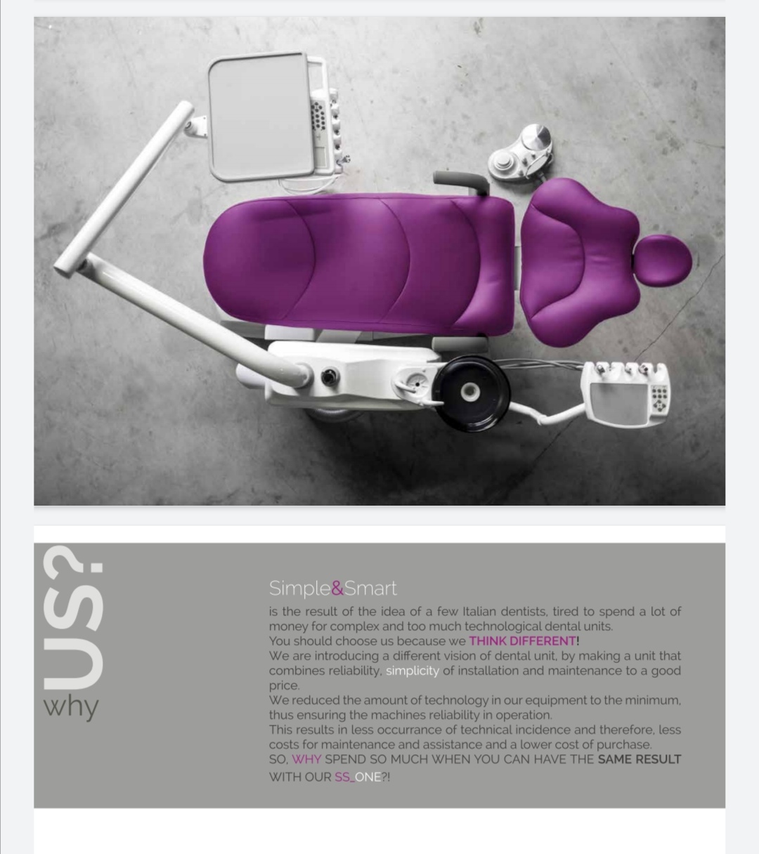 DENTAL TREATMENT UNIT Model Italy
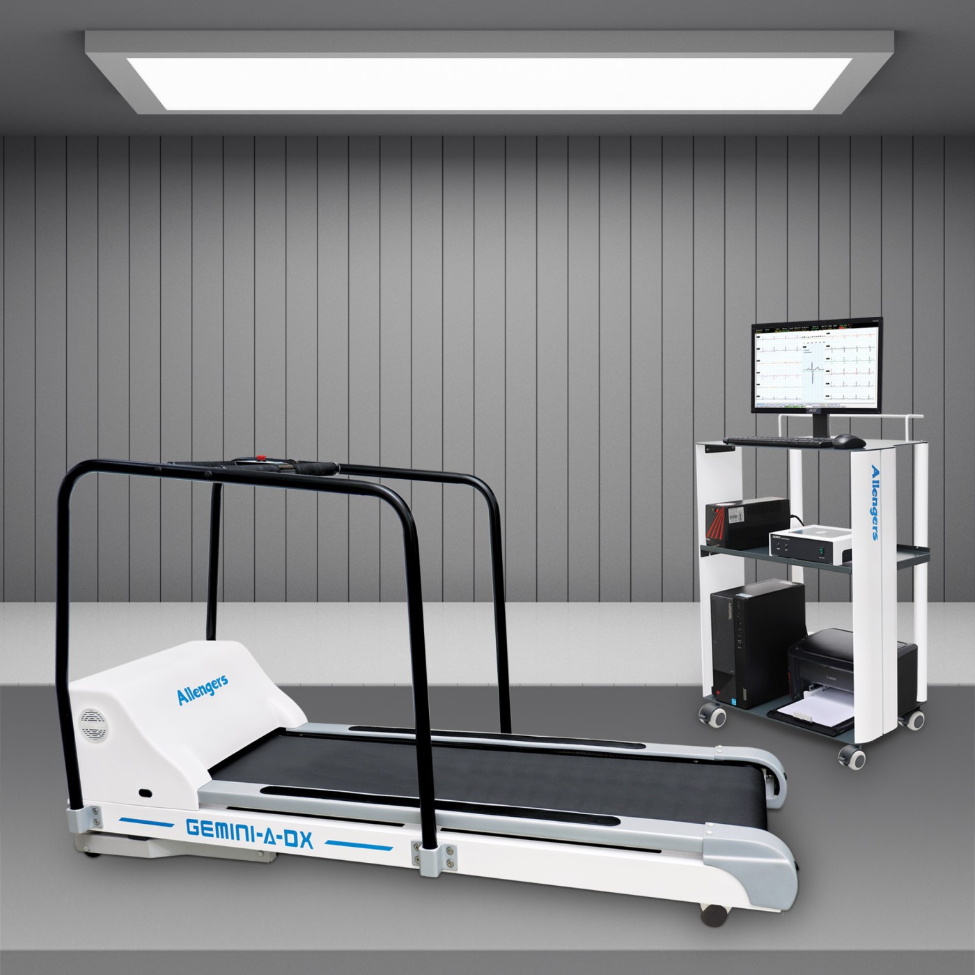 TREADMILL
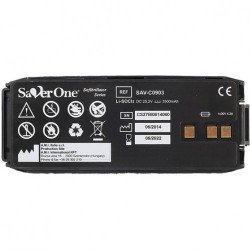 AED battery savor one

